2. A 23 Years old man was admitted to the
emergency department because of severe
abdominal pain located in the right upper
quadrant & beginning 4 hours earlier.
3. Routine lab exam showed the presence of
plenty of erythrocytes in the urine.
The initial diagnosis was a stone in the right ureter.
4. Abdominal ultrasound performed in the
evenings 1 hour after admission, showed the
presence of cystic mass measuring 8 cm located
in the right renal pelvis with dilatation of
the calyceal cavities around this mass.
5. Ultrasound of the right kidney – 1
Large cystic mass in the right upper quadrant
6. Cystic mass of 8 cm located in the right renal pelvis
Ultrasound of the right kidney – 2
7. Ultrasound of the right kidney – 3
Large cystic mass in right renal pelvis
Dilatation of calyceal cavities around the mass
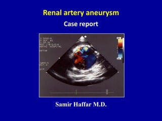
8. Color Doppler US of the cystic mass
Swirling multidirectional flow inside the cystic mass
9. Pulsed Doppler US of the cystic mass
Forward and backward flow inside the cystic mass
10. Communication of cystic mass with RRA
Color Doppler USBlack & white US
Communication of right renal artery with the cystic mass
12. The study of the right renal artery with color
Doppler US clearly showed the communication
of this cystic mass with the right renal artery
and the jet inside it.
13. The diagnosis of right renal artery
aneurysm with threaten rupture was made.
The urologist was informed by telephone
& renal arteriogram was planned for the next day.
14. At 5.00 AM the next day, the patient was in choc.
An urgent laparotomy was done.
The diagnostic was confirmed at operation.
The surgeon was obliged to performed a right
nephrectomy
15. Renal artery aneurysm
Age Most are found in pts 50 - 70 years of age.
Causes Atherosclerosis & fibromuscular disease.
Number 30 % multiple aneurysms in the same side
20% of aneurysms are bilateral
Symptoms Most discovered incidentally
Few pts have symptoms due to aneurysm
Hypertension is the most common symptom
Zubarev AV. Eur Radiol 2001 ; 11 : 1902 – 1915.
16. Location Along the course of the main RA
Color Doppler Effective non-invasive means of dg.
Outpoushing containing color flow
Complications Rupture
Thrombosis
Dissection
Embolization
Renal artery aneurysm
17. Treatment of RAA
Indications Clinical symptoms: back pain – hematuria – HTN
Renal dysfunction: peripheral thromboembolism
Size of aneurysm
Pregnancy
Methods Surgical resection
Percutaneous interventions
Arterial embolization
Expandable stent-graft for fusiform aneurysms
Adopted more frequently Less invasive
Lower cost
Follow-up evaluation
Shonai T et al. J Ultrasound Med 2000 ; 19 : 277 – 280.
18. Treatment of ruptured RAA
Surgery
Midline approach
Supraceliac aortic control (juxtarenal hematoma)
If proximal control of RA obtained: supraceliac clamp removed
Salvaging of kidney Bleeding controlled
Patient hemodynamically stable
Quick bypass of proximal & distal RA
Nephrectomy Required most often
Patient instable
Prolonged ischemia of kidney
Aneurysm extends into renal parenchyma
Rutherford RB – Vascular Surgery – Fifth edition – WB Saunders 2000