Contenu connexe
Similaire à Kumar-Ricker-Poster-mesa_2013_V2
Similaire à Kumar-Ricker-Poster-mesa_2013_V2 (20)
Kumar-Ricker-Poster-mesa_2013_V2
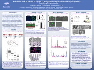
- 1. RESEARCH POSTER PRESENTATION DESIGN © 2012
www.PosterPresentations.com
Embryonic stem cells (ESCs) possess the capacity to differentiate
into multiple cell types and to proliferate indefinitely. Locus-specific
modifications of histones participate in the epigenetic regulation of
genes involved in cell growth, differentiation and cell fate. Histone
H3 lysine 18 acetylation (H3K18ac) plays a major role in cell cycle as
it is abundant in growth-arrested cells and lost in cancer3,4,5.
Furthermore, in growth-arrested cells, depletion of H3K18ac is
sufficient to trigger re-entry into the cell cycle1,2. The H3K18ac-
modification is produced by at least four lysine acetyltransferases
including p300, CBP, PCAF and GCN5. Genetic studies targeting
individual lysine acetyltransferases in mouse indicate redundancy
between these enzymes, e.g. p300 and CBP and possible roles in
early development, e.g. Gcn5 in mesoderm formation. These studies
and others suggest that through H3K18ac, these enzymes may play a
major role in the epigenetic control of cell identity.
In this study, we found that H3K18ac is highly enriched at stem
cell determining loci in ESC and investigate the developmental
consequence of H3K18ac dependency on ESC identity and
pluripotency.
RESULTS
RESULTS (continued) RESULTS (continued)
REFERENCES
ACKNOWLEDGEMENTS
This work was supported by generous grants from the California
Institute for Regenerative Medicine RN2-00908, and institutional funds
to B.D.Y..
Division of Dermatology, Department of Medicine, Stem Cell Program, and Institute for Genomic Medicine,
University of California, San Diego, La Jolla, CA 92093, USA.
Shantanu Kumar, Craig Ricker and Benjamin D. Yu
Functional role of histone H3 lysine 18 acetylation in the maintenance of pluripotency
in embryonic stem cells
CONCLUSION
We demonstrated that upon global reduction of H3K18ac by E1A
oncoprotein, mESCs lose characteristics associated with
pluripotency and fail to activate enhancers that are associated with
stem cell identity. Lineage markers of neuroectoderm and
endoderm were also suppressed by E1A suggesting a functional
role of H3K18ac in lineage specifications. Loss of pluripotency by
E1A requires binding to H3K18 acetyltransferases, P300/CBP, but
does not require pRB- and P400-family interaction.
Fig. 3. Loss of pluripotency in mESCs expressing E1A
(A), Bright field image of mES cells infected with control Ad and Ad E1A
(B), Alkaline phosphatase (AP) staining of mESCs infected with control and Ad E1A
(C), Immunofluorescence for Oct3/4 and E1a expression in MESCs 72h post
transduction. Oct3/4, red; E1a, green
(D), RT-qPCR measuring mRNA levels of pluripotency markers in MESCs expressing
E1A relative to control. Data are represented as mean ± SD
Fig. 5. Change in expression of differentiation markers in
mESCs cells expressing E1A
(A), RT-qPCR measuring mRNA levels of differentiation markers in MESCs
expressing E1a relative to control at 24, 48 and 72 hr post transduction.
Data are represented as mean ± SD
C
E1A
C
E1A
E1A Protein Levels
Cellcounts
0.9 1.2 0.6 0.9 1.0 0.4 1.3 1.2 0.7
H3K18ac
Total H3
24h 48h 72h
Ratio of H3K18ac / Total H3
Ad Control Ad E1A
Loss of pluripotency in mESCs expressing E1A
Figure 1. H3K18ac occupancy in hESCs and derived germ layers.
(A) ChIP-seq occupancy for H3K18ac over POU5F1, SOX2, KLF4 and NANOG
(B) Distribution of ChIP-seq signal near TSS comparing global analysis of ESC
Specific (Top 2% H1 ESC vs. H1 EB) and differentiation specific genes (Bottom 2%).
(C) Box-plot for differences in H3K18ac ChIP-seq density in hESCs and derived
germ layers comparing ESC genes. We found decreasing enrichment of the ESC
related genes in progressively more differentiated cell types. P-values (<2.2e-16),
calculated using two-tailed t-test. ChIP-seq data based on Xie et al. (2013)
Figure 2. Depletion of H3K18ac by E1A oncoprotein in mESCs.
(A) Immunofluorescence for E1a expression in mES cells 24h post transduction.
(B), Flow cytometry analysis for E1a expression.
(C), Western blot analysis for H3K18ac expression at various time point post Ad
control, Ad R2G(Ad mutant does not bind p300/CBP) and Ad E1A transduction
(D), CHIP-qRT PCR for H3K18ac occupancy on pluripotency genes promoters.
Depletion of H3K18ac by Adenovirus E1A oncoprotein
H3K18Ac occupancy in hESCs and derived germ layers
Loss of enhancer activity activity in mESCs expressing E1A
Figure 4. Loss of enhancer reporter in mESCs expressing E1A
(A), mESCs expressing EOS-GFP reporter and loss of GFP expression after
differentiation
(B), Fluorescence and Bright field image of mESCs-EOS reporter cell line transduced
with control and Ad E1a 72h post transduction.
0
1
2
3
4
5
6
7
Pax6 Notch BraT PECAM1 Gata4 Foxa2 Gata6
Control-24h
Control-48
Control-72h
E1a-24h
E1a-48h
E1A-72h
Effect of E1A on lineage differentiation
1. Horwitz, G.A. et al. Adenovirus small e1a alters global patterns of
histone modification. Science 321, 1084-1085 (2008).
2. Ferrari, R. et al. Epigenetic reprogramming by adenovirus e1a.
Science 321, 1086-1088 (2008).
3. Barber, M.F. et al. SIRT7 links H3K18 deacetylation to
maintenance of oncogenic transformation. Nature 487, 114-118
(2012).
4. Seligson, D.B. et al. Global histone modification patterns predict
risk of prostate cancer recurrence. Nature 435, 1262-1266 (2005).
5. Pasqualucci L. et al. Inactivating mutations of acetyltransferases
genes in B-cell lymphoma. Nature 471, 189-95 (2011)
6. Xie W.et al. Epigenomic analysis of multilineage differentiation of
human embryonic stem cells. Cell 153, 1134-48 (2013)
INTRODUCTION
EOS-GFP -LIF (5D) -LIF +RA (4D)
Control E1A
0
1
2
3
4
5
6
Bra Gsc Evx1 Eomes fgf-8 cdx2
FoldchangeFoldchange
CR4
RESULTS (continued)
H1
ESC
H1-derived
mesendoderm
(ME)
H1-derived
neural
(NE)
H1-derived
mesoderm
(MES)
*
*
B
C
A
A B
C
D
A
B
Fig. 6. Loss of pluripotency requires E1A to bind p300/CBP
(A) Schematics of E1A and their mutants.
(B) Flow cytometer analysis of E1A and their mutants expression in mESCs
(C) phase contrasts image of mESCs expressing E1A and their mutants, 72h post
transduction.
Loss of pluripotency requires E1A to bind p300/CBP
A
A B
C
Top 2%
Bot 2%
All
T Gsc Evx1 Eomes Fgf8 Cdx2