SlideShare une entreprise Scribd logo
1  sur  49
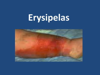
Erysipelas
Definition
Erysipelas is a superficial bacterial
skin infection that is characteristically
extends into cutaneous lymphatics .
It was referred to as :
 Saint Anthony's Fire (= ergotism or
erysipelas or Herpes zoster )
Pathophysiology
      •Bacterial
 inoculation into an
 area of skin trauma
is the initial event in
     developing
     erysipelas.
Pathophysiology
 The source of the bacteria
 in facial erysipelas is often
 the host's naso-
 pharynx, and a history of
 recent streptococcal
 pharyngitis has been
 reported in up to one third
 of cases.
Pathophysiology
Lymphoscintigraphy in patients
with a first-time episode of
lower extremity erysipelas has
documented lymphatic
impairment in both affected
and non affected legs.
regional lymph node
    swelling and
     tenderness
Causative organism
* Streptococci are the primary
cause of erysipelas.
* Most facial infections are
attributed to group A
streptococci,
*lower extremity infections
being caused by non–group A
streptococci.
causes
•Streptococcal toxins are thought to
contribute to the brisk inflammation
that is pathognomonic of this infection.

*No clear proof has emerged that other
bacteria cause typical erysipelas,
although they clearly coexist with
streptococci at sites of inoculation.
causes
Recently, atypical forms reported to be
caused by :
 * Streptococcus pneumoniae,
 *Klebsiella pneumoniae,
 * Haemophilus influenzae,
 *Yersinia enterocolitica,
 *Moraxella species,
they should be considered in cases refractory
to standard antibiotic therapy.
Race
Erysipelas infections affect persons of all
races.
                     Sex
•Erysipelas is common in females.
• at an earlier age it is more in males ( more
activities).
• However predisposing factors, rather than
gender, account for any male/female
differences in incidence.
Age
 All age groups are susiptable.
The peak incidence at 60-80 years
old, especially in patients :
= At high-risk .
= immuno-compromised .
= those with lymphatic drainage
problems (eg, after mastectomy, pelvic
surgery, bypass grafting).
Clinical
symptoms
Prodromal symptoms :
         malaise.
         chills.
         high fever.
often begin before the onset of the skin
lesions and usually are present within 48
hours of cutaneous involvement..
symptoms
      Pruritus .
      burning .
      tenderness.
are typical complaints
Physical
Erysipelas begins
     as a small
   erythematous
     patch that
  progresses to a
       fiery-
  red, indurated
, tense, and shiny
      plaque.
The lesion classically
exhibits raised
sharply demarcated
advancing margins.
     Local signs of
    inflammation
       warmth,
        edema,
      tenderness
 are universal.
Lymphatic
involvement often
 is manifested by
   overlying skin
   streaking and
      regional
lymphadenopathy
More severe
  infections may
 exhibit numerous
vesicles and bullae
    along with
  petechiae and
    even frank
     necrosis.
Erysipelas
    of
 the face
Erysipelas
    Of
  the leg
Erysipelas
   the
 buttock
Erysipelas
     of
the ear Pena
Differential Diagnoses
1) Erythema Annulare Centri-
fugum
2) Stasis Dermatitis
3) Cellulitis
4) Erysipeloid
Erythema Annulare Centrifugum
* Eruptions occur at any
age.
 * begins as small raised
pink-red spot that slowly
enlarges and forms a ring
shape while the central
area flattens and clears.
There may be an inner
rim of scale.
Erythema Annulare Centrifugum

Lesions most often
appear on the
thighs, legs, face, trunk
and arms.
linked to underlying
diseases , viral
, bacterial or even
tumor.
Stasis
Dermatitis
cellulitis
Erysipeloid
 * acute bacterial infection of traumatized skin.
 * caused by Erysipelothrix rhusiopathiae
(gram positive rod-shaped bacterium), which
cause animal and human infections.
 * Direct contact between infected meat and
traumatized human skin results in Erysipeloid.
•more common among
farmers, butchers, cooks, homemakers.
 * Lesions most commonly affect the hands.
Erysipeloid
 * Lesions consist of
         well-
 demarcated, bright
red-to-purple plaques
 with a smooth, shiny
       surface.
  •Lesions are warm
     and tender.
Laboratory Studies
Laboratory Studies
* In classic erysipelas, no
laboratory workup is required
for diagnosis or treatment.
* Cultures are best reserved for
immunocopromized patient in
whom an atypical etiologic agent
is suspected.
Imaging Studies
Imaging studies are not usually
indicated and are of low yield.
MRI and bone scintigraphy are
helpful when early osteoarticular
involvement is suspected.
 In this setting, standard
radiographic findings typically are
normal.
Histological Findings
The histological hallmarks of erysipelas are
*marked dermal edema,
*vascular dilatation,
*streptococcal invasion of lymphatics & tissues.

This bacterial invasion results in a dermal
inflammatory infiltrate consisting of
neutrophils and mononuclear cells.
 The epidermis is often secondarily involved.
Rarely, bacterial invasion of local blood vessels
may be seen.
Treatment
Hospitalization for close monitoring
and IV. antibiotics is recommended
for :
1) severe cases.
2) infants.
3) elderly patients.
4) patients who are immune-
  compromised.
Medical Care
* Elevation and rest of the affected
limb are recommended to reduce
local swelling, inflammation, and pain.
* Saline wet dressings should be
applied to ulcerated and necrotic
lesions and changed every 2-12 hours,
depending on the severity of the
infection.
Medical Care

* penicillin has remained first-line therapy.
 administered orally or IM. for 10-20 days.
Dosing : Adult
Penicillin G procaine: 0.6-1.2 million U IM bid for 10 d
Penicillin VK: 250-500 mg PO qid for 10-14 d
Pediatric : Penicillin G procaine: <30 kg: 300,000 U/d
                             >30 kg: Administer as in adults
            Penicillin VK:
<12 years: 25-50 mg/kg/d PO divided tid/qid;
            not to exceed 3 g/d
>12 years: Administer as in adults
Medical Care
*A first-generation cephalosporin or
macrolide, such as erythromycin or
azithromycin, may be used if the patient has
an allergy to penicillin.
Dosing
Adult
250-500 mg PO qid for 10 d
Pediatric
30-50 mg/kg/d (15-25 mg/lb/d) PO divided
q6-8h; double dose for severe infection.
•Two new drugs:
• roxithromycin & pristinamycin,
 have been reported to be extremely
effective in the treatment of erysipelas.
* Several studies have demonstrated greater
efficacy and fewer adverse effects with
these drugs compared with penicillin.
*Currently, FDA has not approved these

drugs in the United States, but they are in
use in Europe.
Recurrent
erysipelas
Recurrent erysipelas
Patients with recurrent erysipelas
should be educated regarding :
•local antisepstic .
•general wound care.
•Predisposing lower extremity skin
lesions (eg , tineapedis , toe
web intertrigo , stasis ulcers) should
be treated aggressively to prevent
super-infection.
Recurrent erysipelas
•Long-term prophylactic antibiotic therapy
generally is accepted, but no true guidelines are
available.
•Treatment regimens should be tailored to the
patient.

•One reported regimen is benzathine
penicillin G at 2.4 MU IM. every 3 weeks
for up to 2 years . Two-week intervals
have also been used.
Surgical Care
Debridement is necessary
only in severe infections
with necrosis or gangrene.
Complications
Complications
1) The most common
complications of erysipelas are :
* abscess,
* gangrene,
* Thrombophlebitis .
Complications
2) Less common complications
  (<1%) are :
   *acute glomerulonephritis ,
   *endocarditis ,
   *septicemia,
   *streptococcal toxic shock
  syndrome.
Prognosis
* The prognosis for patients
with erysipelas is excellent.
* local recurrence has been
reported in up to 20% of
patients with predisposing
conditions

Contenu connexe

Tendances (20)

Carbuncle
CarbuncleCarbuncle
Carbuncle
 
Skin And Soft Tissue Infections
Skin And Soft Tissue InfectionsSkin And Soft Tissue Infections
Skin And Soft Tissue Infections
 
Ulcer
UlcerUlcer
Ulcer
 
Urticaria
UrticariaUrticaria
Urticaria
 
Complications of malaria
Complications of malariaComplications of malaria
Complications of malaria
 
Paraproctitis
ParaproctitisParaproctitis
Paraproctitis
 
Paronychia
ParonychiaParonychia
Paronychia
 
Bullous diseases
Bullous diseasesBullous diseases
Bullous diseases
 
Warts
WartsWarts
Warts
 
Lichen planus
Lichen planusLichen planus
Lichen planus
 
Hydatid cyst
Hydatid cystHydatid cyst
Hydatid cyst
 
Warts (Verruca) by Dr. Aryan
Warts (Verruca) by Dr. AryanWarts (Verruca) by Dr. Aryan
Warts (Verruca) by Dr. Aryan
 
Varicose vein
Varicose veinVaricose vein
Varicose vein
 
Achalasia cardia
Achalasia cardiaAchalasia cardia
Achalasia cardia
 
15. Chancroid
15. Chancroid15. Chancroid
15. Chancroid
 
Basal cell carcinoma
Basal cell carcinomaBasal cell carcinoma
Basal cell carcinoma
 
Fournier’s gangrene- Surgery
Fournier’s gangrene- SurgeryFournier’s gangrene- Surgery
Fournier’s gangrene- Surgery
 
Cholecystitis
CholecystitisCholecystitis
Cholecystitis
 
Scarlet Fever
Scarlet  FeverScarlet  Fever
Scarlet Fever
 
Gangrene
GangreneGangrene
Gangrene
 

En vedette

Bacterial skin infection jaber
Bacterial skin infection  jaberBacterial skin infection  jaber
Bacterial skin infection jaberJaber Manasia
 
Bacterial infection of the skin
Bacterial infection of the skin Bacterial infection of the skin
Bacterial infection of the skin MEEQAT HOSPITAL
 
Bacterial & Viral Diseases Of The Skin,Mucosa,Eyes
Bacterial & Viral Diseases Of The Skin,Mucosa,EyesBacterial & Viral Diseases Of The Skin,Mucosa,Eyes
Bacterial & Viral Diseases Of The Skin,Mucosa,Eyes000 07
 
Clinical applications of kajjali
Clinical  applications  of kajjaliClinical  applications  of kajjali
Clinical applications of kajjalisomil dubey
 
Cat scratch disease
Cat scratch diseaseCat scratch disease
Cat scratch diseaseahmed esmail
 
Cellulitis And Erysples
Cellulitis And ErysplesCellulitis And Erysples
Cellulitis And Erysplesmohammed sediq
 
Prepatation of arka lavana
Prepatation of arka lavanaPrepatation of arka lavana
Prepatation of arka lavanasomil dubey
 
GEMC- Cutaneous Emergency- Resident Training
GEMC- Cutaneous Emergency- Resident TrainingGEMC- Cutaneous Emergency- Resident Training
GEMC- Cutaneous Emergency- Resident TrainingOpen.Michigan
 
Cat scratch disease
Cat scratch diseaseCat scratch disease
Cat scratch diseaseImad Zoukar
 
Diaper dermatitis
Diaper dermatitisDiaper dermatitis
Diaper dermatitisChandan N
 

En vedette (20)

Erysipelas
ErysipelasErysipelas
Erysipelas
 
Bacterial skin infection jaber
Bacterial skin infection  jaberBacterial skin infection  jaber
Bacterial skin infection jaber
 
Felon
FelonFelon
Felon
 
Bacterial infection of the skin
Bacterial infection of the skin Bacterial infection of the skin
Bacterial infection of the skin
 
Cellulitis
CellulitisCellulitis
Cellulitis
 
Tetanus
TetanusTetanus
Tetanus
 
Bacterial & Viral Diseases Of The Skin,Mucosa,Eyes
Bacterial & Viral Diseases Of The Skin,Mucosa,EyesBacterial & Viral Diseases Of The Skin,Mucosa,Eyes
Bacterial & Viral Diseases Of The Skin,Mucosa,Eyes
 
Tonsillitis
TonsillitisTonsillitis
Tonsillitis
 
Tonsillitis
TonsillitisTonsillitis
Tonsillitis
 
rabies ppt
rabies pptrabies ppt
rabies ppt
 
Economics Assignment Help
Economics Assignment HelpEconomics Assignment Help
Economics Assignment Help
 
Clinical applications of kajjali
Clinical  applications  of kajjaliClinical  applications  of kajjali
Clinical applications of kajjali
 
Cat scratch disease
Cat scratch diseaseCat scratch disease
Cat scratch disease
 
Cgd 2
Cgd 2Cgd 2
Cgd 2
 
Cellulitis And Erysples
Cellulitis And ErysplesCellulitis And Erysples
Cellulitis And Erysples
 
Prepatation of arka lavana
Prepatation of arka lavanaPrepatation of arka lavana
Prepatation of arka lavana
 
GEMC- Cutaneous Emergency- Resident Training
GEMC- Cutaneous Emergency- Resident TrainingGEMC- Cutaneous Emergency- Resident Training
GEMC- Cutaneous Emergency- Resident Training
 
Anthrax 3
Anthrax 3Anthrax 3
Anthrax 3
 
Cat scratch disease
Cat scratch diseaseCat scratch disease
Cat scratch disease
 
Diaper dermatitis
Diaper dermatitisDiaper dermatitis
Diaper dermatitis
 

Similaire à Erysipelas

Erysipelas jainish patel
Erysipelas  jainish patelErysipelas  jainish patel
Erysipelas jainish patelVasyl Sorokhan
 
erysipelas-jainishpatel-121123045440-phpapp02.pdf
erysipelas-jainishpatel-121123045440-phpapp02.pdferysipelas-jainishpatel-121123045440-phpapp02.pdf
erysipelas-jainishpatel-121123045440-phpapp02.pdffatemehbemana1
 
ERYSIPELAS.pptx
ERYSIPELAS.pptxERYSIPELAS.pptx
ERYSIPELAS.pptxMinella4
 
dermatology.Bact .inf 5th.(dr.ali)
dermatology.Bact .inf 5th.(dr.ali)dermatology.Bact .inf 5th.(dr.ali)
dermatology.Bact .inf 5th.(dr.ali)student
 
dermatology.1 eryth, telan, urt & hshp(dr.faraydwn)
dermatology.1 eryth, telan, urt & hshp(dr.faraydwn)dermatology.1 eryth, telan, urt & hshp(dr.faraydwn)
dermatology.1 eryth, telan, urt & hshp(dr.faraydwn)student
 
disorder of skin viji.pptx
disorder of skin viji.pptxdisorder of skin viji.pptx
disorder of skin viji.pptxVijiM14
 
SOFT TISSUE abscess and other....................
SOFT TISSUE abscess and other....................SOFT TISSUE abscess and other....................
SOFT TISSUE abscess and other....................fathyabomuch
 
Bacterial , viral, parasitic infections
Bacterial , viral, parasitic infectionsBacterial , viral, parasitic infections
Bacterial , viral, parasitic infectionsMustafa Al Mously
 
challenge rash
 challenge rash challenge rash
challenge rashEM OMSB
 
10. other important dermatoses
10. other important dermatoses10. other important dermatoses
10. other important dermatosesdthewitt
 
Oral manifestations of infectious diseases in children
Oral manifestations of infectious diseases in childrenOral manifestations of infectious diseases in children
Oral manifestations of infectious diseases in childrenRasha Adel
 
Papulosequamous disorder
Papulosequamous disorder Papulosequamous disorder
Papulosequamous disorder ssuser9127b3
 
SURGICAL INFECTIONS.pptx
SURGICAL INFECTIONS.pptxSURGICAL INFECTIONS.pptx
SURGICAL INFECTIONS.pptxKhansamohamed2
 
common skin condition in children..pptx
common skin condition in children..pptxcommon skin condition in children..pptx
common skin condition in children..pptxMohanapriyaRajendran6
 

Similaire à Erysipelas (20)

Erysipelas jainish patel
Erysipelas  jainish patelErysipelas  jainish patel
Erysipelas jainish patel
 
erysipelas-jainishpatel-121123045440-phpapp02.pdf
erysipelas-jainishpatel-121123045440-phpapp02.pdferysipelas-jainishpatel-121123045440-phpapp02.pdf
erysipelas-jainishpatel-121123045440-phpapp02.pdf
 
ERYSIPELAS.pptx
ERYSIPELAS.pptxERYSIPELAS.pptx
ERYSIPELAS.pptx
 
dermatology.Bact .inf 5th.(dr.ali)
dermatology.Bact .inf 5th.(dr.ali)dermatology.Bact .inf 5th.(dr.ali)
dermatology.Bact .inf 5th.(dr.ali)
 
Dermatology 5th year, 5ht lecture (Dr. Ali El-Ethawi)
Dermatology 5th year, 5ht lecture (Dr. Ali El-Ethawi)Dermatology 5th year, 5ht lecture (Dr. Ali El-Ethawi)
Dermatology 5th year, 5ht lecture (Dr. Ali El-Ethawi)
 
dermatology.1 eryth, telan, urt & hshp(dr.faraydwn)
dermatology.1 eryth, telan, urt & hshp(dr.faraydwn)dermatology.1 eryth, telan, urt & hshp(dr.faraydwn)
dermatology.1 eryth, telan, urt & hshp(dr.faraydwn)
 
dr.farydon DERMATOLOGY
dr.farydon     DERMATOLOGYdr.farydon     DERMATOLOGY
dr.farydon DERMATOLOGY
 
disorder of skin viji.pptx
disorder of skin viji.pptxdisorder of skin viji.pptx
disorder of skin viji.pptx
 
Skin and Soft Tissue Infections
Skin and Soft Tissue InfectionsSkin and Soft Tissue Infections
Skin and Soft Tissue Infections
 
SOFT TISSUE abscess and other....................
SOFT TISSUE abscess and other....................SOFT TISSUE abscess and other....................
SOFT TISSUE abscess and other....................
 
Bacterial , viral, parasitic infections
Bacterial , viral, parasitic infectionsBacterial , viral, parasitic infections
Bacterial , viral, parasitic infections
 
challenge rash
 challenge rash challenge rash
challenge rash
 
10. other important dermatoses
10. other important dermatoses10. other important dermatoses
10. other important dermatoses
 
Oral manifestations of infectious diseases in children
Oral manifestations of infectious diseases in childrenOral manifestations of infectious diseases in children
Oral manifestations of infectious diseases in children
 
Surgical infections
Surgical infectionsSurgical infections
Surgical infections
 
lecture5.pptx
lecture5.pptxlecture5.pptx
lecture5.pptx
 
Papulosequamous disorder
Papulosequamous disorder Papulosequamous disorder
Papulosequamous disorder
 
SURGICAL INFECTIONS.pptx
SURGICAL INFECTIONS.pptxSURGICAL INFECTIONS.pptx
SURGICAL INFECTIONS.pptx
 
Pyodermas.pptx
Pyodermas.pptxPyodermas.pptx
Pyodermas.pptx
 
common skin condition in children..pptx
common skin condition in children..pptxcommon skin condition in children..pptx
common skin condition in children..pptx
 

Plus de Vasyl Sorokhan (20)

Leishmaniasis
LeishmaniasisLeishmaniasis
Leishmaniasis
 
Infectious mononucleosis
Infectious mononucleosisInfectious mononucleosis
Infectious mononucleosis
 
TORCH
TORCHTORCH
TORCH
 
Shigellosis
ShigellosisShigellosis
Shigellosis
 
Botulism
BotulismBotulism
Botulism
 
Lassa fever
Lassa feverLassa fever
Lassa fever
 
Malaria
MalariaMalaria
Malaria
 
Lecture brucellosis 5
Lecture   brucellosis 5Lecture   brucellosis 5
Lecture brucellosis 5
 
Lecture nematodes
Lecture   nematodesLecture   nematodes
Lecture nematodes
 
Lecture 15. hemorrhagic fevers
Lecture 15. hemorrhagic feversLecture 15. hemorrhagic fevers
Lecture 15. hemorrhagic fevers
 
Lecture 14. aids
Lecture 14. aidsLecture 14. aids
Lecture 14. aids
 
Lecture 13.vhbcd
Lecture 13.vhbcdLecture 13.vhbcd
Lecture 13.vhbcd
 
Lecture 12.vha vhe
Lecture 12.vha vheLecture 12.vha vhe
Lecture 12.vha vhe
 
Lecture 11. meningitis
Lecture 11. meningitisLecture 11. meningitis
Lecture 11. meningitis
 
Lecture 10. diphtheria
Lecture 10. diphtheriaLecture 10. diphtheria
Lecture 10. diphtheria
 
Lecture 9. hospital aquired infection
Lecture 9. hospital aquired infectionLecture 9. hospital aquired infection
Lecture 9. hospital aquired infection
 
Lecture 8. anthrex, plague
Lecture 8. anthrex, plagueLecture 8. anthrex, plague
Lecture 8. anthrex, plague
 
Lecture 7. influenza
Lecture 7. influenzaLecture 7. influenza
Lecture 7. influenza
 
Lecture 6. cholera
Lecture 6. choleraLecture 6. cholera
Lecture 6. cholera
 
Lecture 5. typhoid fever 3
Lecture 5. typhoid fever 3Lecture 5. typhoid fever 3
Lecture 5. typhoid fever 3
 

Erysipelas

  • 2. Definition Erysipelas is a superficial bacterial skin infection that is characteristically extends into cutaneous lymphatics . It was referred to as : Saint Anthony's Fire (= ergotism or erysipelas or Herpes zoster )
  • 3. Pathophysiology •Bacterial inoculation into an area of skin trauma is the initial event in developing erysipelas.
  • 4. Pathophysiology The source of the bacteria in facial erysipelas is often the host's naso- pharynx, and a history of recent streptococcal pharyngitis has been reported in up to one third of cases.
  • 5. Pathophysiology Lymphoscintigraphy in patients with a first-time episode of lower extremity erysipelas has documented lymphatic impairment in both affected and non affected legs.
  • 6. regional lymph node swelling and tenderness
  • 7. Causative organism * Streptococci are the primary cause of erysipelas. * Most facial infections are attributed to group A streptococci, *lower extremity infections being caused by non–group A streptococci.
  • 8. causes •Streptococcal toxins are thought to contribute to the brisk inflammation that is pathognomonic of this infection. *No clear proof has emerged that other bacteria cause typical erysipelas, although they clearly coexist with streptococci at sites of inoculation.
  • 9. causes Recently, atypical forms reported to be caused by : * Streptococcus pneumoniae, *Klebsiella pneumoniae, * Haemophilus influenzae, *Yersinia enterocolitica, *Moraxella species, they should be considered in cases refractory to standard antibiotic therapy.
  • 10. Race Erysipelas infections affect persons of all races. Sex •Erysipelas is common in females. • at an earlier age it is more in males ( more activities). • However predisposing factors, rather than gender, account for any male/female differences in incidence.
  • 11. Age All age groups are susiptable. The peak incidence at 60-80 years old, especially in patients : = At high-risk . = immuno-compromised . = those with lymphatic drainage problems (eg, after mastectomy, pelvic surgery, bypass grafting).
  • 13. symptoms Prodromal symptoms : malaise. chills. high fever. often begin before the onset of the skin lesions and usually are present within 48 hours of cutaneous involvement..
  • 14. symptoms Pruritus . burning . tenderness. are typical complaints
  • 16. Erysipelas begins as a small erythematous patch that progresses to a fiery- red, indurated , tense, and shiny plaque.
  • 17. The lesion classically exhibits raised sharply demarcated advancing margins. Local signs of inflammation warmth, edema, tenderness are universal.
  • 18. Lymphatic involvement often is manifested by overlying skin streaking and regional lymphadenopathy
  • 19. More severe infections may exhibit numerous vesicles and bullae along with petechiae and even frank necrosis.
  • 20. Erysipelas of the face
  • 21. Erysipelas Of the leg
  • 22. Erysipelas the buttock
  • 23. Erysipelas of the ear Pena
  • 25. Differential Diagnoses 1) Erythema Annulare Centri- fugum 2) Stasis Dermatitis 3) Cellulitis 4) Erysipeloid
  • 26. Erythema Annulare Centrifugum * Eruptions occur at any age. * begins as small raised pink-red spot that slowly enlarges and forms a ring shape while the central area flattens and clears. There may be an inner rim of scale.
  • 27. Erythema Annulare Centrifugum Lesions most often appear on the thighs, legs, face, trunk and arms. linked to underlying diseases , viral , bacterial or even tumor.
  • 30. Erysipeloid * acute bacterial infection of traumatized skin. * caused by Erysipelothrix rhusiopathiae (gram positive rod-shaped bacterium), which cause animal and human infections. * Direct contact between infected meat and traumatized human skin results in Erysipeloid. •more common among farmers, butchers, cooks, homemakers. * Lesions most commonly affect the hands.
  • 31. Erysipeloid * Lesions consist of well- demarcated, bright red-to-purple plaques with a smooth, shiny surface. •Lesions are warm and tender.
  • 33. Laboratory Studies * In classic erysipelas, no laboratory workup is required for diagnosis or treatment. * Cultures are best reserved for immunocopromized patient in whom an atypical etiologic agent is suspected.
  • 34. Imaging Studies Imaging studies are not usually indicated and are of low yield. MRI and bone scintigraphy are helpful when early osteoarticular involvement is suspected. In this setting, standard radiographic findings typically are normal.
  • 35. Histological Findings The histological hallmarks of erysipelas are *marked dermal edema, *vascular dilatation, *streptococcal invasion of lymphatics & tissues. This bacterial invasion results in a dermal inflammatory infiltrate consisting of neutrophils and mononuclear cells. The epidermis is often secondarily involved. Rarely, bacterial invasion of local blood vessels may be seen.
  • 37. Hospitalization for close monitoring and IV. antibiotics is recommended for : 1) severe cases. 2) infants. 3) elderly patients. 4) patients who are immune- compromised.
  • 38. Medical Care * Elevation and rest of the affected limb are recommended to reduce local swelling, inflammation, and pain. * Saline wet dressings should be applied to ulcerated and necrotic lesions and changed every 2-12 hours, depending on the severity of the infection.
  • 39. Medical Care * penicillin has remained first-line therapy. administered orally or IM. for 10-20 days. Dosing : Adult Penicillin G procaine: 0.6-1.2 million U IM bid for 10 d Penicillin VK: 250-500 mg PO qid for 10-14 d Pediatric : Penicillin G procaine: <30 kg: 300,000 U/d >30 kg: Administer as in adults Penicillin VK: <12 years: 25-50 mg/kg/d PO divided tid/qid; not to exceed 3 g/d >12 years: Administer as in adults
  • 40. Medical Care *A first-generation cephalosporin or macrolide, such as erythromycin or azithromycin, may be used if the patient has an allergy to penicillin. Dosing Adult 250-500 mg PO qid for 10 d Pediatric 30-50 mg/kg/d (15-25 mg/lb/d) PO divided q6-8h; double dose for severe infection.
  • 41. •Two new drugs: • roxithromycin & pristinamycin, have been reported to be extremely effective in the treatment of erysipelas. * Several studies have demonstrated greater efficacy and fewer adverse effects with these drugs compared with penicillin. *Currently, FDA has not approved these drugs in the United States, but they are in use in Europe.
  • 43. Recurrent erysipelas Patients with recurrent erysipelas should be educated regarding : •local antisepstic . •general wound care. •Predisposing lower extremity skin lesions (eg , tineapedis , toe web intertrigo , stasis ulcers) should be treated aggressively to prevent super-infection.
  • 44. Recurrent erysipelas •Long-term prophylactic antibiotic therapy generally is accepted, but no true guidelines are available. •Treatment regimens should be tailored to the patient. •One reported regimen is benzathine penicillin G at 2.4 MU IM. every 3 weeks for up to 2 years . Two-week intervals have also been used.
  • 45. Surgical Care Debridement is necessary only in severe infections with necrosis or gangrene.
  • 47. Complications 1) The most common complications of erysipelas are : * abscess, * gangrene, * Thrombophlebitis .
  • 48. Complications 2) Less common complications (<1%) are : *acute glomerulonephritis , *endocarditis , *septicemia, *streptococcal toxic shock syndrome.
  • 49. Prognosis * The prognosis for patients with erysipelas is excellent. * local recurrence has been reported in up to 20% of patients with predisposing conditions