Muscular system
•
0 j'aime•575 vues

For the candidates who are preparing for N.E.E.T UG exams and PUC.

Recommandé
Recommandé
Contenu connexe
Tendances
Tendances (18)
En vedette
En vedette (20)
Similaire à Muscular system
Similaire à Muscular system (20)
Plus de Srinivasreddy Patil
Plus de Srinivasreddy Patil (14)
Dernier
Dernier (20)
Muscular system
- 1. UNIT 5 – HUMAN PHYSIOLOGY MUSCULAR SYSTEM SUB-UNIT CHAPTER – LOCOMOTION AND MOVEMENT DR.SRINIVAS REDDY PATIL M.Sc Zoology (Gold Medalist).,Ph.D (Reproductive Physiology)., M.Ed.,MBA.,PGDBA.,FMSPI Saturday, February 9, 2013 DR.SRINIVASREDDY PATIL'S 1 BIOLOGY
- 2. • Movement is one of the significant features of living beings. Animals and plants exhibit a wide range of movements. Saturday, February 9, 2013 DR.SRINIVASREDDY PATIL'S 2 BIOLOGY
- 3. • Streaming of protoplasm in the unicellular organisms like Amoeba is a simple form of movement. • Movement of cilia, flagella and tentacles are shown by many organisms. Saturday, February 9, 2013 DR.SRINIVASREDDY PATIL'S 3 BIOLOGY
- 4. • Human beings can move limbs, jaws, eyelids, tongue, etc. Some of the movements result in a change of place or location. Such voluntary movements are called locomotion. Saturday, February 9, 2013 DR.SRINIVASREDDY PATIL'S 4 BIOLOGY
- 5. • Walking, running, climbing, flying, swimming are all some forms of locomotory movements. Locomotory structures need not be different from those affecting other types of movements. Saturday, February 9, 2013 DR.SRINIVASREDDY PATIL'S 5 BIOLOGY
- 6. • Paramoecium, cilia helps in the movement of food through cytopharynx and in locomotion as well. • Hydra can use its tentacles for capturing its prey and also use them for locomotion. • Human beings use limbs for changes in body postures and locomotion as well. DR.SRINIVASREDDY PATIL'S Saturday, February 9, 2013 BIOLOGY 6
- 7. • The observation suggest that movement and locomotion go hand in hand for the purpose of study. The two may be linked by stating that all locomotions are movements but all movements are not locomotions. Saturday, February 9, 2013 DR.SRINIVASREDDY PATIL'S 7 BIOLOGY
- 8. • Methods of locomotion performed by animals vary with their habitats and the demand of the situation. Saturday, February 9, 2013 DR.SRINIVASREDDY PATIL'S 8 BIOLOGY
- 9. Chief aim of locomotion is for the search of food, shelter, mate, suitable breeding grounds, favourable climatic conditions, escape from enemies / predators. Saturday, February 9, 2013 DR.SRINIVASREDDY PATIL'S 9 BIOLOGY
- 10. SYNOPSIS • 1. Types of Movement • 2. Muscle • 3. Skeletal System • 4. Joints • 5. Disorders of Muscular and Skeletal System Saturday, February 9, 2013 DR.SRINIVASREDDY PATIL'S 10 BIOLOGY
- 11. • 1. TYPES OF MOVEMENT - Cells of the human body exhibit three main types of movements namely, amoeboid, ciliary and muscular. Saturday, February 9, 2013 DR.SRINIVASREDDY PATIL'S 11 BIOLOGY
- 12. Some specialised cells in our body like • Macrophages and leucocytes in blood exhibit amoeboid movement. • Cytoskeletal elements like microfilaments are also involved in amoeboid movement. Saturday, February 9, 2013 DR.SRINIVASREDDY PATIL'S 12 BIOLOGY
- 13. • Ciliary movement occurs in most of our internal tubular organs which are lined by ciliated epithelium. • The coordinated movements of cilia in the trachea help us in removing dust particles and some of the foreign substances inhaled along with the atmospheric air. • Passage of ova through the female reproductive tract is also facilitated by the ciliary movement. Saturday, February 9, 2013 DR.SRINIVASREDDY PATIL'S 13 BIOLOGY
- 14. • Movement of our limbs, jaws, tongue,require muscular movement. • The contractile property of muscles are effectively used for locomotion and other movements by human beings and majority of multicellular organisms. • Locomotion requires a perfect coordinated activity of muscular, skeletal and neural systems. Saturday, February 9, 2013 DR.SRINIVASREDDY PATIL'S 14 BIOLOGY
- 15. • The types of muscles, their structure, mechanism of their contraction and important aspects of the skeletal system to under stand locomotion and movements. Saturday, February 9, 2013 DR.SRINIVASREDDY PATIL'S 15 BIOLOGY
- 16. • 2. MUSCLE - Muscle is a specialised tissue of mesodermal origin. • About 40-50 per cent of the body weight is contributed by muscles. • They have special properties like excitability, contractility, extensibility and elasticity. Saturday, February 9, 2013 DR.SRINIVASREDDY PATIL'S 16 BIOLOGY
- 17. All muscles are excitable, contractile, elastic and extensible. This means when nerves stimulate the biceps brachii, it contracts, and the opposing triceps brachii relaxes and extends.
- 19. • Muscles have been classified using different criteria, namely location, appearance and nature of regulation of their activities. Saturday, February 9, 2013 DR.SRINIVASREDDY PATIL'S 19 BIOLOGY
- 21. • Based on their location, three types of muscles are identified : (i) Skeletal, (ii) Visceral and (iii) Cardiac. • Skeletal muscles are closely associated with the skeletal components of the body. They have a striped appearance under the microscope and hence are called striated muscles. As their activities are under the voluntary control of the nervous system, they are known as voluntary muscles too. They are primarily involved in locomotory actions and changes of body postures. Saturday, February 9, 2013 DR.SRINIVASREDDY PATIL'S 21 BIOLOGY
- 27. There is extensive blood and nerve supply to the skeletal muscles
- 29. The psoas is a prime hip flexor. It lifts the front leg as we walk and climb. It assists in thigh rotation and adduction, and in a partnership balance with the large abdominal muscle, rectus abdominus, it maintains the natural lumbar curve, pelvic alignment, and a generally upright stance. It is a muscular support for the lumbar plexus, a network of nerves, which originate in the spinal cord.
- 30. Psoas – the postural muscle Helps in the upright standing pose or Tadasana. We lengthen the psoas before deepening into a posture, draws the abdomen, the rectus abdominus muscle inwards. Lengthens the lumbar spine and creates width across the collarbone. In side bending and twisting postures, this helps to prevent back injury, especially to the tiny facet joints of the spine.
- 31. The psoas maintains vertical alignment in inversions and creates length along the front body in forward bends. Yoga is one of the best ways to lengthen and strengthen the psoas muscle.
- 32. • Visceral muscles are located in the inner walls of hollow visceral organs of the body like the alimentary canal, reproductive tract, etc. They do not exhibit any striation and are smooth in appearance. Hence, they are called smooth muscles (nonstriated muscle). Their activities are not under the voluntary control of the nervous system and are therefore known as involuntary muscles. They assist, for example, in the transportation of food through the digestive tract and gametes through the genital tract. Saturday, February 9, 2013 DR.SRINIVASREDDY PATIL'S 32 BIOLOGY
- 33. Smooth Muscle
- 35. • Cardiac muscles are the muscles of heart. Many cardiac muscle cells assemble in a branching pattern to form a cardiac muscle. Based on appearance, cardiac muscles are striated. They are involuntary in nature as the nervous system does not control their activities directly. Saturday, February 9, 2013 DR.SRINIVASREDDY PATIL'S 35 BIOLOGY
- 36. Cardiac Muscle
- 38. • Each skeletal muscle in our body is made of a number of muscle bundles or fascicles held together by a common collagenous connective tissue layer called fascia. • Each muscle bundle contains a number of muscle fibres. Each muscle fibre is lined by the plasma membrane called sarcolemma enclosing the sarcoplasm. Muscle fibre is a syncitium as the sarcoplasm contains many nuclei. Saturday, February 9, 2013 DR.SRINIVASREDDY PATIL'S 38 BIOLOGY
- 41. • The endoplasmic reticulum, i.e., sarcoplasmic reticulum of the muscle fibres is the store house of calcium ions. A characteristic feature of the muscle fibre is the presence of a large number of parallelly arranged filaments in the sarcoplasm called myofilaments or myofibrils. Saturday, February 9, 2013 DR.SRINIVASREDDY PATIL'S 41 BIOLOGY
- 42. Bon e In general, a skeletal muscle is composed of layers of skeletal muscle fibers (muscle cells), blood vessels, nerves, and connective tissue sheaths that surround the muscle fibers and connect muscle to bone. Note the epimysium, which is dense irregular connective tissue that surrounds the whole muscle. (Muscle cell) Contents of muscle cell
- 43. The perimysium surrounds bundles of muscle cells (fasicles) (Muscle cell) The endomysium is the innermost connective tissue layer and surrounds each individual muscle cell (muscle fiber). (Muscle cell) Contents of muscle cell
- 44. Fascia can be either superficial or deep. It is continuous with the epimysium
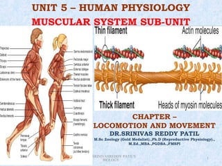
- 45. • Each myofibril has alternate dark and light bands on it. A detailed study of the myofibril has established that the striated appearance is due to the distribution pattern of two important proteins – Actin and Myosin. • The light bands contain actin and is called I-band or Isotropic band, whereas the dark band called ‘A’ or Anisotropic band contains and muscle fibres myosin. Both the proteins are arranged as rod-like structures, parallel to each other and also to the longitudinal axis of the myofibrils. Saturday, February 9, 2013 DR.SRINIVASREDDY PATIL'S 45 BIOLOGY
- 46. Sarcomere
- 47. • Actin filaments are thinner as compared to the myosin filaments, hence are commonly called thin and thick filaments respectively. In the centre of each ‘I’ band is an elastic fibre called ‘Z’ line which bisects it. The thin filaments are firmly attached to the ‘Z’ line. The thick filaments in the ‘A’ band are also held together in the middle of this band by a thin fibrous membrane called ‘M’ line. Saturday, February 9, 2013 DR.SRINIVASREDDY PATIL'S 47 BIOLOGY
- 49. • The ‘A’ and ‘I’ bands are arranged alternately throughout the length of the myofibrils. The portion of the myofibril between two successive ‘Z’ lines is considered as the functional unit of contraction and is called a sarcomere. • In a resting state, the edges of thin filaments on either side of the thick filaments partially overlap the free ends of the thick filaments leaving the central part of the thick filaments. This central part of thick filament, not overlapped by thin filaments is called the ‘H’ zone. Saturday, February 9, 2013 DR.SRINIVASREDDY PATIL'S 49 BIOLOGY
- 50. • a. Structure of Contractile Proteins - Each actin (thin) filament is made of two ‘F’ (filamentous) actins helically wound to each other. Each ‘F’ actin is a polymer of monomeric ‘G’ (Globular) actins. Two filaments of another protein, tropomyosin also run close to the ‘F’ actins throughout its length. Saturday, February 9, 2013 DR.SRINIVASREDDY PATIL'S 50 BIOLOGY
- 51. • A complex protein Troponin is distributed at regular intervals on the tropomyosin. In the resting state a subunit of troponin masks the active binding sites for myosin on the actin filaments. Saturday, February 9, 2013 DR.SRINIVASREDDY PATIL'S 51 BIOLOGY
- 52. • Each myosin (thick) filament is also a polymerised protein. Many monomeric proteins called Meromyosins constitute one thick filament. Each meromyosin has two important parts, a globular head with a short arm and a tail, the former being called the heavy meromyosin (HMM) and the latter, the light meromyosin (LMM). Saturday, February 9, 2013 DR.SRINIVASREDDY PATIL'S 52 BIOLOGY
- 53. • The HMM component, i.e.; the head and short arm projects outwards at regular distance and angle from each other from the surface of a polymerised myosin filament and is known as cross arm. The globular head is an active ATPase enzyme and has binding sites for ATP and active sites for actin. Saturday, February 9, 2013 DR.SRINIVASREDDY PATIL'S 53 BIOLOGY
- 54. • b. Mechanism of Muscle Contraction - Mechanism of muscle contraction is best explained by the sliding filament theory which states that contraction of a muscle fibre takes place by the sliding of the thin filaments over the thick filaments. Saturday, February 9, 2013 DR.SRINIVASREDDY PATIL'S 54 BIOLOGY
- 55. • Muscle contraction is initiated by a signal sent by the central nervous system (CNS) via a motor neuron. A motor neuron alongwith the muscle fibres connected to it constitute a motor unit. The junction between a motor neuron and the sarcolemma of the muscle fibre is called the neuromuscular junction or motor-end plate. Saturday, February 9, 2013 DR.SRINIVASREDDY PATIL'S BIOLOGY 55
- 56. • A neural signal reaching this junction releases a neurotransmitter (Acetyl choline) which generates an action potential in the sarcolemma. This spreads through the muscle fibre and causes the release of calcium ions into the sarcoplasm. Increase in Ca++ level leads to the binding of calcium with a subunit of troponin on actin filaments and thereby remove the masking of active sites for myosin. Saturday, February 9, 2013 DR.SRINIVASREDDY PATIL'S 56 BIOLOGY
- 57. • Utilising the energy from ATP hydrolysis, the myosin head now binds to the exposed active sites on actin to form a cross bridge. This pulls the attached actin filaments towards the centre of ‘A’ band. The ‘Z’ line attached to these actins are also pulled inwards thereby causing a shortening of the sarcomere, i.e., contraction. Saturday, February 9, 2013 DR.SRINIVASREDDY PATIL'S BIOLOGY 57
- 58. • It is clear from the above steps, that during shortening of the muscle, i.e., contraction, the ‘I’ bands get reduced, whereas the ‘A’ bands retain the length. The myosin, releasing the ADP and P1 goes back to its relaxed state. A new ATP binds and the cross- bridge is broken. Saturday, February 9, 2013 DR.SRINIVASREDDY PATIL'S 58 BIOLOGY
- 59. • The ATP is again hydrolysed by the myosin head and the cycle of cross bridge formation and breakage is repeated causing further sliding. The process continues till the Ca++ ions are pumped back to the sarcoplasmic cisternae resulting in the masking of actin filaments. This causes the return of ‘Z’ lines back to their original position, i.e., relaxation. Saturday, February 9, 2013 DR.SRINIVASREDDY PATIL'S BIOLOGY 59
- 60. • The reaction time of the fibres can vary in different muscles. Repeated activation of the muscles can lead to the accumulation of lactic acid due to anaerobic breakdown of glycogen in them, causing fatigue. Muscle contains a red coloured oxygen storing pigment called myoglobin. Myoglobin content is high in some of the muscles which gives a reddish appearance. Such muscles are called the Red fibres. Saturday, February 9, 2013 DR.SRINIVASREDDY PATIL'S BIOLOGY 60
- 61. • These muscles also contain plenty of mitochondria which can utilise the large amount of oxygen stored in them for ATP production. These muscles, therefore, can also be called aerobic muscles. Saturday, February 9, 2013 DR.SRINIVASREDDY PATIL'S 61 BIOLOGY
- 62. • On the other hand, some of the muscles possess very less quantity of myoglobin and therefore, appear pale or whitish. These are the White fibres. Number of mitochondria are also few in them, but the amount of sarcoplasmic reticulum is high. They depend on anaerobic process for energy. Saturday, February 9, 2013 DR.SRINIVASREDDY PATIL'S 62 BIOLOGY
- 63. • 5. DISORDERS OF MUSCULAR AND SKELETAL SYSTEM – • Myasthenia gravis: Auto immune disorder affecting neuromuscular junction leading to fatigue, weakening and paralysis of skeletal muscle. Saturday, February 9, 2013 DR.SRINIVASREDDY PATIL'S 63 BIOLOGY
- 64. • Muscular dystrophy: Progressive degeneration of skeletal muscle mostly due to genetic disorder. • Tetany: Rapid spasms (wild contractions) in muscle due to low Ca++ in body fluid. Saturday, February 9, 2013 DR.SRINIVASREDDY PATIL'S 64 BIOLOGY
- 65. • Arthritis: Inflammation of joints. • Osteoporosis: Age-related disorder characterized by decreased bone mass and increased chances of fractures. A decreased level of estrogen is a common cause. • Gout: Inflammation of joints due to accumulation of uric acid crystals. DR.SRINIVASREDDY PATIL'S Saturday, February 9, 2013 BIOLOGY 65
- 66. QUALITY IS NEVER AN ACCIDENT.IT IS ALWAYS THE RESULT OF HIGH AIM,SINCERE EFFORT,INTELLIGENT DIRECTION AND PERFECT EXECUTION