Cardiopulmonary resuscitation (cpr)
•Télécharger en tant que PPTX, PDF•
151 j'aime•15,326 vues

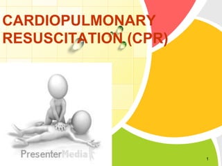
Cardiopulmonary resuscitation (CPR) is a basic life support technique used to manually maintain brain and heart function until further medical help arrives. It involves chest compressions to pump the heart and artificial ventilation to oxygenate the lungs. The steps of CPR include assessing for unresponsiveness, checking for breathing and pulse, calling for help, performing chest compressions at a rate of 100-120 per minute, and giving rescue breaths in a 30:2 ratio. Advanced life support may involve use of an automated external defibrillator, endotracheal intubation, intravenous drugs and fluids, and other emergency medical interventions to restore normal heart rhythm and breathing. Proper and timely CPR can

Recommandé
Contenu connexe
Tendances
Tendances (20)
En vedette
En vedette (20)
Similaire à Cardiopulmonary resuscitation (cpr)
Similaire à Cardiopulmonary resuscitation (cpr) (20)
Dernier
Dernier (20)
Cardiopulmonary resuscitation (cpr)
- 2. What does CPR stands for? • C = Cardio (heart) • P = Pulmonary (lungs) • R = Resuscitation (recover) 2
- 3. DEFINITION Cardio pulmonary resuscitation (CPR) is a technique of basic life support for the purpose of oxygenation to the heart, lungs and brain until and unless the appropriate medical treatment can come and restore the normal cardiopulmonary function. 3
- 4. • Cardio pulmonary resuscitation is a series of steps used to establish artificial ventilation and circulation in the patient who is not breathing and has no pulse. 4
- 5. PURPOSE • Restore cardiopulmonary functioning. • Prevent irreversible brain damage from anoxia. 5
- 6. INDICATION • Cardiac arrest • Respiratory arrest • Combination of both 6
- 7. Definition of Cardiac arrest: It is loss of cardiac function, breathing and loss of consciousness. 7
- 8. Causes of cardiac arrest (6 H & 4 T): 1) 2) 3) 4) 5) 6) Hypoxia. Hypotension. Hypothermia. Hypoglycemia. Acidosis (H+). Hypokalemia (electrolyte disturbance). 1) Cardiac Tamponade. 2) Tension pneumothorax. 3) Thromboembolism (pulmonary, corona ry). 4) Toxicity (eg.digoxin,localanesthetics,i nsecticides). 8
- 9. Diagnosis of cardiac arrest Blood pressure measurement Loss of time !!! Taking the pulse on peripheral arteries Auscultation of cardiac tones 9
- 10. Diagnosis of cardiac arrest (TRIAD): 1) Loss of consciousness. 2) Loss of apical & central pulsations (carotid, femoral). 3) Apnea. 10
- 11. HOW CPR WORKS: The air we breathe in, travels to our lungs were oxygen is picked up by our blood and then pumped by the heart to our tissue and organs. When a person experiences cardiac arrest-whether due to heart failure in adults or the elderly or an injury such as near drowning, or severe trauma in a child-the heart goes from a normal arrhythmic pattern called ventricular fibrillation, and eventually ceases to beat altogether. This prevents oxygen from circulating throughout the body, rapidly killing cells and tissue. 11
- 12. • Inessence, cardio (heart) pulmonary(lung) resuscitation (revive, revitalize) serves as an artificial heartbeat and an artificial respirator. • CPR may not save the victim even when performed properly, but if started within 4 minute of cardiac arrest and defibrillation is provided within 10 minutes, a person has a 40% chance of survival. 12
- 13. EQUIPMENTS • A hard flat surface. • No additional equipment is necessary but in hospital setting, an emergency (crash) cart with defibrillator and cardiac monitor should be brought to the bedside. A crash cart contains: • Airway equipment. • Suction equipment. • Intravenous equipment. • Laboratory tubes and syringes. • Pre packed medication for advanced life support. 13
- 14. PHASES OF THE CARDIO PULMONARY RESUSCITATION: Phases Phase-1 Steps Basic life support C= circulation A= Airway B= Breathing Phase-2 Advance cardiac life support D= Drugs E= ECG F= fibrillation Phase-3 Prolonged life Post resuscitation care support 14
- 15. • What is basic life support (BLS)? It is life support without the use of special equipment. • What is Advanced Life Support (ACLS)? It is life support with the use of special equipment (eg. Airway, endotracheal tube, defibrillator). 15
- 17. ) EARLY RECOGNITION Assessment is of crucial importance. It includes 1) Unresponsiveness • Check the victim for a response. • Shake shoulders gently • Ask “Are you all right 17
- 18. ) No breathing or no normal breathing (i.e, only gasping) 18
- 19. ) No pulse felt within seconds. 19
- 20. ) CPR Sequence A Change From A-B-C to C-A-B 20
- 21. (A) Chest compressions (cardiac massage) The human brain cannot survive more than 3 minutes with lack of circulation. So chest compressions must be started immediately for any patient with absent central pulsations. 21
- 22. TECHNIQUE OF CHEST COMPRESSION - Pt must be placed on a hard surface (wooden board). - The palm of one hand is placed in the concavity of the lower half of the sternum 2 fingers above the xiphoid process. (AVOID xiphisternal junction → fracture & injury). 22
- 23. • The other hand is placed over the hand on the sternum. • Shoulders should be positioned directly over the hands with the elbows locked straight and arms extended. Use your upper body weight to compress. • Sternum must be depressed atleast 5 cm in adults, and 2-4 cm in children, 1-2 cm in infants . 23
- 24. PUSH HARD AND PUSH FAST 24
- 25. • Must be performed at a rate of 100-120/min • During CPR the ratio of chest compressions to ventilation should be as follows: • Single rescuer = 30:2 • In the presence of 2 rescuers chest compressions must not be interrupted for ventilation 25
- 26. + 30 2 26
- 27. Chest compressions in infants (0-12 months) 27
- 28. ► Chest compressions must be continued for 2 minutes before reassessment of cardiac rhythm. ► (2 minutes = equivalent to 5 cycles 30:2). ► Golden rules: • Ensure high quality chest compressions: rate, depth, recoil. • Plan actions before interrupting CPR. • MINIMIZE interruption of chest compressions. • Early defibrillation of shockable rhythm. 28
- 29. Assessment of the adequacy of chest compressions: • Systolic BP: 60-80 mmHg • Diastolic BP: > 40 mmHg • COP = 30% of normal 29
- 30. PROBLEMS AND COMPLICATIONS OF CHEST COMPRESSIONS 1. RIB FRACTURES 2. FRACTURE STERNUM 3. RIB SEPARATION 4. PNEUMOTHORAX 5. HEMOTHORAX 6. LUNG CONTUSIONS 7. LIVER LACERATIONS 8. FAT EMBOLI 9. HIV, HEPATITIS 10.INFECTIONS MANAGE ACCORDINGLY BUT CONTINUE CPR 30
- 31. Airway Loss of consciousness often results in airway obstruction due to loss of tone in the muscles of the airway and falling back of the tongue. CLEAR THE AIRWAY 31
- 32. (A) Basic techniques for airway patency: 1) Head tilt, chin lift: one hand is placed on the forehead and the other on the chin the head is tilted upwards to cause anterior displacement of the tongue 32
- 34. 3) Finger sweep: Sweep out foreign body in the mouth by index finger (in unconscious pt only. This is NOT advised in a conscious or convulsing patient). 34
- 35. 4) Heimlich manoeuvre: if the pt is conscious or the foreign body cannot be removed by a finger sweep. It is done while the pt is standing up or lying down. This is a subdiaphragmatic abdominal thrust that elevates the diaphragm expelling a blast of air from the lungs that displaces the foreign body. In infants his can be done by a series of blows on he back and chest thrusts. 35
- 37. Breathing: Breathe for the person Rescue breathing can be mouth-to-mouth breathing or mouth-to-nose breathing if the mouth is seriously injured or can't be opened. • With the airway open (using the head-tilt, chin-lift maneuver), pinch the nostrils shut for mouth-tomouth breathing and cover the person's mouth with yours, making a seal.
- 38. (A) Basic techniques include: 1) Mouth to mouth breathing: with the airway held open, pinch the nostrils closed, take a deep breath and seal your lips over he patients mouth. Blow steadily into the patients mouth watching the chest rise as if the patient was taking a deep breath. 38
- 39. 2) Mouth to nose breathing: seal the mouth shut and breathe steadily though the nose. 3) Mouth to mouth and nose: is used in infants and small children. 39
- 40. Assessment of restoration of breathing and circulation Contraction of pupil Improved color of the skin Free movement of the chest wall Swallowing attempts Struggling movements 40
- 41. Signs of restored ventilation and circulation include: • • • • Struggling movements Improved color Return of or strong pulse Return of systemic blood pressure 41
- 42. When to terminate BLS • • • • Pulse and respiration returns Emergency medical help arrives Physician declared patient is deceased In a non health setting ,another indication to stop BLS would be that the rescuer was exhausted and physically unable to continue to perform BLS.
- 44. ALS includes: Circulation by cardiac massage Airway management by equipments Breathing by advanced techniques Defibrillation by manual defibrillator Drugs.
- 47. (B) Advanced techniques for airway patency: 1) Face Mask 47
- 50. 4) Laryngeal mask (LMA) 50
- 52. 6) Combitube 52
- 53. 7) Cricothyrotomy (Surgical Airway) 53
- 54. 8) Tracheostomy (Surgical Airway) 54
- 55. BREATHING 55
- 56. Expired air contains 16% O2 so supplemental 100% O2 should be used as soon as possible. Successful breathing is achieved by delivery of a tidal volume of 800-1200 ml in adults at a rate of 10-12 breaths/min in adults. (B) Advanced techniques include: 1) Self-inflating resuscitation bag (Ambu bag) 2) Mechanical ventilator in OR or in ICU 56
- 57. • Expired air = 16% O2 • Ambu Bag (room air) = 21% O2 • Ambu bag + O2 (10-15L) = 45% O2 • Ambu Bag + O2 + Reservoir bag = 85% O2 57
- 58. ) DEFIBRILLATION • Defibrillation consists of delivering a therapeutic dose of electrical energy to the affected heart with a device called a defibrillator 58
- 59. • In cardiac arrest,the associated heart rhythms can be categorised into two groups : 1) Shockable rhythm: VT/VF 2) Non shockable rhythm: asystole and PEA 59
- 60. • The basic difference in the treatment of these two groups of arrythmia is the need for defibrillation in patients with VT/VF 60
- 61. 61
- 62. (C) Defibrillation Position of Paddles: • One paddle is placed in the right infraclavicular region, while the other is placed in the left 5th-6th intercostal space anterior axillary line. 62
- 63. • Alternatively anteroposterior position may be used: one paddle is placed in the left infrascapular region while the other is placed in the left 5th-6th intercostal space anterior axillary line. • 63
- 64. • Action Completely depolarize all myocardial cells so SA node can reestablish as pacemaker • Voltage of electricity discharge High from 150 J to 360J(biphasic) 360 J(monophasic) 64
- 65. Drugs used in CPR ► Adrenaline: - Given as a vasopressor α-1 effect (not as an inotrope). - Dose: 1 mg (0.01 mg/kg) IV every 4 minutes (alternating cycles) while continuing CPR. - Given: 1) Immediately in non-shockable rhythm (non-VT/VF). 2) In VF or VT given after the 3rd shock. -Repeated: in alternate cycles (every 4 minutes). -Once adrenaline → ALWAYS adrenaline. 65
- 66. Amiodarone: - Dose: 300 mg IV bolus (5 mg/kg). - Given: in shockable rhythm after the 3rd shock. - If unavailable give lidocaine 100 mg IV (1-1.5 mg/kg). 66
- 67. ► Vasopressin (ADH): 40 IU single dose once. ► Magnesium: - Dose: 2 g IV. - Given: 1- VF / VT with hypomagnesemia. 2- Torsade de pointes. 3- Digoxin toxicity. 67
- 68. ► Calcium: – Dose: 10 ml of 10% Calcium chloride IV. – Indications: PEA caused by: hyperkalemia, hypocalcemia, hypermagnesemia, and overdose of calcium channel blockers. – Do NOT give calcium solutions and NaHCO3 simultaneously by the same route. 68
- 69. ► IV Fluids: • Infuse fluids rapidly if hypovolemia is suspected. • Use normal saline (0.9% NaCl) or Ringer’s solution. • Avoid dextrose which is redistributed away from the intravascular space rapidly and causes hyperglycemia which may worsen neurological outcome after cardiac arrest. • Dextrose is indicated only if there is documented hypoglycemia. 69
- 70. ► Thrombolytics: • Fibrinolytic therapy is considered when cardiac arrest is caused by proven or suspected acute pulmonary embolism. • If a fibrinolytic drug is used in these circumstances consider performing CPR for at least 60-90 minutes before termination of resuscitation attempts. • Eg: Alteplase, tenecteplase (old generation: steptokinase). 70
- 71. Sodium bicarbonate: ► Used in: 1- Severe metabolic acidosis (pH < 7.1) 2- Life-threatening hyperkalemia. 3- Tricyclic antidepressant overdose. ► Dose: (half correction) 1/2 Base Deficit 1/3 Body weight. 71
- 72. Avoid its routine use due to its complications: 1- Increases CO2 load: 2- Inhibits release of O2 to tissues. 3- Impairs myocardial contractility. 4- Causes hypernatremia. 5- Adrenaline works better in acidic medium. 72
- 73. Atropine: • Its routine use in PEA and asystole is not beneficial and has become obsolete. • Indicated in: sinus bradycardia or AV block causing hemodynamic instability. • Dose: 0.5 mg IV. Repeated up to a maximum of 3 mg (full atropinization). 73
- 74. CONTRAINDICATIONS Do not resuscitate when a decision not to resuscitate has been noted in chart. This order is often abbreviated to DNR (do not resuscitate), is sometime referred to as no code, and is now discussed with the client on admission and is referred to as an advanced directive. 74
- 75. SUMMARY OF STEPS OF CPR COMPONENT RECOMMENDATIONS Recognition unresponsi ve CPR sequence Chest compression, airway, breathing (CA-B) At least 100 per min Compression rate No breathing No pulse felt or no normal within 10 breathing(i.e seconds , only gasping) 75
- 76. Compression depth At least 2 inches (5cm) At least 1/3 AP diameter About 2 inches (5cm) At least 1/3 AP diameter.About 1 and half inches(4cm) Chest wall recoil Allow complete recoil between compressions. Rotate compressors every 2 min. Compression interruptions Minimize interruption in chest compressions. Attempt to limit interruptions to <10 sec Airway Head tilt-chin lift(suspected trauma jaw thrust) Compression to ventilation ratio(until advanced airway placed) Ventilations with advanced airway breath every 6-8 seconds (8-10 breaths/min) Asynchronus with chest compressions.about 1 sec per breath. Visible chest rise Defibrillation Minimize interruptions in chest compressions before and after shock; resume CPR begining with compressions 76 immediately after each shock.
- 77. Summary of Key Issues and Major Changes • Refinements have been made to recommendations for immediate recognition and activation of the emergency response system based on signs of unresponsiveness, as well as initiation of CPR if the victim is unresponsive with no breathing or no normal breathing (i.e victim is only gasping). 77
- 78. • “Look, listen, and feel for breathing” has been removed from the algorithm. • Continued emphasis has been placed on high-quality CPR 78
- 79. Post resuscitation care • • • • Maintain Airway and Breathing Check for Circulation Disability optimising neurological recovery Sedation 79
- 80. • • • • • Control of seizure Temperature control Treatment of hyperpyrexia Treatment of hypothermia Blood glucose level 80
- 81. Managing the Cardiac Arrest Team ► During cardiac arrest the team leader should allocate and assign the various roles and tasks to the team members. Assign one person for each of the following roles: – Airway management & ventilation (Eg.bag & mask. Intubation). – – – Chest compressions . IV drug administration. – Defibrillation (DC shock) – Timing and documentation. 81
- 82. ► The person responsible for the airway may take turns with the person responsible for chest compressions in order to diminish fatigue & exhaustion. ► It is also the responsibility of the team leader to use the 2-minute periods of chest compressions to plan tasks, give orders and eliminate & exclude/ correct the reversible causes of cardiac arrest. 82
- 84. 84
- 85. THANK YOU 85