New from BookNet Canada for 2024: BNC CataList - Tech Forum 2024
3 prr
1. Leading Edge
Review
Pattern Recognition Receptors
and Inflammation
Osamu Takeuchi1,2 and Shizuo Akira1,2,*
1Laboratory of Host Defense, WPI Immunology Frontier Research Center
2Research Institute for Microbial Diseases
Osaka University, 3-1 Yamada-oka, Suita, Osaka 565-0871, Japan
*Correspondence: sakira@biken.osaka-u.ac.jp
DOI 10.1016/j.cell.2010.01.022
Infection of cells by microorganisms activates the inflammatory response. The initial sensing of
infection is mediated by innate pattern recognition receptors (PRRs), which include Toll-like recep-
tors, RIG-I-like receptors, NOD-like receptors, and C-type lectin receptors. The intracellular signaling
cascades triggered by these PRRs lead to transcriptional expression of inflammatory mediators that
coordinate the elimination of pathogens and infected cells. However, aberrant activation of this
system leads to immunodeficiency, septic shock, or induction of autoimmunity. In this Review, we
discuss the role of PRRs, their signaling pathways, and how they control inflammatory responses.
Introduction These PRRs are expressed not only in macrophages and DCs
Inflammation is a protective response by the body to ensure but also in various nonprofessional immune cells. With the
removal of detrimental stimuli, as well as a healing process for exception of some NLRs, the sensing of PAMPs or DAMPs by
repairing damaged tissue (Medzhitov, 2008). Inflammation is PRRs upregulates the transcription of genes involved in inflam-
caused by various factors such as microbial infection, tissue matory responses. These genes encode proinflammatory cyto-
injury, and cardiac infarction. Classically, inflammation is charac- kines, type I interferons (IFNs), chemokines and antimicrobial
terized by five symptoms: redness, swelling, heat, pain, and loss proteins, proteins involved in the modulation of PRR signaling,
of tissue function. These macroscopic symptoms reflect and many uncharacterized proteins. The expression patterns
increased permeability of the vascular endothelium allowing of the inducible genes differ among activated PRRs.
leakage of serum components and extravasation of immune The inflammatory response is orchestrated by proinflamma-
cells. The inflammatory response is then rapidly terminated tory cytokines such as tumor necrosis factor (TNF), interleukin
and damaged tissues are repaired. However, overproduction (IL)-1, and IL-6. These cytokines are pleiotropic proteins that
of cytokines (a cytokine storm) by immune cells to overwhelm regulate the cell death of inflammatory tissues, modify vascular
pathogens can be fatal. A cytokine storm can also be caused endothelial permeability, recruit blood cells to inflamed tissues,
by noninfectious diseases such as graft-versus-host disease and induce the production of acute-phase proteins. Although
(GVHD). Inflammatory responses are also critical for the patho- TNF and IL-6 are mainly regulated at the transcriptional and
genesis of autoimmune diseases. translational levels, the production of IL-1b is regulated by
The innate immune system is the major contributor to acute a two-step mechanism. The first step is the expression of an
inflammation induced by microbial infection or tissue damage IL-1b zymogen, pro-IL-1b, which is regulated by the synthesis
(Akira et al., 2006; Beutler et al., 2006). Furthermore, innate of its mRNA in a TLR signal-dependent manner. However, IL-1b
immunity is also important for the activation of acquired immu- maturation requires cleavage of pro-IL-1b by a protease, cas-
nity. Although innate immune cells including macrophages and pase-1, which is activated independently of TLR signaling. The
dendritic cells (DCs) play important roles, nonprofessional cells complex that activates caspase-1, called the inflammasome, is
such as epithelial cells, endothelial cells, and fibroblasts also composed of NLRs, ASC, and caspase-1 (see Review by
contribute to innate immunity. Germline-encoded pattern recog- K. Schroder and J. Tschopp on page 821 of this issue). Type I
nition receptors (PRRs) are responsible for sensing the presence IFNs, including multiple forms of IFN-a and single forms of
of microorganisms. They do this by recognizing structures IFN-b, IFN-u, etc., are also involved in the modulation of inflam-
conserved among microbial species, which are called path- mation (Honda et al., 2006). Type I IFNs play central roles in anti-
ogen-associated molecular patterns (PAMPs). Recent evidence viral responses by inducing apoptotic cell death in virally infected
indicates that PRRs are also responsible for recognizing endog- cells, rendering cells resistant to virus infection, activating
enous molecules released from damaged cells, termed damage- acquired immunity, and stimulating hematopoietic stem cell
associated molecular patterns (DAMPs). Currently, four different turnover and proliferation. Secreted type I IFNs alert the
classes of PRR families have been identified. These families surrounding cells via type I IFN receptors by triggering a signaling
include transmembrane proteins such as the Toll-like receptors cascade that leads to the phosphorylation and nuclear translo-
(TLRs) and C-type lectin receptors (CLRs), as well as cyto- cation of IFN-stimulated gene factor 3 (ISGF3), a complex
plasmic proteins such as the Retinoic acid-inducible gene composed of Signal Transducers and Activators of Transcription
(RIG)-I-like receptors (RLRs) and NOD-like receptors (NLRs). 1 (STAT1), STAT2, and IFN-regulatory factor (IRF) 9. ISGF3
Cell 140, 805–820, March 19, 2010 ª2010 Elsevier Inc. 805
2. Table 1. PRRs and Their Ligands
PRRs Localization Ligand Origin of the Ligand
TLR
TLR1 Plasma membrane Triacyl lipoprotein Bacteria
TLR2 Plasma membrane Lipoprotein Bacteria, viruses, parasites, self
TLR3 Endolysosome dsRNA Virus
TLR4 Plasma membrane LPS Bacteria, viruses, self
TLR5 Plasma membrane Flagellin Bacteria
TLR6 Plasma membrane Diacyl lipoprotein Bacteria, viruses
TLR7 (human TLR8) Endolysosome ssRNA Virus, bacteria, self
TLR9 Endolysosome CpG-DNA Virus, bacteria, protozoa, self
TLR10 Endolysosome Unknown Unknown
TLR11 Plasma membrane Profilin-like molecule Protozoa
RLR
RIG-I Cytoplasm Short dsRNA, 50 triphosphate dsRNA RNA viruses, DNA virus
MDA5 Cytoplasm Long dsRNA RNA viruses (Picornaviridae)
LGP2 Cytoplasm Unknown RNA viruses
NLR
NOD1 Cytoplasm iE-DAP Bacteria
NOD2 Cytoplasm MDP Bacteria
CLR
Dectin-1 Plasma membrane b-Glucan Fungi
Dectin-2 Plasma membrane b-Glucan Fungi
MINCLE Plasma membrane SAP130 Self, fungi
induces expression of IFN-inducible antiviral genes such as structures and that their cognate ligands interact with internal
protein kinase R (PKR) and 20 50 -oligoadenylate synthase (OAS) pockets formed by the TLR1/TLR2 or TLR6/TLR2 heterodimers
among others. PKR suppresses the proliferation of virus-in- (Jin et al., 2007). Stimulation with TLR2 ligands, such as triacyl
fected cells and 20 50 -OAS activates RNase L, which cleaves viral and diacyl lipoproteins, induces the production of various proin-
nucleotides in order to inhibit virus replication. flammatory cytokines (but not type I IFNs) in macrophages and
In this Review, we discuss how distinct PRRs (with particular DCs. However, another report showed that TLR2 in inflammatory
emphasis on TLRs and RLRs) sense the presence of pathogens monocytes induced type I IFNs in response to viral infection,
and cellular insults and the mechanisms by which these PRR suggesting that the cellular responses to TLR2 ligands differ de-
signals elicit inflammation. pending on the cell types involved (Barbalat et al., 2009). TLR10
is related to TLR1 and TLR6 based on sequence similarity.
TLRs and Their Ligands TLR10 seems to be functional in humans, although mouse
The TLR family is one of the best-characterized PRR families and TLR10 is disrupted by insertion of an endogenous retrovirus.
is responsible for sensing invading pathogens outside of the cell The ligand for TLR10 has not been identified.
and in intracellular endosomes and lysosomes (Akira et al., TLR4 recognizes lipopolysaccharide (LPS) together with
2006). TLRs are characterized by N-terminal leucine-rich repeats myeloid differentiation factor 2 (MD2) on the cell surface. LPS
(LRRs) and a transmembrane region followed by a cytoplasmic is a component derived from the outer membrane of Gram-
Toll/IL-1R homology (TIR) domain. Ten TLRs have been identi- negative bacteria and is known to be a cause of septic shock.
fied in humans and 12 in mice. Different TLRs recognize the The crystal structure of a complex comprising TLR4, MD2, and
different molecular patterns of microorganisms and self-compo- LPS revealed that two complexes of TLR4-MD2-LPS interact
nents (Table 1). symmetrically to form a TLR4 homodimer (Park et al., 2009).
TLR2 senses various components from bacteria, myco- TLR4 is also involved in the recognition of viruses by binding to
plasma, fungi, and viruses. These components include the lipo- viral envelope proteins. In addition, TLR4 modulates the patho-
proteins of bacteria and mycoplasma. TLR2 recognizes its genesis of H5N1 avian influenza virus infection by recognizing
ligands by forming a heterodimer with either TLR1 or TLR6 a DAMP rather than the virus itself (Imai et al., 2008). Acute
(Figure 1). The resulting TLR1/TLR2 and TLR6/TLR2 complexes lung injury caused by avian influenza virus infection produces
recognize distinct ligands (triacyl and diacyl lipoproteins, endogenous oxidized phospholipids, which stimulate TLR4.
respectively). The crystal structures of the extracellular domains Mice lacking TLR4 were found to be resistant to avian flu-
of TLR2, TLR1, and TLR6 revealed that they form M-shaped induced lethality.
806 Cell 140, 805–820, March 19, 2010 ª2010 Elsevier Inc.
3. differentiation of naive T cells into antigen-specific Th17 and
Th1 cells (Uematsu et al., 2008). TLR11, which is present in
mice but not in humans, shows close homology to TLR5.
TLR11 recognizes uropathogenic bacteria and a profilin-like
molecule derived from the intracellular protozoan Toxoplasma
gondii (Yarovinsky et al., 2005).
A set of TLRs, comprising TLR3, TLR7, TLR8, and TLR9,
recognize nucleic acids derived from viruses and bacteria, as
well as endogenous nucleic acids in pathogenic contexts (Akira
et al., 2006). Activation of these TLRs leads to the production of
type I IFNs in addition to proinflammatory cytokines. TLR3
detects viral double-stranded (ds) RNA in the endolysosome.
TLR3 is involved in the recognition of polyinosinic polycytidylic
acid (poly I:C), a synthetic dsRNA analog. Although inoculation
of mice with poly I:C induces the production of cytokines as
well as type I IFNs in mice, TLR3 is essential for the production
of cytokines such as IL-12p40, but not type I IFNs in sera (Kato
et al., 2006). The crystal structure of TLR3 bound to dsRNA
revealed that dsRNA binds to the N-terminal and C-terminal
portions of TLR3 LRRs, and this ligand binding dimerizes two
TLR3 molecules (Choe et al., 2005; Liu et al., 2008). Mouse
TLR7 and human TLR7/8 recognize single-stranded (ss) RNAs
from RNA viruses, as well as small purine analog compounds
(imidazoquinolines). TLR7 also detects RNAs from bacteria
such as Group B Streptococcus in endolysosomes in conven-
tional DCs (cDCs) (Mancuso et al., 2009). TLR9 senses unmethy-
lated DNA with CpG motifs derived from bacteria and viruses.
Although the CpG motif was thought to be essential for TLR9
stimulation, the DNA sugar backbone of 20 deoxyribose also
mediates TLR9 recognition (Haas et al., 2008). In addition to
DNA, TLR9 also recognizes hemozoin, a crystalline metabolite
of hemoglobin produced by the malaria parasite (Coban et al.,
2005). TLR9 directly binds to hemozoin, and a crude extract of
the malaria parasite elicits parasite-antigen-specific immune
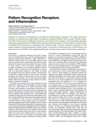
Figure 1. TLR2, TLR3, and TLR4 Signaling Pathways
responses via TLR9 (Coban et al., 2010). However, another
Lipoproteins and LPS are recognized on the cell surface by a heterodimer of report shows that TLR9 recognizes malaria parasite DNA con-
TLR1/6 and TLR2, and by 2 sets of TLR4/MD2 complexes, respectively. tained in purified hemozoin, and that hemozoin only transports
Ligand stimulation recruits MyD88 and TIRAP to the TLR, and a complex of malaria parasite DNA to the endosome, where TLR9 is present
IRAKs and TRAF6 is subsequently formed. TRAF6 acts as an E3 ubiquitin (Parroche et al., 2007). Further studies will clarify the role of
ligase and catalyzes formation of a K63-linked polyubiquitin chain on TRAF6
TLR9 in the recognition of malaria parasite components. TLR7
itself and generation of an unconjugated polyubiquitin chain with an E2 ubiq-
and TLR9, but not TLR3, are highly expressed in plasmacytoid
uitin ligase complex of Ubc13 and Uev1A. Ubiquitination activates a complex
of TAK1, TAB1, and TAB2/3 resulting in the phosphorylation of NEMO and the DCs (pDCs), a cell type that produces large amounts of type I
activation of an IKK complex. Phosphorylated IkB is degraded, and the freed IFNs in response to virus infection.
NF-kB translocates to the nucleus where it drives expression of cytokine Accumulating evidence underscores the importance of the
genes. Simultaneously, TAK1 activates MAP kinase cascades leading to the localization of TLRs in the cell for their recognition by ligand
activation of AP-1, which is also critical for the induction of cytokine genes. (Barton and Kagan, 2009). Given that self-nucleotides are potent
LPS induces translocation of TLR4 to the endosome together with TRAM.
TLR ligands and may facilitate autoimmunity, TLRs that recog-
TLR3 is present in the endosome and recognizes dsRNA. TLR3 and TLR4 acti-
vate TRIF-dependent signaling, which activates NF-kB and IRF3 resulting in nize self-nucleotides are compartmentalized to avoid unwanted
the induction of proinflammatory cytokine genes and type I IFNs. TRAF6 and activation. Although TLR1, TLR2, TLR4, TLR5, and TLR6 are
RIP1 activate NF-kB, whereas TRAF3 is responsible for phosphorylation of present on the plasma membrane, TLR3, TLR7, and TLR9 are
IRF3 by TBK1/IKK-i. NAP1 and SINTBAD are required for the activation of mainly present on the endoplasmic reticulum (ER) membrane.
TBK1/IKK-i. Phosphorylated IRF3 translocates into the nucleus to induce It has been proposed that self-nucleic acids are degraded by
expression of type I IFN genes.
extracellular or endosomal DNases prior to recognition by
TLRs. Nucleic acid-sensing TLRs are recruited from the ER to
TLR5 is highly expressed by DCs of the lamina propria (LPDCs) endolysosomes following stimulation by their ligands (Figure 2).
in the small intestine, where it recognizes flagellin from flagel- The mechanism by which nucleotide-recognizing TLRs are re-
lated bacteria. In response to flagellin, LPDCs induce B cells to cruited from the ER to the endolysosome compartment remains
differentiate into IgA-producing plasma cells and trigger the to be clarified. However, a forward genetics screen in mice
Cell 140, 805–820, March 19, 2010 ª2010 Elsevier Inc. 807
4. whether TLR7 is also cleaved in the endolysosome, although en-
dosomal acidification is required for the sensing of TLR7 ligands.
TLR7 and TLR9 are essential for virus-induced type I IFN
production by pDCs (Kato et al., 2005). Viral nucleotides can
interact with TLR7 and TLR9 in pDCs after they have been endo-
cytosed. Alternatively, once the viruses invade pDCs and virions
are present in the cytoplasm, they can be delivered to the endo-
lysosome where TLR7 and TLR9 are recruited for viral sensing.
pDCs take advantage of a cellular process called autophagy in
which self-proteins and damaged organelles are degraded in
double-membraned vesicles called autophagosomes (Lee
et al., 2007). In the absence of ATG5, a protein essential for au-
tophagosome formation, pDCs fail to produce type I IFNs in
response to virus infection, suggesting that the cytoplasmic
virions are engulfed by autophagosomes and then fuse with lyso-
somes. However, ATG5 is also required for responses to CpG-
DNA. Therefore, autophagy may control either the endosomal
maturation required for CpG-DNA sensing or the TLR9 signaling
pathways in pDCs, or both.
TLR-mediated microbial recognition is very important for host
defense against pathogens. On the other hand, excess re-
sponses to TLR ligands induce lethal septic shock syndrome.
These observations indicate that appropriate activation of
TLRs is vital for eradicating invading pathogens without causing
harmful damage to the host.
TLR Signaling Pathways
Recognition of PAMPs by TLRs leads to transcriptional upregu-
lation of distinct genes, depending on the TLRs and cell types
involved (Figure 1). The difference in the signaling cascades acti-
vated by the individual TLRs can be partly explained by the TIR
domain-containing adaptor molecules recruited to TLRs
(Akira et al., 2006). There are five TIR domain-containing adap-
Figure 2. Nucleic Acid Sensing by TLR7 and TLR9 tors including MyD88, TIR domain-containing adaptor inducing
TLR7 and TLR9 recognize viral ssRNA and CpG DNA, respectively. Stimulation
IFN-b (TRIF; also known as TICAM-1), TIRAP/Mal, TRIF-related
with ligands or infection by viruses induces trafficking of TLR7 and TLR9 from
the ER to the endolysosome via UNC93B1. TLR9 undergoes cleavage by
adaptor molecule (TRAM), and Sterile-alpha and Armadillo
proteases present in the endolysosome. A complex of MyD88, IRAK-4, motif-containing protein (SARM). TLR signaling is roughly
TRAF6, TRAF3, IRAK-1, IKK-a, and IRF7 is recruited to the TLR. Phosphory- divided into two distinct pathways depending on the usage of
lated IRF7 translocates into the nucleus and upregulates the expression of the distinct adaptor molecules, MyD88 and TRIF.
type I IFN genes. Viruses that have entered the cytoplasm are engulfed by The MyD88-Dependent Signaling Pathway
autophagosomes and deliver viral nucleic acids to the endolysosome. An
MyD88 is composed of a death domain (DD) in addition to a TIR
HMGB1-DNA complex released from damaged cells is captured by RAGE.
domain. MyD88 is essential for the downstream signaling of
Autoantibodies recognizing self-DNA or -RNA bind to FcgRIIa. LL37, an anti-
microbial peptide, associates with endogenous DNA. These proteins are various TLRs, with the exception of TLR3. Children with
responsible for the delivery of endogenous nucleic acids to endolyosomes MyD88 deficiency suffer from recurrent pyogenic bacterial infec-
where they are recognized by TLR7 or TLR9. tions. TLR2 and TLR4 signaling requires TIRAP/Mal for bridging
between TLR and MyD88. MyD88 interacts with IL-1R-associ-
ated kinase (IRAK)-4, a serine/threonine kinase with an N-ter-
revealed that UNC93B1 (an ER protein with 12 membrane-span- minal death domain. IRAK-4 activates other IRAK family
ning domains) is responsible for TLR3, TLR7, and TLR9 signaling members, IRAK-1 and IRAK-2 (Kawagoe et al., 2008). The IRAKs
by governing the translocation of these TLRs from the ER to the then dissociate from MyD88 and interact with TNFR-associated
endolysosome (Kim et al., 2008; Tabeta et al., 2006). When TLR9 factor 6 (TRAF6), which acts as an E3 ubiquitin protein ligase.
is recruited from the ER to the endolysosome, it undergoes pro- Together with an E2 ubiquitin-conjugating enzyme complex
cessing by proteases, such as cathepsins, in the endolysosome comprising Ubc13 and Uev1A, TRAF6 catalyzes the formation
(Ewald et al., 2008; Park et al., 2008). The processed form of of a lysine 63 (K63)-linked polyubiquitin chain on TRAF6 itself
TLR9 is responsible for CpG-DNA recognition. It has been shown as well as the generation of an unconjugated free polyubiquitin
that cathepsins B, K, and L and asparagine endopeptidase are chain (Xia et al., 2009). A complex of TGF-b-activated kinase 1
required for TLR9 responses (Asagiri et al., 2008; Matsumoto (TAK1), TAK1-binding protein 1 (TAB1), TAB2, and TAB3 is acti-
et al., 2008; Sepulveda et al., 2009). Currently, it remains unclear vated by the unconjugated free K63 polyubiquitin chain and
808 Cell 140, 805–820, March 19, 2010 ª2010 Elsevier Inc.
5. phosphorylates IkB kinase (IKK)-b and MAP kinase kinase 6. The activation of TBK1 and IKK-i is modulated by various
Subsequently, the IKK complex, composed of IKK-a, IKK-b, proteins. TBK1 and IKK-i interact with TRAF family member-
and NF-kB essential modulator (NEMO), phosphorylates IkBa, associated NF-kB activator (TANK) (also known as I-TRAF),
an NF-kB inhibitory protein. Phosphorylated IkB undergoes NAK-associated protein 1 (NAP1), and the TBK1 adaptor
degradation by the ubiquitin-proteasome system, thereby (SINTBAD), which is similar to NAP1 (Guo and Cheng, 2007;
freeing NF-kB to translocate into the nucleus and activate Ryzhakov and Randow, 2007; Sasai et al., 2006). These mole-
expression of proinflammatory cytokine genes. Activation of the cules contain a TBK1-binding motif and show similarities in their
MAP kinase cascade is responsible for the formation of another coiled-coil domains. However, TANKÀ/À cells do not show
transcription factor complex, AP-1, that targets cytokine genes. impaired type I IFN production in response to dsRNA stimulation
TLR7 and TLR9 signaling induces the production of type I IFNs (Kawagoe et al., 2009). Although knockdown of either NAP1 or
in addition to other NF-kB-dependent cytokines in a MyD88- SINTBAD impairs TRIF signaling, the relationship between these
dependent manner. In pDCs, MyD88 forms a complex with molecules in TRIF signaling is not yet fully understood.
IRAK-1, TRAF6, TRAF3, IKK-a, and IRF7, and phosphorylated
IRF7 translocates to the nucleus to activate the expression RLRs and Virus Recognition
of genes encoding type I IFNs (Figure 2). In cDCs, IRF1, but The RIG-I-like receptor (RLR) family is composed of RIG-I, mela-
not IRF7, is activated downstream of TLR7 and TLR9, resulting noma differentiation-associated gene 5 (MDA5), and LGP2
in the activation of IFN-b gene expression (Negishi et al., 2006; (Takeuchi and Akira, 2009; Yoneyama and Fujita, 2008). RLRs
Schmitz et al., 2007). are composed of two N-terminal caspase recruitment domains
The TRIF-Dependent Signaling Pathway (CARDs), a central DEAD box helicase/ATPase domain, and
In response to stimulation with dsRNA, TLR3 recruits another a C-terminal regulatory domain. They are localized in the cyto-
adaptor protein, TRIF. TLR4 triggers both MyD88-dependent plasm and recognize the genomic RNA of dsRNA viruses and
and TRIF-dependent signaling. TLR4, but not TLR3, requires dsRNA generated as the replication intermediate of ssRNA
another adaptor, TRAM, for activating TRIF. A splice variant of viruses. The expression of RLRs is greatly enhanced in response
TRAM called the TRAM adaptor with GOLD domain (TAG) acts to type I IFN stimulation or virus infection.
as the negative regulator of TRIF-dependent signaling Mouse fibroblasts and cDCs lacking RIG-I are defective in the
(Palsson-McDermott et al., 2009). TRIF associates with TRAF3 production of type I IFNs and inflammatory cytokines in
and TRAF6 through TRAF-binding motifs present in its N-ter- response to various RNA viruses (Kato et al., 2006). These
minal portion. TRIF also contains a C-terminal receptor-interact- include the Paramyxoviridae such as Newcastle disease virus
ing protein (RIP) homotypic interaction motif (RHIM) and (NDV) and Sendai virus (SeV), vesicular stomatitis virus (VSV),
interacts with RIP1 and RIP3 via this motif. In humans, SARM influenza virus, and Japanese encephalitis virus (JEV). In
functions as an inhibitor of TRIF-dependent signaling (Carty contrast, cells lacking MDA5 respond normally to these viruses.
et al., 2006). The TNFR-associated death domain protein Meanwhile, the IFN responses to several Picornaviridae,
(TRADD), an essential adaptor for TNFR signaling, is involved including encephalomyocarditis virus (EMCV), Mengo virus,
in the TRIF-dependent signaling pathway (Ermolaeva et al., and Theiler’s virus, are abrogated in MDA5À/À cells, but not in
2008; Pobezinskaya et al., 2008). TRADD forms a complex cells lacking RIG-I. In addition to JEV, another flavivirus, hepatitis
with FAS-associated death domain-containing protein (FADD) C virus (HCV), is also recognized by RIG-I, whereas both RIG-I
and RIP1, and TRADD mediates ubiquitination of RIP1, an event and MDA5 redundantly recognize Dengue virus and West Nile
required for NF-kB activation. FADD activates caspase-8 or virus (Loo et al., 2008; Sumpter et al., 2005). A double-stranded
caspase-10 in response to poly I:C, and the cleaved form of segmented RNA virus, reovirus, induces IFN production mainly
caspases activates NF-kB (Takahashi et al., 2006). through MDA5. However, the absence of both RIG-I and MDA5
TRAF3 is important for activating two IKK-related kinases, completely abrogates this IFN production, suggesting that
TANK-binding kinase 1 (TBK1) and IKK-i (also known as IKK-3) both RIG-I and MDA5 are involved in the recognition of reovirus
(Hacker et al., 2006; Oganesyan et al., 2006). TRAF3 undergoes (Kato et al., 2008; Loo et al., 2008). Mouse embryonic fibroblasts
K63-linked auto-ubiquitination in response to TLR3 and acts as (MEFs) derived from RIG-IÀ/ÀMDA5À/À mice failed to produce
an E3 ubiquitin ligase. TRAF3 activation is negatively regulated type I IFNs to any of the RNA viruses tested, indicating that
by a deubiquitination enzyme DUBA (Kayagaki et al., 2007), RIG-I and MDA5 are essential and sufficient for evoking type I
and MyD88-dependent signaling triggers K48-linked ubiquitina- IFN production in response to RNA viruses.
tion of TRAF3. Proteasome-mediated degradation of TRAF3 is RIG-I recognizes relatively short dsRNA (up to 1 kb), and the
important for the activation of MAP kinases and the production presence of a 50 triphosphate end greatly enhances its type I
of proinflammatory cytokines (Tseng et al., 2009). A recent study IFN-inducing activity (Figure 3). It has been postulated that 50
identified an E2 ubiquitin ligase, Ubc5, as a molecule required for triphosphate ssRNA synthesized by in vitro transcription is a
IRF3 activation by catalyzing K63-type polyubiquitin chain RIG-I ligand (Hornung et al., 2006; Pichlmair et al., 2006).
formation (Zeng et al., 2009). TBK1 and IKK-i phosphorylate However, recent studies have shown that T7 polymerase
IRF3 and IRF7; IRF3 and IRF7 dimers translocate to the nucleus, commonly produces extended byproducts generating dsRNA
resulting in induction of type I IFNs and expression of IFN-induc- (Schlee et al., 2009; Schmidt et al., 2009). Chemically synthe-
ible genes. IKK-i also phosphorylates STAT1 to facilitate the sized 50 triphosphate ssRNA failed to stimulate RIG-I, indicating
induction of a set of IFN-inducible genes including Adar1, Ifit3, that RNA needs to be double-stranded for activation of RIG-I.
and Irf7 (Tenoever et al., 2007). Regarding a minimum length, the 19-mer or 21-mer dsRNA
Cell 140, 805–820, March 19, 2010 ª2010 Elsevier Inc. 809
6. triphosphate end. HCV genomic RNA has been screened for
RNA sequences that activate RIG-I. Poly(U)- or poly(A)-rich
sequences from the HCV RNA 30 untranslated region (UTR) are
responsible for RIG-I-mediated IFN production, based on
RNAs generated by in vitro transcription using T7 polymerase
(Saito et al., 2008). However, in vitro-transcribed RNAs with
low U/A content also potently activate RIG-I. Therefore, further
studies are required to clarify whether particular RNA sequences
are important for RIG-I-mediated recognition.
VSV produces dsRNA in infected cells (Kato et al., 2008).
Disruption of dsRNA among RNAs from VSV-infected cells
reduces the IFN-b-inducing activity, suggesting that the pres-
ence of dsRNA in VSV-infected cells is important for recognition
by RIG-I. Interestingly, the dsRNA fragments produced by VSV
infection are about 2.0–2.5 kb, and much shorter than the size
of the VSV genomic RNA. It has been reported that defective
interfering (DI) particles are generated in VSV-infected cells,
and that the sizes of DI particle dsRNAs are about 2.2 kb
(Pattnaik et al., 1995). Thus, the dsRNA generated during the
course of VSV replication may be derived from DI particles,
although further studies are needed to clarify the source of this
dsRNA. As DI particles are known to strongly induce type I
IFNs, RIG-I may play a role in detecting the presence of dsRNA
in DI particles. In contrast, it is difficult to detect dsRNA in cells
infected with influenza virus. Genomic RNA from influenza virus
lost its IFN-inducing activity after a phosphatase treatment that
removed the 50 triphosphate end. The sequences of the 50 and
30 ends of the viral RNA are partially complementary to each
other, and it has been suggested that a panhandle structure
can form (Hsu et al., 1987). Thus, it is tempting to speculate
that short panhandle dsRNA with a 50 triphosphate is responsible
for recognition by RIG-I.
In contrast to RIG-I, MDA5 detects long dsRNA (more than
2 kb) such as poly I:C. MDA5À/À mice show severely reduced
production of type I IFNs in response to poly I:C inoculation
in vivo, whereas their production of IL-12p40 is not impaired
(Kato et al., 2006). Shortening the length of the poly I:C by treat-
ment with a dsRNA-specific nuclease converts poly I:C from an
MDA5 ligand to a RIG-I ligand, indicating that long, but not short,
dsRNA is recognized by MDA5. EMCV produces high levels of
Figure 3. Recognition of RNA Viruses by RLRs
dsRNA in infected cells, and the 50 end of its genomic RNA is
RIG-I and MDA5 recognize different RNA viruses by detecting short dsRNAs
with 50 triphosphate ends and long dsRNAs, respectively. LGP2 functions as
covalently linked to a small protein, VPg. Investigation of RNA
a positive regulator in RIG-I-mediated and MDA5-mediated virus recognition. from EMCV-stimulated cells has revealed that higher-order
Activation of RIG-I is positively and negatively regulated by ubiquitin ligases structure RNA containing both dsRNA and ssRNA, but not
TRIM25 and RNF125, respectively. RIG-I and MDA5 interact with IPS-1 simple dsRNA as the replication intermediate, has MDA5-stimu-
through homophilic interactions between CARD domains. IPS-1 then activates lating activity (Pichlmair et al., 2009).
signaling cascades leading to the expression of type I IFN genes via EYA4,
LGP2, the third member of the RLR family, lacks a CARD, and
TRAF3, NAP1/SINTBAD, TBK1/IKK-i, and IRF3/7. RLR signaling mediates
in vitro studies have suggested that LGP2 functions as a negative
polyubiquitination of TRAF3, which is removed by a deubiquitinase, DUBA.
Simultaneously, IPS-1 signaling induces nuclear translocation of NF-kB via regulator of RIG-I and MDA5 responses by sequestering dsRNA
TRADD and FADD and caspase-8/-10. A cleaved fragment of caspase-8/-10 or inhibiting RIG-I conformational changes (Rothenfusser et al.,
is responsible for the activation of NF-kB. 2005; Saito et al., 2007; Yoneyama et al., 2005). However, the
generation of LGP2À/À mice and mice with a point mutation,
with a 50 triphosphate end is able to potently induce type I IFNs. D30A, that disrupts the ATPase activity of LGP2 revealed that
In contrast, a 50 triphosphate end is not always necessary, as LGP2 positively regulates production of type I IFNs in response
chemically synthesized dsRNAs with a 50 monophosphate end to RNA viruses recognized by both RIG-I and MDA5 (Satoh
or those without a 50 phosphate can potently activate RIG-I et al., 2010). Nevertheless, LGP2 is dispensable for type I IFN
(Takahasi et al., 2008; Kato et al., 2008). The amount of IFNs production following transfection by synthetic RNAs. These
produced by these RNAs is low compared to dsRNA with a 50 results suggest that LPS2 may modify viral RNA by removing
810 Cell 140, 805–820, March 19, 2010 ª2010 Elsevier Inc.
7. proteins from viral ribonucleoprotein (RNP) complexes or autophagy-related proteins, such as ATG5 or ATG16L1, dam-
unwinding complex RNA structures to facilitate MDA5-mediated aged mitochondria accumulate together with IPS-1, and reactive
and RIG-I-mediated recognition of dsRNA. oxygen species associated with dysfunctional mitochondria are
RLRs contain a C-terminal regulatory domain, which is responsible for the overproduction of type I IFNs.
responsible for the binding to dsRNAs. The recent solution of
RLR C-terminal regulatory domain structures revealed that the Recognition of Cytoplasmic DNA
C-terminal domains of LGP2 and RIG-I have a large basic Although DNA with a CpG motif is sensed by TLR9, introduction
surface forming an RNA-binding loop (Cui et al., 2008; Takahasi of dsDNA into cells evokes type I IFN responses in a TLR9-
et al., 2008, 2009). The RIG-I and LGP2 C-terminal domains bind independent manner. Right-handed spiral B-DNA, but not left-
to the termini of dsRNA. Although the MDA5 C-terminal domain handed Z-DNA, activates these responses (Ishii et al., 2006).
also has a large basic surface, it is extensively flat because of the Although the sequence specificity has not been clearly
open conformation of the RNA-binding loop. Therefore, the observed, synthetic poly (dA:dT) is more potent for induction of
RNA-binding activity of MDA5 is much weaker than that of IFN responses than poly (dI:dC) or poly (dG:dC). Transfection
RIG-I and LGP2. RLRs catalyze ATP via the DExD/H helicase of dsDNA leads to the activation of IRF3 via TBK1 and IKK-i,
domain, and the ATPase activity is essential for RLRs to induce but activation of NF-kB is barely detected in response to DNA.
type I IFN production. Although RIG-I has helicase activity, it is It is believed that cytoplasmic DNA recognition is important for
not clear if the unwinding of dsRNA by RIG-I is required for trig- the production of type I IFNs to infection with DNA viruses. Infec-
gering the signaling pathway. RIG-I might change its conforma- tion with intracellular bacteria such as Listeria monocytogenes
tion to expose the CARDs and dimerize by catalyzing ATP, or and Legionella pneumophila induces type I IFN production
alternatively, RIG-I may act as a translocase for dsRNA. in response to bacterial DNA in the cytoplasm (Stetson and
Medzhitov, 2006). In addition, the adjuvant effect of DNA
The RLR Signaling Pathways vaccines can be explained by TLR9-independent cytoplasmic
The RIG-I conformation is known to be modulated by ubiquitina- recognition of DNAs that activates the TBK1-IRF3-dependent
tion. TRIM25 and Riplet (also known as RNF135) act as E3 ubiq- pathway (Ishii et al., 2008).
uitin ligases that mediate the K63-linked polyubiquitination of Two studies have shown that poly (dA:dT) can be transcribed
RIG-I (Gack et al., 2007; Pichlmair et al., 2009). This modification into dsRNA by polymerase III and that dsRNA is recognized in
is required for the activation of RLR signaling. On the other hand, a RIG-I-IPS-1-dependent manner in human cells or transformed
K48-type polyubiquitination of RIG-I by RNF125 leads to RIG-I mouse MEFs (Figure 4) (Ablasser et al., 2009; Chiu et al., 2009;
degradation by the proteasome and inhibition of RIG-I signaling Choi et al., 2009). However, primary DCs prepared from RIG-I-
(Arimoto et al., 2007). deficient mice do not show a defect in IFN production, suggest-
The CARDs of RLRs are responsible for triggering signaling ing that cytoplasmic DNAs are sensed in both a polymerase
cascades by interacting with the N-terminal CARD-containing III-RIG-I-dependent and -independent manner depending on
adaptor IFN-b-promoter stimulator 1 (IPS-1) (also known as the species or cell types involved. A cytoplasmic DNA-binding
MAVS, CARDIF, or VISA) (Kawai and Akira, 2006) (Figure 3). protein, DNA-dependent activator of IRF (DAI, also known as
IPS-1 is localized on the mitochondrial membrane, and cleavage Zbp1 or DLM1), interacts with TBK1 and activates type I IFN
of IPS-1 by an HCV NS3/4A protease, which dislodges it from the responses (Takaoka et al., 2007). Nevertheless, Zbp1À/À MEFs
mitochondrial membrane, results in abrogation of RLR signaling. are still capable of producing type I IFNs in response to cyto-
NLRX1 (also known as NOD9), an NLR family member, is local- plasmic DNA, suggesting that cytoplasmic DNA is redundantly
ized on the mitochondrial membrane and acts as an inhibitor of recognized by as-yet unidentified receptors (Ishii et al., 2008).
IPS-1 signaling (Moore et al., 2008). However, another report Expression cloning of a gene inducing IFN-b promoter activa-
showed that NLRX1 is a mitochondrial matrix protein responsible tion identified STING (also known as MITA, ERIS, or TMEM173),
for the generation of reactive oxygen species. Therefore, further a transmembrane protein expressed in the ER (Ishikawa and
studies are required to clarify the role of NLRX1 in RLR signaling Barber, 2008; Sun et al., 2009; Zhong et al., 2008). Cells and
(Arnoult et al., 2009). IPS-1 activates TRAF3 and TRADD mice lacking STING show impaired IFN production in response
(Michallet et al., 2008). The downstream signaling molecules to both RNA and DNA stimulation. STING associates with
for the expression of IFN-inducible genes are shared between a member of the translocon-associated protein (TRAP) complex
the IPS-1 and TRIF signaling pathways. The recently identified that is required for protein translocation across the ER in resting
protein Eyes absent 4 (EYA4) associates with IPS-1 and cells. In response to transfection of dsDNA, STING translocates
NLRX1 and stimulates the expression of IFN-inducible genes in from the ER to the Golgi apparatus and subsequently to cyto-
response to DNA stimulation (Okabe et al., 2009). EYA4 is a phos- plasmic punctate structures where it colocalizes with TBK1
phatase for both phosphotyrosine and phosphothreonine, and (Ishikawa et al., 2009). The exocyst complex is involved in secre-
the threonine phosphatase activity is essential for enhancing tory vesicle sorting and is especially required for post-Golgi
IFN-b gene activation. Identification of molecules that are de- transport to the plasma membrane. Sec5, a component of the
phosphorylated by EYA4 will be important for understanding exocyst complex, colocalizes with STING. Activation of the
the mechanism for how EYA4 regulates IFN responses. RalB GTPase promotes the assembly of TBK1 and Sec5, leading
In contrast to pDCs, autophagy negatively regulates IFN to phosphorylation of Sec5 by TBK1. STINGÀ/À mice are highly
responses in fibroblasts and cDCs by suppressing RLR signal- susceptible to HSV-1 or VSV infection, indicating a role for
ing (Jounai et al., 2007; Tal et al., 2009). In the absence of STING in host defense against virus infection. Interestingly,
Cell 140, 805–820, March 19, 2010 ª2010 Elsevier Inc. 811
8. High-mobility group box (HMGB) proteins were originally iden-
tified as nuclear proteins with DNA-binding capacity. HMGB1 is
also known to be secreted in response to cell damage and
evokes inflammatory responses. Recently, HMGB proteins
present in the cytoplasm were found to act as the initial sensors
for cytoplasmic nucleic acids that lead to the activation of down-
stream receptors such as RLRs, TLRs, and unknown DNA recep-
tors (Yanai et al., 2009).
Although stimulation with TLR ligands alone does not lead to
activation of the inflammasome, the introduction of dsDNA
induces not only the production of pro-IL-b but also its cleavage
via caspase-1. Sensing of cytoplasmic DNA by absent-in-mela-
noma 2 (AIM2) activates the inflammasome via ASC and cas-
pase-1, leading to the production of IL-1b (see Review by
K. Schroder and J. Tschopp on page 821).
NLR- and CLR-Mediated Pathogen Recognition
The NLR family consists of cytoplasmic pathogen sensors that
are composed of a central nucleotide-binding domain and
C-terminal leucine-rich repeats (Inohara et al., 2005). The N-ter-
minal portions of most NLRs harbor protein-binding motifs, such
as CARDs, a pyrin domain, and a baculovirus inhibitor of
apoptosis protein repeat (BIR) domain. NLRs harboring a pyrin
domain or a BIR domain in their N terminus are not involved in
the transcriptional activation of inflammatory mediators and
are components of the inflammasome that regulates caspase-
1 activation. NOD1 and NOD2, which harbor CARDs in addition
to NOD and LRR domains, activate NF-kB via an adaptor, RIP2/
RICK. NOD1 and NOD2 induce transcriptional upregulation of
proinflammatory cytokine genes. NOD1 and NOD2 recognize
the structures of bacterial peptidoglycans, g-D-glutamyl-meso-
diaminopimelic acid (iE-DAP) and muramyl dipeptide (MDP),
respectively. As TLRs also recognize bacterial peptidoglycan
components, TLRs and NODs synergistically activate proinflam-
matory cytokine production. In addition to bacteria, a recent
Figure 4. Recognition of Cytoplasmic DNA and IFN Production
study found that the expression of NOD2 is involved in
Double-stranded DNA (dsDNA) accumulates in the cytoplasm after infection
by viruses or bacteria or through failure to process endogenous DNA. The 50 -triphosphate RNA-induced type I IFN production and host
intracellular DNA is captured by HMGB1 and recognized by an unidentified defense against respiratory syncytial virus infection (Sabbah
cytoplasmic receptor or DAI. Alternatively, dsDNA is transcribed into dsRNA et al., 2009).
by polymerase III in a species- and cell type-specific manner. Generated CLRs comprise a transmembrane receptor family character-
dsRNA is recognized by RIG-I and production of type I IFNs is induced. ized by the presence of a carbohydrate-binding domain. CLRs
Following DNA stimulation, an ER protein STING translocates from the ER to
recognize carbohydrates on microorganisms such as viruses,
the cytoplasmic punctate structure, where STING colocalizes with TBK1.
bacteria, and fungi. CLRs either stimulate the production of
proinflammatory cytokines or inhibit TLR-mediated immune
autophagy-related gene 9a (ATG9a) colocalizes with STING complexes. The functions of CLRs are described in detail else-
(Saitoh et al., 2009). Loss of ATG9a, but not another autoph- where (Geijtenbeek and Gringhuis, 2009), and here we give just
agy-related gene (ATG7), greatly enhances the assembly of a few examples of CLR-mediated microbial recognition. Dec-
STING with TBK1 in response to dsDNA, followed by increased tin-1 and dectin-2 are immunoreceptor tyrosine-based activa-
production of type I IFNs. Notably, the introduction of dsRNA did tion motif (ITAM)-coupled CLRs responsible for sensing
not induce the translocation of STING; it will be interesting to b-glucans from fungi. DCs activated by dectin-1 or dectin-2
explore how STING regulates the IFN response to dsRNA. It are able to instruct T cells to confer protective immunity against
has been shown that TBK1 activation by RalB is involved in Candida albicans (Robinson et al., 2009). The macrophage
cell transformation and tumor progression (Chien et al., 2006). C-type lectin MINCLE (also known as Clec4e and Clecsf9)
Furthermore, a recent study revealed that TBK1 is required for senses infection by fungi such as Malassezia and Candida
KRAS-driven cancer formation, probably by activating the (Yamasaki et al., 2009). In addition, MINCLE is responsible for
NF-kB-induced antiapoptotic gene BCL-xL (Barbie et al., the detection of an endogenous protein, spliceosome-associ-
2009). Thus, TBK1 is a pleiotropic kinase involved in both innate ated protein 130 (SAP130), which is a component of U2 snRNP
immunity and tumorigenesis. from necrotic host cells (Yamasaki et al., 2008). CLEC9A is
812 Cell 140, 805–820, March 19, 2010 ª2010 Elsevier Inc.
9. expressed on CD8+ DCs in the spleen and also recognizes
necrotic cells for cross-priming. CLRs activate intracellular
signaling either via the ITAM domain of CLRs or via adaptors
harboring an ITAM domain, such as FcRg, DAP10, or DAP12.
The Syk tyrosine kinase is activated by ITAM-containing proteins
either directly or indirectly via ITAM-containing adaptors.
Engagement of Syk with CLRs activates MAP kinases, the
transcription factor NF-AT, and NF-kB through CARD9. As
a result, proinflammatory cytokines are produced. However,
the signaling mechanism for the production of proinflammatory
cytokines is not well understood.
Transcriptional Regulation of Inflammatory Mediators
Activation of PRR signaling pathways leads to the nuclear trans-
location of a set of transcription factors, including NF-kB, AP-1,
IRFs, and C/EBPb. These factors cooperatively regulate the tran-
scription of their target genes. Furthermore, remodeling of chro-
matin is important for controlling the transcriptional regulation of
a set of TLR-inducible genes (Ramirez-Carrozzi et al., 2006). In
addition, epigenetic controls of a set of TLR-inducible genes
are critical for induction of tolerance to LPS, a well-known
phenomenon rendering cells refractory to a second exposure
to LPS (Foster et al., 2007) (see Review by S.T. Smale on page
833 of this issue). Here, we focus on the TLR-inducible proteins
that modulate inflammatory responses. Besides cytokines, che-
mokines, and IFNs, TLR stimulation upregulates the expression
of hundreds of genes in macrophages. These genes are poten-
tially involved in antimicrobial defense (e.g., defensins, lipoca-
line), metabolic changes, and tissue repair (e.g., secretory leuko-
cyte peptidase inhibitor). Nevertheless, the functions of various
TLR-inducible genes remain largely unexplored. Some gene
products are associated with the positive and negative regula-
tion of inflammatory responses by controlling TLR-signaling
pathways. The genes immediately induced by TLR ligands
potentially regulate TLR signaling or responses after their
expression.
It is well known that the components of NF-kB are TLR-
induced genes that undergo positive feedback regulation by
NF-kB (Figure 5). In addition, IkBz and activating transcription
factor 3 (ATF3) are nuclear factors that are rapidly induced by
the TLRs (Gilchrist et al., 2006; Yamamoto et al., 2004). IkBz Figure 5. TLR-Inducible Proteins Regulate Inflammation
harbors C-terminal ankyrin repeat motifs that bind to the TLR signaling initially induces genes encoding TNF, Pro-IL-1b, IkBz, ATF3,
Zc3h12a, TTP, and so on. Proteins generated from these genes modulate
NF-kBp50 subunit, and expressed IkBz positively regulates the
inflammatory protein expression by various mechanisms. IkBz positively regu-
induction of a set of genes including IL-6 and IL-12p40, which lates transcription of genes including IL-6 and IL-12p40 by associating with
are transcribed later. In contrast, ATF3 negatively regulates the NF-kBp50. In contrast, ATF3 negatively regulates transcription of these genes
transcription of genes encoding IL-6 and IL-12p40 by increasing by activating HDAC, which suppresses gene expression by epigenetic regula-
histone deacetylase activity. Epigenetic changes can be regu- tion. Zc3h12a acts as an RNase degrading IL-6 and IL-12p40 mRNA, whereas
lated by TLR-inducible proteins. Trimethyl-histone 3 lysine 27 TTP binds to TNF mRNA and recruits a deadenylase, which induces its degra-
demethylase, Jmjd3, is expressed in response to TLR stimula- dation through an exosome.
tion via NF-kB and is recruited to the transcription start sites of
LPS-inducible genes (De Santa et al., 2007). Given that Jmjd3 (Matsushita et al., 2009). TTP harbors two CCCH-type Zf
is recruited to the transcription start site of various LPS-inducible domains and associates with mRNA via AU-rich elements
genes, it may be responsible for the fine-tuning of LPS-induced present in the 30 UTR, leading to removal of the poly(A) tail by
gene expression in macrophages (De Santa et al., 2009). recruitment of a deadenylase (Carrick et al., 2004). The deadeny-
In response to TLR ligand stimulation, genes encoding lated mRNAs traffic to exosomes where the RNA is degraded.
RNA-binding proteins with a CCCH-type zinc finger motif (Zf) TTP is required to prevent the generation of autoimmune arthritis
are rapidly induced. These proteins include Zc3h12a, Zc3h12c, in mice through controlling mRNAs encoding TNF. Zc3h12a and
Zc3hav1, and tristetraprolin (TTP; also known as Zfp36) Zc3h12c are composed of a CCCH-type Zf domain and an
Cell 140, 805–820, March 19, 2010 ª2010 Elsevier Inc. 813
10. RNase domain (Matsushita et al., 2009). Zc3h12a is responsible such as HMGB1, RNPs, antimicrobial peptides, and autoanti-
for degrading mRNAs for TLR-inducible proteins such as IL-6 bodies, endocytosis of the resulting nucleic acid-protein
and IL-12p40 in macrophages. Zc3h12a controls its target complexes is facilitated and induces TLR7- and TLR9-mediated
mRNAs via the 30 UTR independently of AU-rich elements; the type I IFN production (Figure 2). For instance, HMGB1 interacts
RNase activity is essential for mRNA degradation. Zc3h12aÀ/À with the receptor for advanced glycation end-products (RAGE)
mice spontaneously develop a fatal inflammatory disease char- on the cell surface, and HMGB1-DNA-containing immune
acterized by highly elevated immunoglobulin levels and autoan- complexes released from damaged cells stimulate TLR9 and
tibody production. RLR signaling induces another gene encod- RAGE, which cooperate to stimulate pDCs and B cells (Tian
ing a CCCH-type Zf protein, ZAP (also known as Zc3hav1). et al., 2007). Endogenous RNA-protein complexes, such as U1
ZAP contains four clusters of CCCH-type Zf domains and snRNP, can activate autoreactive B cells and DCs via TLR7
directly binds to viral RNAs as well as exosomes where viral (Vollmer et al., 2005). Furthermore, nucleic acids bound to auto-
RNA is degraded (Gao et al., 2002). ZAP overexpression is re- antibodies are endocytosed through B cell receptors and
ported to confer resistance against various viruses such as retro- FcgRIIA in B cells and DCs (Means et al., 2005; Viglianti et al.,
viruses and alphaviruses. Zcchc11 is a CCHC-type Zf domain- 2003). Recognition of immune complexes by these receptors
containing RNA-binding protein with a nucleotidyltransferase leads to the activation of autoreactive B cells as well as the
domain. A recent study found that Zcchc11 adds terminal production of type I IFNs, thus exacerbating the causes of auto-
uridines to miR-26 family microRNAs, which target IL-6 mRNA immune diseases. Similarly, the cationic antimicrobial peptide
(Jones et al., 2009). Uridylated miR-26 fails to repress IL-6 LL37 forms a complex with self-DNA or self-RNA, thereby facil-
mRNA, and Zcchc11 thereby potentiates IL-6 production in itating endocytosis by pDCs and stimulating TLR-mediated IFN
response to TNF stimulation. It was reported that a set of mRNAs responses (Ganguly et al., 2009; Lande et al., 2007). LL37 is
rapidly expressed in response to TNF stimulation are critically highly expressed in psoriasis skin lesions, and the production
controlled through mRNA stability. These mRNAs tend to have of IFNs contributes to the pathogenesis of this disease. Taken
abundant AU-rich elements in their 30 UTRs compared with together these data show that the responses of nucleic acid-
mRNAs expressed at later time points (Hao and Baltimore, sensing TLRs are positively regulated by endogenous proteins
2009). Therefore, control of mRNA decay may be as important that facilitate endocytosis of nucleic acids. Type I IFNs and
as control of transcription in terms of the regulation of innate cytokines produced by the TLRs cause inflammation, and this
immune responses. positive feedback mechanism is involved in the exacerbation
TLR signaling induces the expression not only of protein- of autoimmune diseases.
coding mRNAs but also of noncoding RNAs (Guttman et al., The contributions of nucleic acid-sensing TLRs to autoimmu-
2009). Some of the noncoding RNAs produce microRNAs nity have been examined using mouse models that develop
including miR-146a/b, miR-147, and miR-155 (Taganov et al., autoimmune disease spontaneously. Duplication of the Tlr7
2006). These microRNAs interact with their target mRNAs and gene accounts for the autoimmune phenotypes associated
fine-tune their expression to modulate the inflammatory with Y chromosome-linked autoimmune accelerator (Yaa) mice
response. One of the best characterized microRNAs is (Pisitkun et al., 2006; Subramanian et al., 2006). Reciprocally,
miR-155, which is rapidly induced in response to TLR ligands TLR7 deficiency in lupus-prone MRL lpr/lpr mice results in
and modulates innate and adaptive immune responses by reduced levels of autoantibodies recognizing RNA-containing
modulating the expression of multiple target mRNAs encoding antigens and abrogation of disease (Christensen et al., 2006).
PU.1, IKK-i, SHIP1, and so on (Faraoni et al., 2009). A recent But the relationship between TLR9 and autoimmune disease
report shows that miR-21 negatively regulates TLR4 signaling mouse models is complicated. Although CpG-DNA has been
by the tumor suppressor PDCD4, a protein required for NF-kB used as an adjuvant to generate organ-specific autoimmune
activation (Sheedy et al., 2009). disease in mouse models, the lack of TLR9 exacerbated the
severity of disease in the MRL lpr/lpr murine lupus model.
PRR-Dependent Recognition of Self-Nucleotides Although the TLR7 and TLR9 signaling pathways are indistin-
in Autoimmunity guishable, there is a clear difference in the roles of TLR7 and
Under physiological conditions, PRRs strictly discriminate self TLR9 in the establishment of systemic autoimmune disease. A
and microbial components to prevent the development of auto- mutation of amino acid D34 in UNC93B1 that changes its ability
immune disease. A hallmark of autoimmune disease is the to bind to TLR7 and TLR9 has been identified (Fukui et al., 2009).
production of antibodies recognizing self-antigens. In human The mutant UNC93B1 increases TLR7 responses but causes
autoimmune diseases such as systemic lupus erythematosus hyporesponsiveness to TLR9. Given that TLR7 is important for
(SLE), immune complexes are formed by autoantibodies and the generation of autoimmune disease models in mice, it is
deposited in the kidneys, causing glomerular nephritis. It has possible that UNC93B1 suppresses RNA sensing by skewing
been shown that serum type I IFN levels in SLE patients posi- the response toward DNA.
tively correlate with disease severity. Appropriate clearance of nucleic acids is essential for prevent-
TLRs, especially TLR7 and TLR9, have been implicated in the ing autoimmune disease. DNase II is located in the lysosomes of
production of autoantibodies in various mouse models. Self- macrophages and is required for the degradation of DNA from
nucleic acids released from damaged cells are rapidly degraded apoptotic cells. In the absence of DNase II, undigested DNA
by serum nucleases and do not encounter TLR7 or TLR9. accumulates in macrophages, and the resulting production of
However, when self-nucleic acids interact with cellular proteins IFN-b and TNF causes embryonic lethality (Kawane et al.,
814 Cell 140, 805–820, March 19, 2010 ª2010 Elsevier Inc.
11. 2003). The type I IFNs are produced independently of TLR9, sug- et al., 2008). In the absence of A20, mice develop multiorgan
gesting that recognition of DNA by intracellular receptors is inflammatory disorders that lead to premature death. Another
responsible for the embryonic lethality of DNase II-deficient example of a TLR negative regulator is TANK. Generation of
mice (Okabe et al., 2005). Similarly, deficiency in DNase I, a major mice lacking TANK revealed that TANK is critical for controlling
DNase present in serum, results in the generation of SLE-like TLR signaling in macrophages as well as antigen receptor
autoimmune disease in mice; a mutation in DNase I has been signaling in B cells, by inhibiting the activation of TRAF6
identified in human SLE patients (Lee-Kirsch et al., 2007). (Kawagoe et al., 2009). TANKÀ/À mice spontaneously develop
`
Aicardi-Goutieres syndrome (AGS) is a human disorder charac- autoimmune glomerular nephritis depending on the presence
terized by fatal encephalopathy owing to overproduction of of MyD88 or IL-6. These results suggest that aberrant activation
IFN-a. Recent studies have revealed that AGS is caused by of innate immune cells and B cells is responsible for the genera-
a mutation in 30 repair exonuclease 1 (Trex1) (Crow et al., tion of autoimmune diseases resembling human SLE. These
2006). It has been shown that DNA derived from endogenous ret- studies indicate that negative regulation of PRRs, especially
roelements accumulates in Trex1À/À cells and stimulates the TLR signaling, is important for coordinated innate immune
induction of IFN-inducible genes (Stetson et al., 2008). In addi- responses.
tion, mutations in components of RNase H2 are also involved It is well known that TLR-dependent inflammation contributes
in AGS (Rice et al., 2009). Nucleic acid accumulation because significantly to the pathogenesis of ischemia-reperfusion
of a deficiency in these nucleases probably leads to recognition myocardial injury. Mice lacking TLR2, TLR4, or MyD88 show
of the nucleic acids by cytoplasmic PRRs resulting in the produc- reduced infarcted regions in response to cardiac as well as cere-
tion of type I IFNs. Interestingly, mutations in MDA5 that disrupt bral ischemic-reperfusion injury (Arumugam et al., 2009). In
its signaling ability correlate with resistance to type I diabetes, contrast, pretreatment of mice with TLR ligands such as LPS
although the mechanism for the contribution of MDA5 to this induces tolerance and reduces the infarct size after ischemic-
disease remains unclear (Nejentsev et al., 2009). Future studies reperfusion injury. The effects of TLR agonists may be explained
will clarify how cytoplasmic PRRs contribute to the generation by tolerance induction through initial stimulation of TLRs. TLRs
of autoimmune disease. are similarly involved in the pathogenesis of cerebral ischemia-
reperfusion injury models. Other extensive studies have shown
PRR Activation in Acute and Chronic Inflammation that TLR2 and TLR4 are required for the production of athero-
PRRs recognizing non-nucleic acid ligands are also involved in sclerotic lesions in mice with hyperlipidemia.
various inflammatory and autoimmune diseases. It is well known Given that TLR2 and TLR4 are clearly responsible for the
that activation of TLR signaling by bacterial components leads to severity of inflammation induced by nonmicrobial agents, it has
acute inflammation, culminating in septic shock in some cases. been postulated that endogenous molecules directly activate
TLR ligands also are commonly used as adjuvants to generate these TLRs. It has been shown that HMGB1 released from
organ-specific autoimmune disease models in mice for arthritis necrotic cells stimulates TLR2 and TLR4 to induce the produc-
or encephalitis, for example. tion of proinflammatory cytokines (Lotze and Tracey, 2005).
A mutation in NOD2 that generates a truncated form of the Furthermore, heat shock proteins (HSPs) such as HSP60,
protein is frequently found in patients with Crohn’s disease, an HSP70, gp96, and HSP22 are also reported to be recognized
inflammatory bowel disorder (Cho, 2008). This disease may arise by TLR2 and TLR4 (Asea et al., 2002; Ohashi et al., 2000; Roelofs
through impairment of proper responses to intestinal bacteria et al., 2006; Warger et al., 2006). However, most studies
and aberrant bacterial growth causing increased inflammatory describing endogenous protein ligands for TLR2 and TLR4
responses in the intestine. Mutations in the autophagy-related used recombinant proteins generated in an Escherichia coli
gene ATG16L1 are also implicated in susceptibility to Crohn’s expression system. We need to interpret the data carefully as it
disease. In the absence of ATG16L1 in mice, overactivation of is very difficult to completely eliminate the possibility of contam-
the inflammasome occurs, and increased production of IL-1b ination by E. coli components that stimulate TLRs. Nevertheless,
and IL-18 contributes to inflammation in the intestine (Saitoh it is apparent that these TLRs contribute to inflammatory
et al., 2008). Furthermore, ATG16L1 is critical for the normal diseases caused by nonmicrobial agents. Further studies will
function of intestinal Paneth cells (Cadwell et al., 2008). uncover how these agents activate TLR-induced inflammation.
Lack of negative regulators for TLRs can cause autoimmune
disease based on various mouse models. Loss of the protein Future Perspectives
tyrosine phosphatase SHP1 in mice induced by N-ethyl-N-nitro- In this Review, we have discussed how the sensing of pathogens
sourea (ENU) mutagenesis leads to the generation of inflamma- and cellular components by PRRs triggers inflammation and its
tory lesions together with overactivation of macrophages in consequences. The cellular mechanisms for individual PRRs to
response to TLR stimulation (Croker et al., 2008). The inflamma- induce pleiotropic outcomes are complex, and we are far from
tion is suppressed by loss of MyD88, suggesting that microbe- predicting the entire immune response, which is mediated by
induced hyperactivation of TLR signaling is responsible for the crosstalk among the various PRRs. Furthermore, we do not
generation of this inflammatory disease. A20 is a protein with know much about the dynamic regulation of immune cell activa-
dual enzyme domains, comprising an E3 ubiquitin ligase and tion or behavior. Investigations into how inflammation is physio-
a deubiquitinase that removes K63-linked polyubiquitin chains. logically controlled to cause appropriate outcomes are in their
A20 negatively regulates NF-kB activation downstream of infancy. We believe that one direction for future research is to
TLR2 and NOD1/NOD2 (Boone et al., 2004; Hitotsumatsu define the dynamics of immune cell activation and behavior
Cell 140, 805–820, March 19, 2010 ª2010 Elsevier Inc. 815
12. in vivo by using imaging techniques, as immune cell activation is Barbalat, R., Lau, L., Locksley, R.M., and Barton, G.M. (2009). Toll-like
differentially regulated depending on the cell type involved. A receptor 2 on inflammatory monocytes induces type I interferon in response
to viral but not bacterial ligands. Nat. Immunol. 10, 1200–1207.
second important approach is to integrate accumulating knowl-
edge using a systems biology strategy. Indeed, there have been Barbie, D.A., Tamayo, P., Boehm, J.S., Kim, S.Y., Moody, S.E., Dunn, I.F.,
Schinzel, A.C., Sandy, P., Meylan, E., Scholl, C., et al. (2009). Systematic
several attempts to incorporate systems biology into immu-
RNA interference reveals that oncogenic KRAS-driven cancers require
nology research. Using a systems biology approach, Aderem TBK1. Nature 462, 108–112.
and colleagues identified ATF3 and C/EBPg as a suppressor Barton, G.M., and Kagan, J.C. (2009). A cell biological view of Toll-like receptor
and amplifier of TLR-mediated gene expression, respectively function: regulation through compartmentalization. Nat. Rev. Immunol. 9,
(Gilchrist et al., 2006; Litvak et al., 2009). There have also been 535–542.
attempts to comprehensively understand the transcription Beutler, B., Jiang, Z., Georgel, P., Crozat, K., Croker, B., Rutschmann, S., Du,
networks activated in response to PRR stimulation in DCs. X., and Hoebe, K. (2006). Genetic analysis of host resistance: Toll-like receptor
Recently, Regev and coauthors selected 125 transcription regu- signaling and immunity at large. Annu. Rev. Immunol. 24, 353–389.
lators involved in TLR-mediated gene expression based on gene Boone, D.L., Turer, E.E., Lee, E.G., Ahmad, R.C., Wheeler, M.T., Tsui, C., Hur-
expression profiles and constructed a network model by knock- ley, P., Chien, M., Chai, S., Hitotsumatsu, O., et al. (2004). The ubiquitin-modi-
ing down each of them in turn (Amit et al., 2009). However, fying enzyme A20 is required for termination of Toll-like receptor responses.
Nat. Immunol. 5, 1052–1060.
multiple PRRs are activated simultaneously in the course of
Cadwell, K., Liu, J.Y., Brown, S.L., Miyoshi, H., Loh, J., Lennerz, J.K., Kishi, C.,
microbial infection. Thus, the dynamic changes in transcriptional
Kc, W., Carrero, J.A., Hunt, S., et al. (2008). A key role for autophagy and the
networks activated in response to inflammatory stimuli are likely autophagy gene Atg16l1 in mouse and human intestinal Paneth cells. Nature
to be highly complex. In the future, the merging of imaging, 456, 259–263.
systems biology, and immunology will uncover the dynamics of Carrick, D.M., Lai, W.S., and Blackshear, P.J. (2004). The tandem CCCH zinc
PRR-mediated inflammatory responses and their role in autoim- finger protein tristetraprolin and its relevance to cytokine mRNA turnover and
mune disease. arthritis. Arthritis Res. Ther. 6, 248–264.
Carty, M., Goodbody, R., Schroder, M., Stack, J., Moynagh, P.N., and Bowie,
A.G. (2006). The human adaptor SARM negatively regulates adaptor protein
ACKNOWLEDGMENTS
TRIF-dependent Toll-like receptor signaling. Nat. Immunol. 7, 1074–1081.
We thank D.M. Standley for critical reading of this manuscript and E. Kamada Chien, Y., Kim, S., Bumeister, R., Loo, Y.M., Kwon, S.W., Johnson, C.L., Bala-
for secretarial assistance. This work was supported by the Special Coordina- kireva, M.G., Romeo, Y., Kopelovich, L., Gale, M., Jr., et al. (2006). RalB
tion Funds of the Japanese Ministry of Education, Culture, Sports, Science and GTPase-mediated activation of the IkappaB family kinase TBK1 couples
Technology, the 21st Century Center of Excellence Program of Japan, and the innate immune signaling to tumor cell survival. Cell 127, 157–170.
NIH (P01 AI070167). Chiu, Y.H., Macmillan, J.B., and Chen, Z.J. (2009). RNA polymerase III detects
cytosolic DNA and induces type I interferons through the RIG-I pathway. Cell
138, 576–591.
REFERENCES
Cho, J.H. (2008). The genetics and immunopathogenesis of inflammatory
bowel disease. Nat. Rev. Immunol. 8, 458–466.
Ablasser, A., Bauernfeind, F., Hartmann, G., Latz, E., Fitzgerald, K.A., and
Hornung, V. (2009). RIG-I-dependent sensing of poly(dA:dT) through the Choe, J., Kelker, M.S., and Wilson, I.A. (2005). Crystal structure of human toll-
induction of an RNA polymerase III-transcribed RNA intermediate. Nat. Immu- like receptor 3 (TLR3) ectodomain. Science 309, 581–585.
nol. 10, 1065–1072. Choi, M.K., Wang, Z., Ban, T., Yanai, H., Lu, Y., Koshiba, R., Nakaima, Y.,
Akira, S., Uematsu, S., and Takeuchi, O. (2006). Pathogen recognition and Hangai, S., Savitsky, D., Nakasato, M., et al. (2009). A selective contribution
innate immunity. Cell 124, 783–801. of the RIG-I-like receptor pathway to type I interferon responses activated
by cytosolic DNA. Proc. Natl. Acad. Sci. USA 106, 17870–17875.
Amit, I., Garber, M., Chevrier, N., Leite, A.P., Donner, Y., Eisenhaure, T.,
Christensen, S.R., Shupe, J., Nickerson, K., Kashgarian, M., Flavell, R.A., and
Guttman, M., Grenier, J.K., Li, W., Zuk, O., et al. (2009). Unbiased reconstruc-
Shlomchik, M.J. (2006). Toll-like receptor 7 and TLR9 dictate autoantibody
tion of a mammalian transcriptional network mediating pathogen responses.
specificity and have opposing inflammatory and regulatory roles in a murine
Science 326, 257–263.
model of lupus. Immunity 25, 417–428.
Arimoto, K., Takahashi, H., Hishiki, T., Konishi, H., Fujita, T., and Shimotohno,
Coban, C., Ishii, K.J., Kawai, T., Hemmi, H., Sato, S., Uematsu, S., Yamamoto,
K. (2007). Negative regulation of the RIG-I signaling by the ubiquitin ligase
M., Takeuchi, O., Itagaki, S., Kumar, N., et al. (2005). Toll-like receptor 9 medi-
RNF125. Proc. Natl. Acad. Sci. USA 104, 7500–7505.
ates innate immune activation by the malaria pigment hemozoin. J. Exp. Med.
Arnoult, D., Soares, F., Tattoli, I., Castanier, C., Philpott, D.J., and Girardin, 201, 19–25.
S.E. (2009). An N-terminal addressing sequence targets NLRX1 to the mito-
Coban, C., Igari, Y., Yagi, M., Reimer, T., Koyama, S., Aoshi, T., Ohata, K., Tsu-
chondrial matrix. J. Cell Sci. 122, 3161–3168.
kui, T., Takeshita, F., Sakurai, K., et al. (2010). Immunogenicity of whole para-
Arumugam, T.V., Okun, E., Tang, S.C., Thundyil, J., Taylor, S.M., and site vaccines against plasmodium falciparum involves malarial hemozoin and
Woodruff, T.M. (2009). Toll-like receptors in ischemia-reperfusion injury. host TLR9. Cell Host Microbe 7, 50–61.
Shock 32, 4–16.
Croker, B.A., Lawson, B.R., Rutschmann, S., Berger, M., Eidenschenk, C.,
Asagiri, M., Hirai, T., Kunigami, T., Kamano, S., Gober, H.J., Okamoto, K., Blasius, A.L., Moresco, E.M., Sovath, S., Cengia, L., Shultz, L.D., et al.
Nishikawa, K., Latz, E., Golenbock, D.T., Aoki, K., et al. (2008). Cathepsin (2008). Inflammation and autoimmunity caused by a SHP1 mutation depend
K-dependent toll-like receptor 9 signaling revealed in experimental arthritis. on IL-1, MyD88, and a microbial trigger. Proc. Natl. Acad. Sci. USA 105,
Science 319, 624–627. 15028–15033.
Asea, A., Rehli, M., Kabingu, E., Boch, J.A., Bare, O., Auron, P.E., Stevenson, Crow, Y.J., Hayward, B.E., Parmar, R., Robins, P., Leitch, A., Ali, M., Black,
M.A., and Calderwood, S.K. (2002). Novel signal transduction pathway utilized D.N., van Bokhoven, H., Brunner, H.G., Hamel, B.C., et al. (2006). Mutations
by extracellular HSP70: role of toll-like receptor (TLR) 2 and TLR4. J. Biol. in the gene encoding the 30 -50 DNA exonuclease TREX1 cause Aicardi-Gou-
Chem. 277, 15028–15034. tieres syndrome at the AGS1 locus. Nat. Genet. 38, 917–920.
816 Cell 140, 805–820, March 19, 2010 ª2010 Elsevier Inc.