24 lec composition of plasma & plasma protein
•Télécharger en tant que PPT, PDF•
34 j'aime•8,697 vues

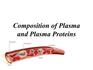
Plasma is the liquid component of blood that remains after red blood cells, white blood cells, and platelets are removed. It is composed primarily of water, proteins, electrolytes, nutrients, wastes, and blood gases. The major proteins in plasma include albumin, globulins, and immunoglobulins. Albumin maintains osmotic pressure and transports molecules like hormones, fatty acids, and bilirubin throughout the body. Globulins such as alpha-1 antitrypsin regulate enzymes and transport metals. Immunoglobulins act as antibodies to help fight infection.

Recommandé
Contenu connexe
Tendances
Tendances (20)
En vedette
En vedette (20)
Similaire à 24 lec composition of plasma & plasma protein
Similaire à 24 lec composition of plasma & plasma protein (20)
Plus de Dr UAK
Plus de Dr UAK (20)
Dernier
Dernier (20)
24 lec composition of plasma & plasma protein
- 1. Composition of Plasma and Plasma Proteins
- 2. Composition of whole blood
- 3. • Respiratory – Transport O2 from lungs to tissues – Transport CO2 from tissues to lungs • Nutrition – Transport “food” from gut to tissues (cells) • Excretory – Transport waste from tissues to kidney (urea, uric acid, water) Blood - Functions
- 4. • Regulatory – Water Content of Tissues & regulation of water balance • Water exchanged through vessel walls to tissue • Regulation of Body Temperature • Protective/defense – Antibodies, antitoxins, white blood cells (WBC) • Acid-base balance – pH 7.35~7.45, NaHCO3/H2CO3 • Coagulation • Transport of hormones ti regulate metabolism
- 5. Blood composition • Blood composition –70 mL/kg of body weight –5 L (average) in an adult –Suspension of cells in a carrier fluid (plasma) • Cells - 45% by volume • Plasma - 55% by volume • Cells –Red cells (erythrocytes): 5x10⁶ /µL –White cells (leukocytes): 7x10³ /µL –Platelets (thrombocytes):3x10⁵ /µL
- 6. Centrifuged Blood Sample Add anticoagulants (heparin, potassium oxalate)
- 8. Separation of Components Plasma = Less Dense RBCs More Dense Platelets / WBCs
- 9. 2. Plasma vs. serum Plasma is the liquid, cell-free part of blood, that has been treated with anti- coagulants. Anticoagulated Serum is the liquid part of blood AFTER coagulation, therfore devoid of clotting factors as fibrinogen. serum= plasma - fibrinogen Clotted
- 10. Components of Plasma Blood plasma Consists of: Water 90% Plasma Proteins 6-8 % Electrolytes (Na+ & Cl- ) 1% Other components: Nutrients (e.g. Glucose and amino acids) Hormones (e.g. Cortisol, thyroxine) Wastes (e.g. Urea) Blood gases (e.g. CO2, O2)
- 11. A large number of dissolved proteins of the plasma includes simple proteins, conjugated proteins carry out a number of different functions. 3. Plasma proteins
- 12. Separation 1. Electrophoresis 2. Ultra-centrifuge A/G=1.5~2.5
- 13. Plasma proteins
- 14. General characteristics of plasma proteins 1. They are synthesized in liver except immunoglobulin. 2. Almost all plasma proteins are glycoproteins. 3. Many plasma proteins exhibit polymorphism such as α1-antitrypsin, transferrin, haptoglobin 4. Each plasma protein has a characteristic half- life in the circulation. 5. Acute Phase Proteins, APP
- 15. 3.1 Albumin Albumin (69 kDa), single polypeptide chain having 585 aa with 17 disulfide bonds, is the most abundant protein (60%) in the blood plasma. (3.5-5.0 g/dl) Synthesis of albumin: – Liver produced about 12g albumin per day which represent 25% of total hepatic protein synthesis and 50% of secreted protein. half-life: 20 days – For this reason, measurement of serum albumin concentration is used to assays liver function test.
- 16. Structure 1. Shape: ellipsoid. 2. Charge: pI=4.0. 3. Domain: three major domains. Human Serum Albumin
- 17. Albumin bind and transport many diverse molecules and serve as low-specificity transport protein, which include: Metal ions: such as calcium and copper. Free fatty acid: albumin binds to free fatty acid released by adipose tissue and facilitates their transfer to other tissue Bilirubin: this protects from the toxic side effects of unconjugated bilirubin. Bile acid: albumin carries the bile acids that are recycle from the intestine to the liver in the hepatic portal Functions of Albumin: TRANSPORT
- 18. Functions of Albumin: Maintainance of osmotic pressure Colloid osmotic pressure, is a form of osmotic pressure exerted by proteins in blood plasma that usually tends to pull water into the circulatory system. Because large plasma proteins cannot easily cross through the capillary walls. In conditions where plasma proteins are reduced, e.g (proteinuria) or (malnutrition), there will be a reduction in osmotic pressure, leading to fluid retention in tissue
- 19. Colloid osmotic pressure Low albumin, causing edema.
- 20. Clinical aspects 1. Albumin binds different drugs and strongly affects the pharmacokinetics of these drugs. E.g sulfonamides can cause the release of unconjugated bilirubin from albumin by competitive binding. If given to infants, sulfonamides may lead to kernicterus. 2. In cases of liver disease or starvation, albumin synthesis decreases. This lead to edema.
- 21. Clinical aspects 3. Hypoalbuminemia – lowered plasma albumin – in malnutrition, nephrotic syndrome and cirrhosis of liver. 4. Albuminuria – albumin is excreted into urine – in nephrotic syndrome and certain inflammatory conditions of urinary tract. 5. Albumin is therapeutically useful for the treatment of burns and hemorrhage.
- 22. Globulins
- 23. 3.2.1 α1–Antitrypsin /α1–Antiproteinase(α1–AT or AAT) It (52 kDa) is a glycoprotein with 394 aa. It is a major constituent of α1globulin fraction of plasma protein, normal concentration about 200mg/dl. It is a serine protease inhibitor and can combines with trypsin, elastase and other protease and inhibits them.
- 24. Clinical significance Emphysema: used to represent the abnormal distension of lungs. –About 5% is due to the deficiency of α1–AT. –This is associated with lung infection and increase the activity of macrophage to release elastase that damage lungs tissue. Smoking can cause oxidation of Met358 to methionine sulfoxide and inactivate α1–AT.
- 25. α1 –antitrypsin deficiency liver disease due to mutant α1 –antitrypsin accumulates and aggregates to form polymers, by unknown mechanism, cause liver damage followed by accumulation of collagen resulting in fibrosis (cirrhosis).
- 26. α2 –Macroglobulin (α2 –MG) It (720 kDa) is a glycoprotein with 4 identical subunits, a major constituent of α2 fraction. It is a panprotease inhibitor and can combine and inhibit many protease. It can bind cytokines such as PDGF and TGFβ and target them to particular cells to affect on cell growth or function.
- 27. Clinical significance α2 -MG levels are increased in nephrotic syndrome a condition wherein the kidneys start to leak out some of the smaller blood proteins. Because of its size, α2 -MG is retained in the bloodstream. This increase has little adverse effect on the health, but is used as a diagnostic clue. nephrotic syndromenormal
- 28. Hepatoglobin (Hp) It (90 kDa) is a glycoprotein. It can bind with the free hemoglobin (extra-corpuscular Hb) in a tight noncovalent complex Hp-Hb during hemolysis. Hp-Hb(155 kDa) cannot pass through glomeruli of kidney while free Hb(65kDa) can and Hp prevent the loss of free Hb into urine. ※Low levels of plasma concentration of Hp can diagnose
- 29. Ceruloplasmin(CER) It (160 kDa) is a blue-coloured, copper- containing α2 fraction. It can carry 90% of plasma copper tightly so that copper is not readily exchangeable. It processes copper- dependent oxidase activity. Albumin carries the other 10% , which is the major supplier of copper to
- 30. Clinical significance Low level of ceruloplasmin is associated with Wilson’s disease (hepatolenticular degeneration) Wilson's disease is an inherited disorder in which there is too much copper in the body's tissues. The excess copper damages the liver and nervous system .Treatment: penicillamine is the first treatment used. This binds copper (chelation) and leads to excretion of copper in the urine.
- 31. Transferrin (Tf) It (76 kDa) is a glycoprotein, part of β fraction. It can transport iron in plasma as ferric ions (Fe3+ ) and protect the body against the toxic effects of free iron.
- 32. The levels of certain plasma proteins change during inflammation, infection, injury, cancer etc. These proteins are “Acute Phase Proteins, APP ” Include C-reactive protein, CRP, α1-acid glycoprotein, fibrinogen, haptoglobin ,α1-antitrypsin, albumin and transferrin. APP are believed to play a role in the bodys’ response to inflammation, changes in their plasma concentrations are generally regarded as being sensitive, although non-specific, indicators if inflammation. Acute Phase Proteins, APP
- 33. C-reactive protein, CRP • A major component of acute phase protein. • It reacts with the C polysaccharide of pneumococci. • Involved in the promotion of immune system through the activation of complement cascade. • Estimation of CRP in serum is important for the evaluation of acute phase response. – CRP rises up to 50,000-fold in acute inflammation, such as infection. It rises above normal limits within 6 hours, and Complement: rupturing membranes of foreign cells, cell swells and bursts. Complement activation
- 34. Immunoglobulins
- 35. Immunoglobulin(Ig)/antibod y(Ab): Glycoprotein molecules that are produced by plasma cells in response to an immunogen and which function as antibodies, mostly associated with γ fraction. But γ-globulin and Ig are not synonymous. Ig is a functional term γ-globulin is physical term. Amountofprotein Mobility albumin globulins +-
- 36. General Functions of Immunoglobulin 1. Antigen(Ag) binding - Ig binds to a specific antigenic determinant 2. Effector functions - Complement activation - Binding to various cells such as phagocytic cells, lymphocytes, mast cells: antibody- mediated phagocytosis or antibody-dependent cell-mediated cytotoxicity (ADCC).
- 37. Two Forms of Ig mIg 1. Membrane Ig, mIg It confers antigenic specificity on B cells. 2. Secreted Ig, SIg It can circulate in the blood and serve as the effectors of humoral immunity by searching out and neutralizing antigens or marking them for elimination. SIg
- 38. Basic Structure 1. four chains (H2L2): Y shape two identical light chains (L): 23 kDa two identical heavy chains (H): 53-75 kDa 2. Disulfide bonds and such noncovalent interactions as salt linkages, hydrogen bonds and hydrophobic bonds to form heterodimer (H-L).
- 39. 1. Variable region (V): VL&VH 2. Constant region (C): CL&CH 3. Hinge region: flexibility Hinge region Light Chain: – VL (110 aa) + CL (110 aa) Heavy Chain: – VH (110 aa) + CH1-CH3 (or CH4) (330-440 aa)