08448380779 Call Girls In Friends Colony Women Seeking Men
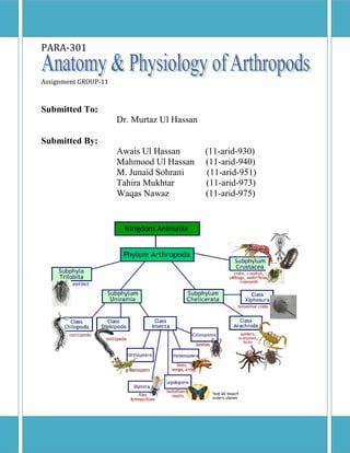
Anatomy & physiology of arthropods
1. PARA-301
Assignment GROUP-11
Submitted To:
Dr. Murtaz Ul Hassan
Submitted By:
Awais Ul Hassan
Mahmood Ul Hassan
M. Junaid Sohrani
Tahira Mukhtar
Waqas Nawaz
(11-arid-930)
(11-arid-940)
(11-arid-951)
(11-arid-973)
(11-arid-975)
2. OUTLINE
Unit 1: GENERAL
1.1 Introduction
1.2 Classification
Unit 2: EXTERNAL ANATOMY & PHYSIOLOGY
2.1 Arthropods Anatomy
2.2 Exoskeleton
2.3 Head
2.4 Molting/Ecdysis
2.4.1 Hardening of cuticle
2.4.2 Hormonal Control
2.5 Antennae (Its Function)
2.5.1 Antennae types
2.6 Mouth Parts
2.7 Wings
2.8 Thorax & Abdomen
2.9 Legs
Unit 3: INTERNAL ANATOMY & PHYSIOLOGY
3.1 Circulatory System
3.2 Respiratory System
3.2.1 Trachea & Oxygen Diffusion
3.2.2 Spiracles
3.3 Nervous System
3.4 Digestive System
3.5 Excretory System
3.6 Reproductive System
3.6.1 Male Reproductive System
3.6.2 Female Reproductive System
3.6.3 Life Cycle
3. Unit1: GENERAL
1.1 Introduction:
Phylum Arthropoda includes an enormous assemblage of both fossil and extant species
that far outnumbers all other known animals put together. Nearly a million species of insects
have been described, and more than a quarter of these are beetles. There are over 50,000 species
of arachnids and another 30,000 of crustaceans.
Arthropods are well represented in the geological record, revealing that all extant classes
appeared during the Paleozoic. Chelicerate and crustacean fossils are present in Cambrian-age
rocks. Most of the so-called “key innovations” leading to insect success, including internal
fertilization, mandibles, and wings, all occurred before the end of the Devonian, and complete
metamorphosis was present in early Carboniferous species. Several modern orders, including
Hymenoptera, Diptera, and Coleoptera, were present by the end of the Paleozoic, and roaches
(order Blattaria) go back at least to the middle Carboniferous. Much current evolutionary
research focuses on the origin of arthropod diversity and includes efforts to establish the
evolutionary role of hormones, especially juvenile hormone (JH) because of its effect on
postembryonic development. Similarly, the action of homeobox genes is now a very active area
of evolutionary research, especially because of the known influence these genes have on
segmental development.
Two structural features contribute significantly to arthropod success: relatively small size and
a chitinous exoskeleton. Although some species such as lobsters and king crabs are quite large
as adults, the vast majority of arthropods are less than 1 cm in length. The planet provides many
places for small organisms to occupy: spaces between sand grains, for example, or cracks in tree
bark and, of course, the bodies of other animals. As a general rule, complex environments
support relatively diverse faunas and floras, and on a small scale Earth is an exceedingly
complex environment. Small species, especially parasitic ones, therefore have a rich supply of
potential ecological niches.
1.2 Classification:
4. Unit 2: EXTERNAL ANATOMY & PHYSIOLOGY
2.1 Arthropods Anatomy:
There are two major classes of arthropods of veterinary importance, namely the Insecta and
Arachnida. The two major classes can be differentiated by the following general characteristics:
Insecta
3 pairs of legs
Body is divided into 3 parts (head, thorax and abdomen)
Head has 6 fused segments and a single pair of antenna which helps in direction
Larval stages
2 pairs of wings (only one pair is functional and of larger size, the second being reduced
to small knob-like sensory structures called halteres, which apparently have a balancing
function.
e.g. lice, bugs, flies, fleas
Arachnida
4 pairs of legs
Body is divided into 2 parts (cephalo-thorax and abdomen)
No antenna
Nymph stages
No wings
e.g. ticks, mites
5.
Ticks active in summer season
Ticks have creeping movement while Fleas have jumping movement
Ticks are larger than fleas
Sand fly causes Leishmaniasis
2.2 Exoskeleton:
The success of the arthropods is related to their hard exoskeleton, segmentation, and jointed
,
appendages.
The exoskeleton provides more support and better protection of internal organs than the
covering of other invertebrates. The cuticle (non cellular protective covering) in arthropods
(non-cellular
forms a rigid exoskeleton, composed mainly of chitin, which is periodically shed as the animal
,
grows. The exoskeleton's middle zone is made of both protein and chitin and is responsible for
the strength of the exoskeleton. It may be additionally strengthened by minerals, such as calcium.
The innermost zone is flexible at the joints allowing free movement. The outer zone is non
nonchitinous and is a complex of proteins and lipids. It provides moisture proofing and protection.
The exoskeleton takes the form of plates ca
called sclerites on the segments, plus rings on the
appendages that divide them into segments separated by joints. This is what differentiates
arthropods from their very close relatives, the Onychophora and Tardigrada.
2.3 Head:
The insect head is a strongly sclerotized capsule joined to the thorax by a flexible, membranous
neck. It bears the mouthparts, comprising the labrum, mandibles, maxillae and labium, and also
thparts,
xillae
the antennae, compound eyes and ocelli.
nd
(1)
(2)
(3)
6.
The compound eyes are often the most prominent structures on the head of
the insects that possess them, as is shown in the dragonfly above (photo 1).
The compound eye is made up of thousands of sensory units called
ommatidia, each of which has an hexagonal lens and 6-8 light sensitive cells.
In the photo 2 above, the ommatidia can be clearly seen as individual
hexagonal units.
Each omnatidium has a limited field of view, but sensory information from
adjacent ommatidia combines to allow for an image to be 'compiled' in the
optic lobe of the insect brain.
The head of an insect generally compromises six fused
segments with a single pair of antenna. There is great
variation in the structure of the mouthparts, depending on
feeding habits, with adaptations for chewing-biting, sponging
or piercing-sucking
2.4 Molting or Ecdysis:
1. Epidermal cells divide mitotically
2. Space develops between epidermis and cuticle
3. Molting fluid, containing proteinases and chitinase, secreted into space. This fluid will
digest the endocuticle but the enzymes are inactive when first secreted.
4. Secretion of cuticulin for epicuticle
5. When cuticulin complete, epidermal cells begin to lay down pro-cuticle
6. Molting fluid activated. Enzymes digest endocuticle.
7. Ecdysial lines (lines of little or no exo-cuticle, e.g. Y shaped line on head of grasshopper
and down dorsal mid line of thorax) become discontinuities in cuticle.
8. Insect removes itself from old cuticle (epicuticle and exocuticle).
7. 2.4.1 Hardening of Cuticle:
The insect swallows air or water and muscles force blood into
the head and thorax. This increased pressure causes the cuticle to split along the ecdysial lines.
The insect suspends itself on a support and helped by gravity, draws itself out, head and thorax
first.
The new cuticle is soft and the insect expands it by swallowing more air or water. Then
hardening of the cuticle occurs, after which, no further expansion of the cuticle can occur. As the
cuticle hardens, it also darkens. Old cuticle is called exuviae and often includes old tracheae that
is continuous with the cuticle.
2.4.2 Hormonal Control:
Molting is controlled by neurosecretory cells in the brain which in turn stimulate
Corpora allata (small glands behind the brain) which produce juvenile hormone (JH)
Prothoracic glands which produce molting hormones (ecdysteroids)
Hormonal Control of Molting.
Ecdysteroids stimulate the epithelial cells in the cuticle to begin the molting process.
The outcome of a molt is determined by the level of juvenile hormone. Juvenile
hormone suppresses adult characters.
8. Large amounts of juvenile hormone = larva => larva
Small amounts of juvenile hormone = larva => pupa
No juvenile hormone = pupa => adult
2.5 Antennae:
Insect antennae vary morphologically. Antennae can detect very low levels of
chemicals and are used in insect communication, finding host plants or mates. (Absent in
arachnids). The antennae are the primary site of olfactory reception and also serve as active
sensors in many insects.
1) The first antennal segment (closest to the head) is called the scape.
2) The second antennal segment is called the pedicel.
3) The remainder of the antenna is collectively called the flagellum
2.5.1 Antennae Types
Aristate antennae are
pouch-like with a
lateral bristle.
Example: House flies.
Aristate Antenna
Capitate antennae are
abruptly clubbed at the
end.
Example: Butterflies.
Capitate Antenna
Clavate antennae are
gradually clubbed at
the end.
Example: Carrion beetles (a family of
beetles in which the adults generally
feed on decaying animal matter or on
the maggots that feed on carrion.)
Clavate Antenna
Example: Ground beetles and
cockroaches. (Ground beetles are
so called because many species
do not fly and lack hind wings).
Filiformis antennae have
a thread-like shape.
Filiformis Antenna
9. Geniculate antennae are
hinged or bent like an
elbow.
Example: Bees and ants.
Geniculate Antenna
Monoliform antennae are
bead-like in shape.
Example: Termites.
Monoliform Antenna
Pectinate antennae have a
comb-like shape.
Example: Fire-coloured beetles
and glow-worms
Pectinate Antenna.
Plumose antennae have a
brush or feather-like
shape.
Example: Moths and
mosquitoes.
Plumose Antenna
Serrate antennae
have a sawtoothed
shape
Example: Click beetles (they derive their name
from the clicking noise produced by a hingelike
structure on their elytra. The click is produced
when the beetle rights itself after falling on its
back).
Serrate Antenna
Setaceous antennae have a
bristle-like shape.
Example: Dragonflies.
Setaceous Antenna
10. 2.6 Mouth Parts:
The 4 mouthparts are the labrum, mandibles, maxillae and labium.The labrum is a simple
fused sclerite, often called the upper lip, and moves longitudinally. It is hinged to the clypeus.
The mandibles, or jaws, are highly sclerotised paired structures that move at right angles to the
body. They are used for biting, chewing and severing food.The maxillae are paired structures
that can move at right angles to the body and possess segmented palps.The labium (often called
the lower lip), is a fused structure that moves longitudinally and possesses a pair of segmented
palps.
Arachnids:
Body is oval
covered with rounded discs
Hard plate (scutum) is absent
Mouthparts not visible from above
Festoons lacking
Take intermittent blood meals
There is great variation in the structure of the mouthparts, depending on feeding habits, with
adaptations for chewing-biting, sponging or piercing-sucking. The labrum or upper lip is a
hinged plate attached to the face or clypeus. The paired mandibles and maxillae or jaws have
areas of their surfaces adapted for cutting, slashing or grinding. The maxillae may also carry
maxillary palps which are sensory in function and used in the monitoring of food. A
hypopharynx, which arises from the floor of the mouth, bears the external opening of the
salivary glands and is similar to a tongue.A labium or lower lip, which may be extensively
modified, especially in the flies, and sometimes bears two sensory labial palps.
11. 2.7 Wings:
Wings are absent in Arachnids. Most adult insect possess wings. Some have shortened
(brachypterous) wings while others may be wingless (apterous). Wings have a network of
veins which give rigidity and support. Air, nerves and blood also pass through the wing veins.
There are several major longitudinal veins. These are the costa, subcosta, radius, median, cubitus
and anal veins. Primitive wings have many, short cross veins. Such wings are called reticulate.
Grasshoppers and cockroaches have leathery forewings which are termed tegmina (sing.
tegmen). Many plant bugs have forewings that are thickened at the base but
membranous distally. These are called hemelytra (sing. hemelytron). Most beetles have very
hardened forewings called elytra(sing. elytron). In flies, (Diptera), the hind wings have become
modified to form small balance organs, called halteres. In more advanced insects the wings are
fastened together. Hamuli are tiny hooks on the anterior margin of the hind wing. These hooks
engage a vein on the posterior margin of the forewing.
A frenulum is a bristle on the hind wing of many butterflies and moths. The frenulum fits
into a hook, (or retinaculum), on the forewing rather like a safety pin. The three segments in the
thorax (pro-, meso- and meta-thorax) each bear a pair of jointed legs. The thorax of many
insects also bears two pairs of wings, but in the winged insects of veterinary significance, i.e.
the Diptera only one pair is functional, the second being reduced to small knob-like sensory
structures, called halteres, which apparently have a balancing function. Wings are outgrowths
of the thoracic tegument supported by hollow tubes called veins which run longitudinally and
crosswise, the intervening areas of tegument being known as cells. The arrangement of the veins
and the shape of the cells are important in identification.
2.8 Thorax & Abdomen:
The insect thorax is box-like with dorsal, ventral and lateral sclerites. The dorsal sclerites
are collectively called the notum or tergum. The ventral sclerites are called the sternum and the
lateral sclerites are called the pleuron. This construction allows attachment and contraction of
muscles used in the movement of the wings and legs.
The thorax is further subdivided into 3 segments, the prothorax, mesothorax and metathorax.
Each of these segments bears a pair of legs. In addition, the mesothorax may bear a pair of fore
wings and the metathorax may bear a pair of hind wings.
12. The insect abdomen has a tergum (never called a notum) and sternum but has no pleuron since it
does not bear legs or wings. Terminally the abdomen bears the external genitalia. In some female
insects there is a very obvious ovipositor for depositing eggs. A pair of cerci is also present at
the end of the abdomen. These have a sensory function. In some orders there may also be
additional terminal appendages. The abdomen of insects consists of up to 11 segments with
terminal modifications to form the genitalia.
2.9 Legs:
The legs, named from the anterior, are the fore, mid and hind legs. Each leg has several
segments:
The coxa articulates with the body.
The trochanter is usually quite small.
The femur is usually the longest and strongest segment.
The tibia is usually long and slender.
The tarsus is collectively composed of 2 to 5 smaller tarsomeres. The last tarsomere
usually has a pair of claws and often 2 or 3 tarsal pads.
Unmodified legs are used for walking and are called ambulatory.
Legs modified for running are called cursorial.
Digging legs are called fossorial.
Swimming legs are called natatorial.
Jumping legs are called saltatorial.
Grasping legs are called raptorial.
13. Unit 3: INTERNAL ANATOMY & PHYSIOLOGY
3.1 Circulatory System:
The arthropod coelom is greatly reduced, its remnants being found in excretory organ or
gonad spaces. The main body cavity of arthropods is thus a secondary space—the hemocoel—
filled with fluid (hemolymph) containing a variety of cell types. Muscles, sometimes very large
ones, are bathed in this fluid, which is circulated through an open circulatory system by means of
a dorsal tubular heart. Hemolymph enters the heart from the surrounding pericardial
sinusthrough pairs of lateral openings, the ostia.Ostia are one-way valves; when the heart
contracts, ostia close, forcing hemolymph anteriorly into the arteries and finally into a system of
tissue spaces, or sinuses. Hemolymph works its way back to the heart through these sinuses,
often aided by body movements. Formed elements of hemolymph are mostly amebocytes.
Parasites, especially larval stages, may penetrate the gut and come to lie in the hemocoel, as in
the case of acanthocephalan or tapeworm larvae. And malarial sporozoites escape from their
oocyst on the gut and migrate through the hemocoel to the mosquito vector’s salivary glands.
Arthropods have an open circulatory system. Haemolymph, a copper-based blood analogue, is
propelled by a series of hearts into the body cavity where it comes in direct contact with the
tissues. Arthropods are protostomes. There is a coelom (body cavity), but it is reduced to a tiny
cavity around the reproductive and excretory organs, and the dominant body cavity is a
hemocoel, filled with hemolymph that bathes the organs directly. The arthropod body is divided
into a series of distinct segments, plus a presegmental acron that usually supports compound and
simple eyes and a postsegmental telson (the last body division in crustaceans, but not a true
segment). These are grouped into distinct, specialized body regions called tagmata. Each
segment at least primitively supports a pair of appendages.
14. 3.2 Respiratory System:
Gas exchange takes place directly through the body wall in very small arthropods that may
lack specialized respiratory organs and even a heart. Larger Crustacea have gills,which are
extensive folds of the epidermis, covered with thin cuticle, through which hemolymph circulates.
Most insects, as well as many Acari, have a tracheal system, a branching network of tubes. The
tracheal system opens at spiracles and ramifies through the body into a large number of very fine
tracheoles. The cuticle of tracheae but not that of tracheoles is shed at ecdysis. Ventilation of the
tracheal system is accomplished by pressure of body muscles on the walls of elastic tracheae, on
tracheal air sacs, or both. Arachnid tracheal systems are thought to have evolved from book
lungs,membranous folds inside a chamber that opens through a slit or spiracle. Book lungs occur
in several arachnid orders but not in Acari.
3.2.1 Trachea & Oxygen diffusion:
Insects, in general, do not have an oxygen-carrying chemical in their blood so oxygen reaches
cells by other means. Most insects have a waterproof cuticle but some insects live in moist areas
and are sedentary. Their cuticle is permeable to water and they obtain sufficient oxygen by
diffusion across their cuticle. However, most insects have a special respiratory system
comprising a system of internal tubes, called trachea, which branch and re-branch. Very fine
branches, tracheoles, penetrate individual cells. Trachea has spiral stiffening - like vacuum
cleaner hose - to prevent collapse. Air enters from the outside through a series of
openings, spiracles. Typically there are 2 pair of spiracles laterally on the thorax and 8 pair
laterally on the abdomen.
O2 from spiracles => tracheae => tracheoles => cells
15. 3.2.2 Spiracles:
Insects need to avoid water loss through their spiracles and also to prevent
contamination by dust etc.
Spiracles are therefore usually equipped with opening and closing devices and filtering lobes or
hairs in an atrium before the beginning of the trachea. Very active insects have internal air sacs,
as extra reservoirs, as part of their tracheal system. They also may employ mechanical ventilation
along the larger trachea. Bees and wasps may extend and telescope their abdomens to pump air
along.
3.3 Nervous System:
The arthropod central nervous system consists of a dorsal ganglionic mass, the brain, lying
above the stomodaeum (anterior most part of the digestive system); nerves that supply cephalic
sense organs; nerve trunks or commissures surrounding the esophagus and connecting the brain
to a sub-esophageal ganglion; and a ventral nerve trunk that lies beneath the digestive tract. The
ventral trunk consists of a double cord connecting segmental ganglia. However, in many if not
most arthropods this fundamental structure is modified by postembryonic compression and
shortening of the nerve trunk, fusion of ganglia, and lengthening of fibers to the posterior part of
the animal. The brain itself consists of three major regions: protocerebrum, deuterocerebrum,
which in crustacea supplies nerves to the first antennae; and tritocerebrum. On the basis of
evidence from comparative anatomy and embryological studies, the tritocerebrum consists of
segmental ganglia incorporated by fusion into the brain. Evidence for homology of arthropod
anterior appendages is found in the fact that nerve centers of crustacean second antennae, the
chelicerae of chelicerates, and the antennae of insects are all located in the tritocerebrum. The
peripheral nervous system includes axons that innervate muscles and glands and bi- or multipolar
16. neurocytes, their distal processes, and axons. Sensory neurocytes are connected to a variety of
sense organs, including tactile hairs and bristles and chemo-receptors.
The three pairs of fused ganglia of the head region, form the brain. The three pairs of ganglia
from the segments bearing the mouthparts have coalesced to form the sub-esophageal ganglion.
Brain and ventral nerve cord
Cells that detect and transmit sensations of pain (Nociceptors), but not proven that insects feel
pain consciously.
3.4 Digestive System:
3.4.1 ALIMNTARY CANAL: The alimentary canal or gut of insects can be divided into 3
sections
Foregut (stomodeam)
Midgut (mesenteron)
Hindgut (proctodeam)
17. 3.4.1.1 Foregut:
The foregut consists of the mouth (oral cavity) The salivary glands provide fluids and enzymes
to the mouth for lubrication and to begin food breakdown.
Throat (pharynx)
Esophagus
Crop, for storage of food
Proventriculus, (or gizzard) where present, sometimes armed with teeth for grinding
3.4.1.2 Mid-gut
The midgut consists of the;
Ventriculus, where most digestion is carried out
Gastric caeca (sing. caecum) which , if present provide greater area for digestion
3.4.1.3 Hind-gut
The hindgut consists of the;
Anterior hindgut
Rectum
Both these areas reabsorb water and salts.
Valves are present to prevent back-flow of material within the gut cardiac (stomodeal) valve
between fore and midgut pyloric valve between mid and hindgut In insects the products
of excretion are emptied from the Malpighian tubules into the alimentary canal at the beginning
of the hindgut.
3.4.2 SPECIALISATION: THE FILTER CHAMBER
The most obvious specialization in gut structure occurs in many of the Hemiptera or true bugs.
These insects ingest large quantities of often dilute fluid and have an unusual arrangement of the
18. gut called a filter chamber whereby the gut is looped upon itself. This allows contact of the
anterior and hind guts so that water can be absorbed across the hind gut and gut contents
concentrated by passing through several loops before major absorption of nutrients.
3.4.3 FAT BODY
The fat body is diffuse tissue lying usually in the abdomen. It is important in the storage of fat,
protein and glycogen and as such, grows in size in larvae, and is reduced during pupation and
metamorphosis. It is quite small in the adult when reserves are used for egg production. The fat
body is also involved in intermediate metabolism and in this way, resembles the vertebrate liver.
The fat body is rich in enzymes. Fats may be synthesized, released by the fat body into the
haemocoel or be broken down. It is also important in detoxification and symbiotic are present in
some insect fat bodies and aid in the synthesis of various vitamins and amino acids for protein
synthesis.
3.5 Excretory System:
Crustacean excretory organs are pairs of antennal and maxillary glands opening to the
outside on or near the bases of antennae or maxillae, respectively. Both pairs are often present in
larvae; adults normally retain only one or the other. The principal nitrogenous excretory products
are ammonia with some amines and small amounts of urea and uric acid. Considerable excretion
of ammonia also takes place across the gills. Almost all insects have Malpighian tubules, ranging
in number from 4 to over 100. These thin-walled tubules are closed at their distal ends but open
into the mid-gut near its junction with the hindgut. Uric acid is excreted, usually as an
ammonium, potassium, or sodium salt. Water in the urine is reabsorbed by the proximal
Malpighian tubules or by the rectal wall; sodium and potassium are resorbed as bicarbonates,
leaving virtually insoluble free uric acid as a precipitate. Thus, water and cations are recycled, as
part of the overall water conservation mechanism of insects. Bloodsucking forms, however,
produce large amounts of fluid urine after a meal, an event that rids the animal of excess water.
Excretory coxal glands are found in some mites and other arachnids. These glands open to the
outside at the bases of one or more pairs of appendages. Most ticks and mites also have
Malpighian tubules. Waste from the hemocoel is taken up by tubule walls and excreted into the
lumen as guanine, the main excretory product. In those Prostigmata and Metastigmata whose
ventriculus does not connect with the hindgut, an anteriorly directed excretory canal is joined to
the hindgut, and guanine is excreted by this organ through the “anus” (uropore).
19. The excretory system maintains a constant internal environment by
Regulating water and ionic balance
Eliminating nitrogenous wastes
Nitrogenous wastes are eliminated either as
Ammonia - as in aquatic insects, meat-eating maggots and aphids
Urea - as in clothes moths (and humans)
Uric acid - as in most insects
The choice of nitrogenous excretory product is dependent upon the need to conserve water.
Ammonia, NH3, is simple, easy to make but quite toxic. It needs to be dissolved in large
quantities of water so is suitable for insects in moist environments.
H : N = 3 : 1 so it requires a lot of water to make.
Urea is moderately toxic and also needs to be eliminated in water, but H : N = 2 : 1 so
less water is needed for its manufacture than ammonia.
Uric acid is fairly harmless and insoluble. It crystallizes out of solution and can be
excreted as a solid or retained in special body cells in the insect. H : N = 1 : 1 so it
requires the least amount of water for its manufacture.
20. Malpighian Tubules: These are the main excretory organs of the insect body. They are fine,
(one cell thick), lying free within the body cavity or attached to the outside of the gut. They
absorb wastes from the haemocoel either by diffusion across a concentration gradient or by
active transport. The number of Malpighian tubules varies and they enter the alimentary canal at
the junction of the mid and hind guts.
Faeces: Re-absorption of salts and water occurs in the rectum. The faeces, (or urine if liquid),
contains wastes from both the alimentary canal and the Malpighian tubules. The texture is
variable depending upon the diet and ranges from a clear liquid in aphids to hard pellets or a dry,
powdery material in borers.
3.6 Reproductive System:
3.6.1 Male Reproductive System:
The male reproductive system consists of paired testes (where sperm are produced), vas
deferens (tubes from the testes), seminal vesicles, (where sperm are stored), accessory glands,
(which provide seminal fluid and the spermatophore) and a common ejaculatory duct .
3.6.2 Female Reproductive System:
The female reproductive system includes a pair of ovaries, lateral oviducts, a common
oviduct and a vagina. Generally each ovary is composed of several ovarioles to produce multiple
21. eggs (ova). Most female insects also have one or more spermathecae where sperm can be stored
for some time and can be nourished by secretions from the spermathecal glands.
Accessory glands add various coatings to eggs before they are laid. These usually aid in the
adhesion of the eggs to a substrate.
3.6.2.1 MATING:
Many insects use displays or dancing to entice females to mate. Males may fight each
other or decide the victor by comparing size.
Some female insects will not mate unless the male is in possession of a suitable territory
or food source.
Butterflies of both sexes are known to congregate on hill tops or other geographic
protrusions where they will select a mate. Other insects form swarms.
Female scorpion flies require a nuptial gift of food from the male before mating. The
male mates with her while she eats it. Males are selected by the size and quality of the
gift.
3.6.2.2 FERTILIZATION:
Aquatic insect ancestors could discharge sperm into water but terrestrial insects must
transfer sperm to the female without allowing it to dry out. This is often done via
spermatophores, packets of sperm. In some insects the spermatophores also have edible
portions for the female.
Most insect eggs have a coating to protect the embryo from desiccation. However, they
also possess micropyles, small holes to allow the entry of sperm. (There are also other
holes - aeropyles - connecting to air bubbles within the egg shell membranes).
Fertilization occurs within the oviduct. As the fertilized egg moves down the oviduct, it is
coated with secretions from various accessory organs.
3.6.3 Life Cycle:
In insects the sexes are separate and after fertilization either eggs or larvae arc produced.
Development often involves three or more larval stages followed by the formation of a pupa and
a marked transformation or metamorphosis to the adult stage as in all the flies and fleas, i.e. a
holometabolous life cycle. In other insects development occurs from the egg through several
nymphal stages which resemble the adult, as in lice, i.e. a hemimetabolous life cycle.
The different developmental stages in the life cycle are known as instars OR it is the time space
between 1st and 2nd larval stage.
Holometabolous life cycle = complete metamorphosis
Hemimetabolous life cycle = incomplete metamorphosis
Males of the Hypodermatidae family die after mating
22. Generally, insects have large females and small males
Ticks Life cycles may be one-host, two-host or three-host ticks life cycle.
ONE-HOST TICK: (Boophilus)