SlideShare une entreprise Scribd logo
1  sur  42
Promoters and enhancers
21.1 Introduction

I   nitiation of transcription requires the enzyme RNA polymerase and transcription factors. Any
protein that is needed for the initiation of transcription, but which is not itself part of RNA
polymerase, is defined as a transcription factor. Many transcription factors act by recognizing
c/s-acting sites on DNA. However, binding to DNA is not the only means of action for a
transcription factor. A factor may recognize another factor, or may recognize RNA polymerase, or
may be incorporated into an initiation complex only in the presence of several other proteins. The
ultimate test for membership of the transcription apparatus is functional: a protein must be needed
for transcription to occur at a specific promoter or set of promoters.

A significant difference between the transcription of eukaryotic and prokaryotic mRNAs is that
initiation at a eukaryotic promoter involves a large number of factors that bind to a variety of ci's-
acting elements. The promoter is defined as the region containing all these binding sites, that is,
which can support transcription at the normal efficiency and with the proper control. So the major
feature defining the promoter for a eukaryotic mRNA is the location of binding sites for
transcription factors. RNA polymerase itself binds around the startpoint, but does not directly
contact the extended upstream region of the promoter. By contrast, the bacterial promoters
discussed in 9 Transcription are largely defined in terms of the binding site for RNA polymerase in
the immediate vicinity of the startpoint. Transcription in eukaryotic cells is divided into three
classes. Each class is transcribed by a different RNA polymerase:

• RNA polymerase I transcribes rRNA
• RNA polymerase IT transcribes mRNA
• RNA polymerase III transcribes tRNA and other small RNAs.
Transcription factors are needed for initiation, but are not required

subsequently. For the three eukaryotic enzymes, the factors, rather than the RNA polymerases
themselves, are principally responsible for recognizing the promoter. This is different from bacterial
RNA polymerase,where it is the enzyme that recognizes the promoter sequences. For all
eukaryotic RNA polymerases, the factors create a structure at the promoter to provide the target that
is recognized by the enzyme. For RNA polymerases I and III, these factors are relatively simple,
but for RNA polymerase II they form a sizeable group collectively known as the basal factors. The
basal factors join with RNA polymerase II to form a complex surrounding the startpoint, and they
determine the site of initiation. The basal factors together with RNA polymerase constitute the basal
transcription apparatus.The promoters for RNA polymerases I and II are (mostly) upstream of
thestartpoint, but some promoters for RNA polymerase III lie downstream of the startpoint. Each
promoter contains characteristic sets of short conserved sequences that are recognized by the
appropriate class of factors. RNA polymerases I and III each recognize a relatively restricted
set of promoters, and rely upon a small number of accessory factors. Promoters utilized by RNA
polymerase II show more variation in sequence, and have a modular organization. Short sequence
elements that are recognized by transcription factors lie upstream of the startpoint. These cis-acting
sites usually are spread out over a region of >200 bp. Some of these elements and the factors that
recognize them are common: they are found in a variety of promoters and are used constitutively.
Others are specific: they identify particular classes of genes and their use is regulated. The elements
occur in different combinations in individual promoters. All RNA polymerase II promoters have
sequence elements close tothe startpoint that are bound by the basal apparatus and that establish the
site of initiation. The sequences farther upstream determine whether the promoter is expressed in all
cell types or is specifically regulated. Promoters that are constitutively expressed (their genes are
sometimes called housekeeping genes) have upstream sequence elements that are recognized by
ubiquitous activators. No element/factor combination is an essential component of the promoter,
which suggests that initiation by RNA polymerase II may be sponsored in many different ways.
Promoters that are expressed only in certain times or places have sequence elements that require
activators that are available only at those times or places.

Sequence components of the promoter are defined operationally by the demand that they must be
located in the general vicinity of the startpointand are required for initiation. The enhancer is
another type of site involved in initiation. It is identified by sequences that stimulate initiation, but
that are located a considerable distance from the startpoint. Enhancer elements are often targets for
tissue-specific or temporal regulation.

Figure 21.1 illustrates the general properties of promoters and enhancers.

The components of an enhancer resemble those of the promoter; they consist of a variety of
modular elements. However, the elementsare organized in a closely packed array. The elements in an
enhancerfunction like those in the promoter, but the enhancer does not need to benear the startpoint. However,
proteins bound at enhancer elements interactwith proteins bound at promoter elements. The distinction
betweenpromoters and enhancers is operational, rather than implying a fundamental
difference in mechanism. This view is strengthened by the fact that some types of element are found in both
promoters and enhancers. Eukaryotic transcription is most often under positive regulation: a transcription
factor is provided under tissue-specific control to activate a promoter or set of promoters that contain a
common target sequence. Regulation by specific repression of a target promoter is less common.


Figure 21.1 A typical gene transcribed by RNA polymerase II has a promoter that extends upstream from the
site where transcription is initiated. The promoter contains several short (<10 bp) sequence
elements that bind transcription factors, dispersed over >200 bp. An enhancer containing a more closely
packed array of elements that also bind transcription factors may be located several kb distant. (DNA may be
coiled or otherwise rearranged so that transcription factors at the promoter and at the enhancer interact
to form a large protein complex.)
21.2 Eukaryotic RNA polymerases consist of
many subunits
: Key Concepts
• • RNA polymerase I synthesizes rRNA in the nucleolus.
: • RNA polymerase II synthesizes mRNA in the nucleoplasm.
: • RNA polymerase III synthesizes small RNAs in the nucleoplasm.
: • All eukaryotic RNA polymerases have — 12 subunits and are
• aggregates of >500 kD.
: • Some subunits are common to all three RNA polymerases.
: • The largest subunit in RNA polymerase II has a CTD (carboxyi
terminal domain) consisting of multiple repeats of a septamer.


T    he three eukaryotic RNA polymerases have different locations in the nucleus, corresponding with the
genes that they transcribe. The most prominent activity is the enzyme RNA polymerase I, which resides in the
nucleolus and is responsible for transcribing the genes coding for rRNA. It accounts for most cellular RNA
synthesis (in terms of quantity).
The other major enzyme is RNA polymerase II, located in the nucleoplasm (the part of the nucleus excluding
the nucleolus). It represents most of the remaining cellular activity and is responsible for synthesizing
heterogeneous nuclear RNA (hnRNA), the precursor for mRNA. RNA polymerase III is a minor enzyme
activity. This nucleoplasmicenzyme synthesizes tRNAs and other small RNAs. All eukaryotic RNA
polymerases are large proteins, appearing as aggregates of >500 kD. They typically have ~12 subunits. The
purified enzyme can undertake template-dependent transcription of RNA, but is not able to initiate selectively
at promoters. The general constitution of a eukaryotic RNA polymerase II enzyme as typified in S.
cerevisiaeis illustrated in Figure 21.2. The two largest subunits are homologous to the β and $' subunits of
bacterial RNA polymerase. Three of the remaining subunits are common to all the RNA polymerases, that is,
they are also components of RNA polymerases I and III. The largest subunit in RNA polymerase II has a
carboxy-terminaldomain (CTD), which consists of multiple repeats of a consensussequence of 7 amino acids.
The sequence is unique to RNA polymeraseII. There are ~26 repeats in yeast and ~50 in mammals. The
number ofrepeats is important, because deletions that remove (typically) more thanhalf of the repeats are
lethal (in yeast). The CTD can be highly phosphorylatedon serine or threonine residues; this is involved in the
initiationreaction (see 21.11 Initiation is followed by promoter clearance).

The RNA polymerases of mitochondria and chloroplasts are smaller, and resemble bacterial RNA polymerase
rather than any of the nuclearenzymes. Of course, the organelle genomes are much smaller, the resident
polymerase needs to transcribe relatively few genes, and the control of transcription is likely to be very
much simpler (if existing at all). So these enzymes are analogous to the phage enzymes that do not need
the ability to respond to a more complex environment. A major practical distinction between the
eukaryotic enzymes is drawn from their response to the bicyclic octapeptideα-amanitin. In basically all
eukaryotic cells the activity of RNA polymerase II is rapidly inhibited by low concentrations of α-
amanitin. RNA polymerase I is not inhibited. The response of RNA polymerase III to α-amanitin is
less well conserved; in animal cells it is inhibited by high levels, but in yeast and insects it is not
inhibited.
21.3 Promoter elements are defined by
mutations and footprinting
Key Concepts
• Promoters are defined by their ability to cause transcription of an
attached sequence in an appropriate test system in vitro or in vivo.


T    he first step in characterizing a promoter is to define the overall length of DNA that contains all the
necessary sequence elements. To do this, we need a test system in which the promoter is responsible
for the production of an easily assayed product. Historically, several types of systems have been used:

• In the oocyte system, a DNA template is injected into the nucleus of the X. laevisoocyte. The RNA
transcript can be recovered and analyzed. The main limitation of this system is that it is restricted to the
conditions that prevail in the oocyte. It allows characterization of DNA sequences, but not of the factors
that normally bind them.

• Transfection systems allow exogenous DNA to be introduced into a cultured cell and expressed. (The
procedure is discussed in 18.17 Transfectionintroduces exogenous DNA into cells.) The system is
genuinely
in vivo in the sense that transcription is accomplished by the same apparatus responsible for expressing
the cell's own genome. However, it differs from the natural situation because the template consists of a
gene that would not usually be transcribed in the host cell. The usefulness of the system may be
extended by using a variety of host cells.

• Transgenic systems involve the addition of a gene to the germline of an animal. Expression of the
transgene can be followed in any or all of the tissues of the animal. Some common limitations apply to
transgenic systems and to transfection: the additional gene often is present in multiple copies, and is
integrated at a different location from the endogenous gene. Discrepancies between the expression of a
gene in vitro and its expression as a transgene can yield important information about the role of the
genomic context of the gene.
• The in vitro system takes the classic approach of purifying all the components and manipulating
conditions until faithful initiation is seen. "Faithful" initiation is defined as production of an RNA
starting at the site corresponding to the 5' end of mRNA (or rRNA or tRNA precursors). Ultimately this
allows us to characterize the individual sequence elements in the promoter and the transcription factors
that bind to them. When a promoter is analyzed, it is important that only the promoter sequence changes.
Figure 21.3 shows that the same long upstream sequence is always placed next to the promoter to ensure
that it is always inthe same context. Because termination does not occur properlyin the in vitro
systems, the template is cut at some distancefrom the promoter (usually ~500 bp downstream), to
ensure that all polymerases "run off" at the same point, generating an identifiable transcript.
We start with a particular fragment of DNA that can initiate transcription in one of these systems.
Then theboundaries of the sequence constituting the promoter can be determined by reducing the
length of the fragment from either end, until at some point it ceases to be active, as illustrated in
Figure 21.4. The boundary upstream can be identified by progressively removing material from this
end until promoter function is lost. To test the boundary downstream, it is necessary to reconnect
the shortened promoter to the sequence to be transcribed (since otherwise there is no product to
assay). Once the boundaries of the promoter have been defined, the importance of particular bases
within it can be determined by introducing point mutations or other rearrangements in the sequence.
As with bacterial RNA polymerase, these can be characterized as up or down mutations. Some of
these rearrangements affect only the rate of initiation; others influence the site at which initiation
occurs, as seen in a change of the startpoint. To be sure that we are dealing with comparable
products, in each case it is necessary to characterize the 5' end of the RNA. We can apply several
criteria in identifying the sequence components of a promoter (or any other site in DNA):

• Mutations in the site prevent function in vitro or in vivo. (Many techniques now exist for
introducing point mutations at particular base pairs, and in principle every position in a promoter
can be mutated, and the mutant sequence tested in vitro or in vivo.)

• Proteins that act by binding to a site may be footprinted on it. There should be a correlation
between the ability of mutations to prevent promoter function and to prevent binding of the factor.

• When a site recognized by a particular factor is present at multiple promoters, it should be possible
to derive a consensus sequence that is bound by the factor. A new promoter should become
responsive to this factor when an appropriate copy of the element is introduced.
Figure 21.3 A promoter is tested by modifying the sequence that is connected to a constant upstream
sequence and a constant downstream transcription unit.


Figure 21.4 Promoter boundaries can be determined by making deletions that progressively remove more
material from one side. When one deletion fails to prevent RNA synthesis but the next stops
transcription, the boundary of the promoter must lie between them.
21.4 RNA polymerase I has a bipartite
promoter
Key Concepts
• The RNA polymerase I promoter consists of a core promoter and
an upstream control element.
• The factor UBF1 binds to both regions and enables the factor SL1
to bind.
• SL1 includes the factor TBP that is involved in initiation by all
threeRNA polymerases.
• RNA polymerase binds to the UBF1-SL1 complex at the core
promoter.


R    NA polymerase I transcribes only the genes for ribosomal RNA, from a single type of
promoter. The transcript includes the sequencesof both large and small rRNAs, which are later
released by cleavages and processing. There are many copies of the transcription unit, alternating
with nontranscribedspacers, and organized in a cluster as discussed in 4.8Genes for rRNAform
tandem repeats. The organizationof the promoter, and the events involved in initiation,
are illustrated in Figure 21.5. The promoter consists of two separate regions.The core promoter
surrounds the startpoint, extendingfrom -45 to +20, and is sufficient for transcription
to initiate. It is generally G-C-rich (unusual for a promoter) except for the only conserved sequence
element, a short A-T-rich sequence around the startpointcalled the Inr. However, its efficiency is
very much increased by the upstream promoter element (UPE), another G-C-rich sequence, related
to the core promoter sequence, which extends from -180 to -107. This type of organization is
common to pol I promoters in many species, although the actual sequences vary widely. RNA
polymerase I requires two ancillary factors.

The factor that binds to the core promoter consists of 4 proteins. (It is called SL1, TIF-IB,
Ribl in different species). One of its components, called TBP, is a factor that is required also for
initiation by RNA polymerases II and III (see 21.8TBP is a universal factor). TBP does not bind
directly to G-C-rich DNA, so DNA-binding is probably the responsibility of the other components
of the core-binding factor. It is likely that TBP interacts with RNA polymerase, possibly with a
common subunit or a feature that has been conserved among polymerases. Core-binding factor
enables RNA polymerase I to initiate from the promoter at a low basal frequency. The core-binding
factor has primary responsibility for ensuring that the RNA polymerase is properly localized at the
startpoint. We see shortly that a comparable function is provided for RNA polymerases II and III by
a factor that consists of TBP associated with other proteins. So a common feature in initiation by all
three polymerases is a reliance on a "positioning" factor that consists of TBP associated with
proteins that are specific for each type of promoter.

For high frequency initiation, the factor UBF is required. This is a single polypeptide that binds to a
G-C-rich element in the upstream promoter element. One indication of how UBF interacts with the
corebindingfactor is given by the importance of the spacing between the upstream promoter element
and the core promoter. This can be changed by distances involving integral numbers of turns of
DNA, but not by distances that introduce half turns. This implies that UBF and corebindingfactor
need to be bound on the same face of DNA in order to interact. In the presence of UBF, core-
binding factor binds more efficiently to the core promoter.
Figure 21.5 Transcription units for RNA polymerase I have a core promoter separated by —70 bp from the
upstream promoter element. UBF binding to the UPE increases the ability of core-binding factor
to bind to the core promoter. Core-binding factor positions RNA polymerase I at the startpoint.

21.5 RNA polymerase III uses both
downstream and upstream promoters
Key Concepts
• RNA polymerase III has two types of promoters.
• Internal promoters have short consensus sequences located
within the transcription unit and cause initiation to occur a fixed
distance upstream.
• Upstream promoters contain three short consensus sequences
upstream of the startpoint that are bound by transcription factors.


R    ecognition of promoters by RNA polymerase III strikingly illustrates the relative roles of
transcription factors and the polymerase enzyme. The promoters fall into two general classes that
are recognized in different ways by different groups of factors. The promoters for 5S and tRNA
genes are internal; they lie downstream of thestartpoint. The promoters for snRNA (small nuclear
RNA) genes lie upstream of the startpoint in the more conventional manner of other promoters. In
both cases, the individual elements that are necessary for promoter function consist exclusively of
sequences recognized by transcription factors, which in turn direct the binding of RNA polymerase.

Before the promoter of 5S RNA genes was identified inX. laevis, all attempts to identify promoter
sequences assumed that they would lie upstream of the startpoint. But deletion analysis showed that
the 5S RNA product continues to be synthesized when the entire sequence upstream of the gene is
removed! When the deletions continue into the gene, a product very similar in size to the usual 5S
RNA continues to be synthesized so long as the deletion ends before base +55. Figure 21.6 shows
that the first part of the RNA product corresponds to plasmid DNA; the second part represents
the segment remaining of the usual 5S RNA sequence. But when the deletion extends past +55,
transcription does not occur. So the promoter lies downstream of position +55, but causes RNA
polymerase III to initiate transcription a more or less fixed distance upstream. When deletions
extend into the gene from its distal end, transcription is unaffected so long as the first 80 bp remain
intact. Once the deletion cuts into this region, transcription ceases. This places the downstream
boundary position of the promoter at about position +80. So the promoter for 5S RNA transcription
lies between positions+55 and +80 within the gene. A fragment containing this region can sponsor
initiation of any DNA in which it is placed, from a startpoint~55 bp farther upstream. (The wild-
type startpoint is unique; in deletions that lack it, transcription initiates at the purine base nearest to
the position 55 bp upstream of the promoter.)

The structures of three types of promoters for RNA polymerase III are summarized in Figure 21.7.
There are two types of internal promoter. Each contains a bipartite structure, in which two short
sequence elements are separated by a variable sequence. Type 1 consists of a boxA sequence
separated from a boxC sequence, and type 2 consists of a boxA sequence separated from a boxB
sequence. The distance between boxA and boxB in a type 2 promoter can vary quite extensively,
but the boxes usually cannot be brought too close together without abolishing function. Type 3
promoters have three sequence elements all located upstream of the startpoint.
21.6 TF|||B is the commitment factor for pol III
promoters
Key Concepts
• TF|MA and TFmC bind to the consensus sequences and enable
TFM|B to bind at the startpoint.
• TFmB has TBP as one subunit and enables RNA polymerase to
bind.


T   he detailed interactions are different at the two types of internal promoter, but the principle is
the same. TFniC binds downstream of the startpoint, either independently (type 2 promoters) or in
conjunction with TFIUA (type 1 promoters). The presence of TFITICenables the positioning factor
TFmBto bind at the startpoint. Then RNA polymerase is recruited.Figure 21.8 summarizes the
stages of reaction at type 2 internal promoters. TF[][C binds to both boxA and boxB. This enables
TFmB to bind at the startpoint. Then RNA polymerase III can bind. The difference at type 1 internal
promoters is that TFmA must bind at boxA to enable TFmC to bind at boxC. Figure 21.9 shows that,
once TFU1C has bound, events follow the same course as at type 2 promoters, with TFJJJB binding at
the startpoint, and RNA polymerase III joining the complex. Type 1 promoters are found only in the
genes for 5S rRNA.

TFmA and TFmC are assembly factors, whose sole role is to assist the binding of TFIHB at the right
location. Once TFmB has bound, TFUIA and TFTljC can be removed from the promoter (by high salt
concentration in vitro) without affecting the initiation reaction. TFn]Bremains bound in the vicinity
of the startpoint and its presence is sufficientto allow RNA polymerase 111 to bind at the startpoint.
So TFniB is the only true initiation factor required by RNA polymerase III. This sequence of events
explains how the promoter boxes downstream can cause RNA polymerase to bind at the startpoint,
farther upstream. Although the ability to transcribe these genes is conferred by the internal
promoter, changes in the region immediately upstream of the startpointcan alter the efficiency of
transcription. TFmC is a large protein complex (>500 kD), comparable in size to RNA polymerase
itself, and containing 6 subunits. TFinA is a member of an interesting class of proteins containing a
nucleic acid-binding motif called a zinc finger (see 22.9 A zinc finger motif is a DNA-binding
domain). The positioning factor, TFmIB, consists of three subunits.

 It includes the same protein, TBP, that is present in the core-binding factor for pol I promoters, and
also in the corresponding transcription factor (TFnD) for RNA polymerase II. It also contains Brf,
which is related to the factor TFnB that is used by RNA polymerase II. The third subunit is called
B"; it is dispensable if the DNA duplex is partially melted, which suggests that its function is to
initiate the transcription bubble. The role of B " may be comparable to the role played by sigma
factor in bacterial RNA polymerase (see 9.16 Substitution of sigma factors maycontrol initiation).
The upstream region has a conventional role in the third class of polymerase III promoters. In the
example shown in Figure 21.7, there are three upstream elements. These elements are also found in
promoters for snRNA genes that are transcribed by RNA polymerase II. (Genes for some snRNAs
are transcribed by RNA polymerase II, while others are transcribed by RNA polymerase III.) The
upstream elements function in a similar manner in promoters for both polymerases II and III.

Initiation at an upstream promoter for RNA polymerase III can occur on a short region that
immediately precedes the startpoint and contains only the TATA element. However, efficiency of
transcription is much increased by the presence of the PSE and OCT elements. The factors that bind
at these elements interact cooperatively. (The PSE element may be essential at promoters used by
RNA polymerase II, whereas it is stimulatory in promoters used by RNA polymerase III; its
name stands for proximal sequence element.)
The TATA element confers specificity for ftie type of polymerase (II or III) that is recognized by
ansnRNA promoter. It is bound by a factor that includes the TBP, which actually recognizes the
sequence in DIVA. The TBP is associated with other proteins, which are specific for the type of
promoter. The function of TBP and its associated proteins is to position the RNA polymerase
correctly at the startpoint. We discuss thisin more detail for RNA polymerase II (see 21.8 TBP is a
universal factor).
The factors work in the same way for both types of promoters forRNA polymerase III. The factors
bind at the promoter before RNA polymerase itself can bind. They form a preinitiation complex that
directs binding of the RNA polymerase. RNA polymerase III does not itself recognizes the
promoter sequence, but binds adjacent to factors that are themselves bound just upstream of the
startpoint. For the type 1 and type 2 internal promoters, the assembly factors ensure that TFmB
(which includes TBP) is bound just upstream of the startpoint, to provide the positioning
information. For the upstream promoters, TFmB binds directly to the region including the TATA
box. So irrespective of the location of the promoter sequences, factor(s) are bound close to the
startpoint in order to direct binding of RNA polymerase III.
21.7 The startpoint for RNA polymerase II
Key Concepts
• RNA polymerase II requires general transcription factors (calledTF||X) to initiate transcription.
• RNA polymerase II promoters have a short conserved sequence
Py2CAPy5 (the initiator InR) at the startpoint.
• The TATA box is a common component of RNA polymerase
II promoters and consists of an A-T-rich octamer located —25 bp
upstream of the startpoint.
• The DPE is a common component of RNA polymerase II promoters
that do not contain a TATA box.
• A core promoter for RNA polymerase II includes the InR and either
a TATA box or a DPE.


T    he basic organization of the apparatus for transcribing proteincodinggenes was revealed by the
discovery that purified RNA polymerase II can catalyze synthesis of mRNA, but cannot initiate
transcription unless an additional extract is added. The purification of this extract led to the
definition of the general transcription factors—a group of proteins that are needed for initiation by
RNA polymerase II at all promoters. RNA polymerase II in conjunction with these factors
constitutes the basal transcription apparatus that is needed to transcribe any promoter. The general
factors are described as TFj|X, where "X" is a letter that identifies the individual factor. The
subunits of RNA polymerase II and the general transcription factors are conserved among
eukaryotes. Our starting point for considering promoter organization is to define the core promoter
as the shortest sequence at which RNA polymerase II can initiate transcription. A core promoter can
in principle be expressed in any cell. It comprises the minimum sequence that enables the general
transcription factors to assemble at the startpoint.

 They are involved in the mechanics of binding to DNA and enable RNA polymerase II to initiate
transcription. A core promoter functions at only a low efficiency. Other proteins, called activators,
are required for a proper level of function (see 21.13 Short sequence elements bind activators). The
activators are not described systematically, but have casual names reflecting their histories of
identification. We may expect any sequence components involved in the binding of RNA
polymerase and general transcription factors to be conserved at most or all promoters. As with
bacterial promoters, when promotersfor RNA polymerase II are compared, homologies in the
regions near the startpoint are restricted to rather short sequences. These elements
correspond with the sequences implicated in promoter function by mutation. Figure 21.10 shows
the construction of a typical pol II core promoter.

At the startpoint, there is no extensive homology of sequence, but there is a tendency for the first
base of mRNA to be A, flanked on either side by pyrimidines. (This description is also valid for the
CAT start sequence of bacterial promoters.) This region is called the initiator(Inr), and may be
described in the general form Py2CAPy5. The Inr is contained between positions -3 and +5.
Many promoters have a sequence called the TATA box, usually located -25 bp upstream of the
startpoint. It constitutes the only upstream promoter element that has a relatively fixed location with
respect to the startpoint. The core sequence is TATAA, usually followed by three more A-T base
pairs. The TATA box tends to be surrounded by G-C-rich sequences, which could be a factor in its
function. It is almost identical with the -10 sequence found in bacterial promoters; in fact, it could
pass for one except for the difference in its location at -25 instead of-10. Single base substitutions in
trie TATA box act as strong down mutations. Some mutations reverse the orientation of an A-T
pair, so base composition alone is not sufficient for its function. So the TATA box comprises an
element whose behavior is analogous to our concept of the bacterial promoter: a short, well-defined
sequence just upstream of the startpoint, which is necessary for transcription. Promoters that do not
contain a TATA element are called TATA-lesspromoters. Surveys of promoter sequences suggest
that 50% or more of promoters may be TATA-less. When a promoter does not contain a TATA
box, it usually contains another element, the DPE (downstream promoter element) which is located
at +28 - +32. A core promoter can consist either of a TATA box plus InR or of an InR plus DPE.


21.8 TBP is a universal factor
Key Concepts
• TBP is a component of the positioning factor that is required for
each type of RNA polymerase to bind its promoter.
• The factor for RNA polymerase II is TFMD, which consists of TBP
and 11 TAFs, with a total mass - 8 0 0 kD.


T    he first step in complex formation at a promoter containing a TATA box is binding of the
factor TF|,Dto a region that extends upstream from the TATA sequence. TFnD contains two types of
component. Recognition of the TATA box is conferred by the TATA-bindingprotein (TBP), a small
protein of ~30 kD. The other subunits are called TAFs (for TBP-associated factors). Some TAFs are
stoichiometric with TBP; others are present in lesser amounts. TFnDs containing different
TAFs could recognize different promoters. Some (substoichiometric) TAFs are tissue-specific. The
total mass of TFnD typically is ~800 kD, containing TBP and 11 TAFs, varying in mass from 30-
250 kD. The TAFs in TFnD are named in the form TAFn00, where "00" gives the molecular mass of
the subunit.

Positioning factors that consist of TBP associated with a set of TAFs are responsible for identifying
all classes of promoters. TFmB (for pol III promoters) and SL1 (for pol I promoters) may both be
viewed asconsisting of TBP associated with a particular group of proteins that substitute for the
TAFs that are found in TFnD. TBP is the key component,and is incorporated at each type of
promoter by a different mechanism. In the case of promoters for RNA polymerase II, the key
feature in positioning is the fixed distance of the TATA box from the startpoint. Figure 21.11
shows that the positioning factor recognizes the promoter in a different way in each case. At
promoters for RNA polymerase III, TFinB binds adjacent to TFIIIC. At promoters for RNA
polymerase I, SL1 binds in conjunction with UBF. TFnD is solely responsible for recognizing
promoters for RNA polymerase II. At a promoter that has a TATA element, TBP binds specifically
to DNA, but at other promoters it may be incorporated by association with other proteins that bind
to DNA. Whatever its means of entry into the initiation complex, it has the common purpose of
interaction with the RNA polymerase. TFnD is ubiquitous, but not unique. All multicellular
eukaryotes also express an alternative complex, which has TLF (TBP like factor) instead of TBP. A
TLF is typically -60% similar to TBP. It probably initiates complex formation by the usual set of
TFU factors. However, TLF does not bind to the TATA box, and we do not yet know how it works.
Drosophila also has a third factor, TRF1, which behaves in the same way as TBP and binds its own
set of TAFs, to form a complex that functions as an alternative to TFnD at a specific set of
promoters.
T   BP has the unusual property of binding to DNA in the minor groove. (Virtually all known
DNA-binding proteins bind in the major groove.) The crystal structure of TBP suggests a detailed
model for its binding to DNA. Figure 21.12 shows that it surrounds one face of DNA, forming a
"saddle" around the double helix. In effect, the inner surface of TBP binds to DNA, and the larger
outer surface is available to extend contacts to other proteins. The DNA-binding site consists of a
C-terminal domain that is conserved between species, while the variable N-terminal tail is exposed
to interact with other proteins. It is a measure of the conservation of mechanism in transcriptional
initiation that the DNA-binding sequence of TBP is 80% conserved between yeast and Man.
Binding of TBP may be inconsistent with the presence of nucleosomes. Because nucleosomes form
preferentially by placing A-T-rich sequences with the minor grooves facing inward, they could
prevent binding of TBP. This may explain why the presence of nucleosomes prevents initiation of
transcription. TBP first binds to the minor groove, and then bends the DNA by ~80°, as illustrated
in Figure 21.13. The TATA box bends towards the major groove, widening the minor groove. The
distortion is restricted to the 8 bp of the TATA box; at each end of the sequence, the minor groove
has its usual width of ~5 A, but at the center of the sequence the minor groove is >9 A. This is a
deformation of the structure, but does not actually separate the strands of DNA, because base
pairing is maintained.
This structure has several functional implications. By changing the spatial organization of DNA on
either side of the TATA box, it allows the transcription factors and RNA polymerase to form a
closer association than would be possible on linear DNA. The bending at the TATA box
corresponds to unwinding of about 1/3 of a turn of DNA, and is compensated by a positive writhe.
We do not know yet how this relates to the initiation of strand separation.

The presence of TBP in the minor groove, combined with other proteins binding in the major
groove, creates a high density of protein- DNA contacts in this region. Binding of purified TBP to
DNA invitro protects ~1 turn of the double helix at the TATA box, typically extending from -37 to -
25; but binding of the TFnD complex in the initiation reaction regularly protects the region from -45
to -10, and also extends farther upstream beyond the startpoint. TBP is the only general
transcription factor that makes sequence-specific contacts with DNA.

Within TFJJD as a free protein complex, the factor TAFn230 binds to TBP, where it occupies the
concave DNA-binding surface. In fact, the structure of the binding site, which lies in the N-terminal
domain of TAFu230, mimics the surface of the minor groove in DNA. This molecular mimicry
allows TAFn230 to control the ability of TBP to bind to DNA; the N-terminal domain of TAFn230
must be displaced from the DNA-binding surface of TBP in order for TFnD to bind to
DNA.Some TAFs resemble histones; in particular TAFn42 and TAFn62appear to be (distant)
homologues of histones H3 and H4, and they form a heterodimer using the same motif (the histone
fold) that histones use for the interaction. (Histones H3 and H4 form the kernel of the histone
octamer—the basic complex that binds DNA in eukaryotic chromatin; see 20.8 Organization of the
histone octamer.) Together with other TAFs, TAFn42 and TAFn62 may form the basis for a
structure resembling a histone octamer; such a structure may be responsible for the nonsequence-
specific interactions of TFUD with DNA.

Histone folds are also used in pairwise interactions between other TAFns. Some of the TAFns may
be found in other complexes as well as in TFHD. In particular, the histone-like TAFns are found also
in protein complexes that modify the structure of chromatin prior to transcription (see 23.7
Acetylases are associated with activators).

21.10 The basal apparatus assembles at the
promoter
Key Concepts
• Binding of TFMD to the TATA box is the first step in initiation.
• Other transcription factors bind to the complex in a defined order,
extending the length of the protected region on DNA.
• When RNA polymerase II binds to the complex, it initiates
transcription.


I    nitiation requires the transcription factors to act in a defined order to build a complex that is
joined by RNA polymerase. The series of events can be followed by the increasing size of the
protein complex associated with DNA. Footprinting of the DNA regions protected by each complex
suggests the model summarized inFigure 21.14. As each TF1I factor joins the complex, an
increasing length of DNA is covered. RNA polymerase is incorporated at a late
stage. Commitment to a promoter is initiated when TFnD binds the TATA box. (TFnD also
recognizes the InR sequence at the startpoint.) When TFj[A joins the complex, TFnD becomes able
to protect a region extending farther upstream. TFnA may activate TBP by relieving the
repression that is caused by the TAFu230.
Addition of TFnB gives partial protection of the region of the templatestrand in the vicinity of the
startpoint, from —10 to +10. This suggests that TFnB is bound downstream of the TATA box,
perhaps loosely associated with DNA and asymmetrically oriented with regard to the two DNA
strands. The crystal structure shown in Figure 21.15 extends this model. TFnB binds adjacent to
TBP, extending contacts along one face of DNA. It makes contacts in the minor groove downstream
of the TATA box, and contacts the major groove upstream of the TATA box, in a region called the
BRE. In archaea, the homologue of TFnB actually makes sequence-specific contacts with the
promoter in the BRE region. TFUB may provide the surface that is in turn recognized by RNA
polymerase, so that it is responsible for the directionality of the binding of
the enzyme.

The factor TFnF is a heterotetramer consisting of two types of subunit. The larger subunit (RAP74)
has an ATP-dependent DNA helicase activity that could be involved in melting the DNA at
initiation. The smaller subunit (RAP38) has some homology to the regions of bacterial sigma factor
that contact the core polymerase; it binds tightly to RNA polymerase II. TFnF may bring RNA
polymerase II to the assembling transcription complex and provide the means by which it binds.
The complex of TBP and TAFs may interact with the CTD tail of RNA polymerase, and interaction
with TFnB may also be important when TFIIF/polymerase joins the complex.

Polymerase binding extends the sites that are protected downstream to +15 on the template strand
and +20 on the nontemplate strand. The enzyme extends the full length of the complex, since
additional protection is seen at the upstream boundary. What happens at TATA-less promoters? The
same general transcription factors, including TFnD, are needed. The Inr provides the positioning
element; TFnD binds to it via an ability of one or more of the TAFs to recognize the Inr directly.
Other TAFs in TFnD also recognize the DPE element downstream from the startpoint. The function
of TBP at these promoters is more like that at promoters for RNA polymerase I and at internal
promoters for RNA polymerase III.

Assembly of the RNA polymerase II initiation complex provides an interesting contrast with
prokaryotic transcription. Bacterial RNA polymerase is essentially a coherent aggregate with
intrinsic ability to bind DNA; the sigma factor, needed for initiation but not for elongation,
becomes part of the enzyme before DNA is bound, although it is later released. But RNA
polymerase II can bind to the promoter only after separate transcription factors have bound. The
factors play a role analogous to that of bacterial sigma factor—to allow the basic polymerase to
recognize DNA specifically at promoter sequences—but have evolved more independence. Indeed,
the factors are primarily responsible for the specificity of promoter recognition. Only some of the
factors participate in protein-DNA contacts (and only TBP makes sequence-specific contacts); thus
protein-protein interactions are important in the assembly of the complex.

When a TATA box is present, it determines the location of the startpoint. Its deletion causes the site
of initiation to become erratic, although any overall reduction in transcription is relatively small.
Indeed, some TATA-less promoters lack unique startpoints; initiationoccurs instead at any one of a
cluster of startpoints. The TATA box aligns the RNA polymerase (via the interaction with TFUD
and other factors) so that it initiates at the proper site. This explains why its location
is fixed with respect to the startpoint. Binding of TBP to TATA is the predominant feature in
recognition of the promoter, but two large TAFs (TAFn250 andTAFn150) also contact DNA in the
vicinity of the startpoint and influence the efficiency of the reaction. Although assembly can take
place just at the core promoter in vitro,this reaction is not sufficient for transcription in vivo, where
interactions with activators that recognize the more upstream elements are required. The activators
interact with the basal apparatus at various stages during its assembly (see 22.5 Activators interact
with the basal
apparatus).


21.11 Initiation is followed by promoter
clearance
Key Concepts
• TFME and TFMH are required to melt DNA to allow polymerase
movement.
• Phosphorylation of the CTD may be required for elongation to
begin.
• Further phosphorylation of the CTD is required at some promoters
to end abortive initiation.
• The CTD may coordinate processing of RNA with transcription.


M      ost of the transcription factors are required solely to bind RNA polymerase to the promoter,
but some act at a later stage. Binding of TFnE causes the boundary of the region protected
downstream to be extended by another turn of the double helix, to +30. Two further
factors, TFnH and TFnJ, join the complex after TFnE. They do not change the pattern of binding to
DNA. TFUH is the only general transcription factor that has independent enzymatic activities. Its
several activities include an ATPase, helicases of both polarities, and a kinase activity that can
phosphorylate the CTD tail of RNA polymerase II. TFHH is an exceptional factor that may play a
role also in elongation. Its interaction with DNA downstream of the startpoint is required for RNA
polymerase to escape from the promoter. TFUH is also involved in repair of damage to DNA (see
next section).
The initiation reaction, as defined by formation of the first phosphodiesterbond, occurs once RNA
polymerase has bound. Figure 21.16proposes a model in which phosphorylation of the tail is
needed to release RNA polymerase II from the transcription factors so that it can make the
transition to the elongating form. Most of the transcription factors are released from the promoter at
this stage. On a linear template, ATP hydrolysis, TFnE, and the helicase activity of TFnH (provided
by the XPB subunit) are required for polymerasemovement. This requirement is bypassed with a
supercoiled template. This suggests that TFUE and TFUH are required to melt DNA to allow
polymerase movement to begin. The helicase activity of the XPB subunit of TFnH is responsible for
the actual melting of DNA.

RNA polymerase II stutters at some genes when it starts transcription. (The result is not dissimilar
to the abortive initiation of bacterial RNA polymerase discussed in 9.11 Sigma factor controls
binding to DNA, although the mechanism is different.) At many genes, RNA polymerase
IIterminates after a short distance. The short RNA product is degraded rapidly.
To extend elongation into the gene, a kinase called P-TEFb is required. This kinase is a member of
the cdk family that controls the cell cycle (see 29 Cell cycle and growth regulation). P-TEFb acts on
the CTD, to phosphorylate it further. We do not yet understand why this effect is required at some
promoters but not others or how it is regulated. The CTD may also be involved, directly or
indirectly, in processing RNA after it has been synthesized by RNA polymerase II. Figure 21.17
summarizes processing reactions in which the CTD may be involved. The capping enzyme
(guanylyltransferase), which adds the G residue to the 5' end of newly synthesized mRNA, binds to
the phosphorylated CTD: this may be important in enabling it to modify the 5' end as soon as it is
synthesized. A set of proteins called SCAFs bind to the CTD, and they may in turn bind to splicing
factors. This may be a means of coordinating transcription and splicing. Some components of the
cleavage/polyadenylationapparatus also bind to the CTD. Oddly enough, they do so at the
time of initiation, so that RNA polymerase is all ready for the 3' end processing reactions as soon as
it sets out! All of this suggests that the CTD may be a general focus for connecting other processes
with transcription.
In the cases of capping and splicing, the CTD functions indirectly to promote formation of the
protein complexes that undertake the reactions. In the case of 3' end generation, it may participate
directly in the reaction. The general process of initiation is similar to that catalyzed by bacterial
RNA polymerase. Binding of RNA polymerase generates a closed complex, which is converted at a
later stage to an open complex in which the DNA strands have been separated. In the bacterial
reaction, formation of the open complex completes the necessary structural change to DNA; a
difference in the eukaryotic reaction is that further unwinding of the template is needed after this
stage.
21.12 A connection between transcription and
repair
Key Concepts
• Transcribed genes are preferentially repaired when DNA damage
occurs.
• TFMH provides the link to a complex of repair enzymes.
• Mutations in the XPD component of TFMH cause three types of
human diseases.


I   n both bacteria and eukaryotes, there is a direct link from RNA polymerase to the activation of
repair. The basic phenomenon was first observed because transcribed genes are preferentially
repaired. Then it was discovered that it is only the template strand of DNA that is the target—the
nontemplate strand is repaired at the same rate as bulk DNA. In bacteria, the repair activity is
provided by the uvrexcision-repair system (see 15.21 Excision repair systems in E. coli).
Preferential repair is abolished by mutations in the gene mfd, whose product provides the
link from RNA polymerase to the Uvr enzymes. Figure 21.18 shows a model for the link between
transcription and repair. When RNA polymerase encounters DNA damage in the template
strand, it stalls because it cannot use the damaged sequences as a template to direct complementary
base pairing. This explains the specificity of the effect for the template strand (damage in the
nontemplate strand does not impede progress of the RNA polymerase). The Mfd protein has two
roles. First, it displaces the ternary complex of RNA polymerase from DNA. Second, it causes the
UvrABCenzyme to bind to the damaged DNA. This leads to repair of DNA by the excision-repair
mechanism (see Figure 15.40). After the DNA has been repaired, the next RNA polymerase to
traverse the gene is able to produce a normal transcript.

A similar mechanism, although relying on different components, is used in eukaryotes. The
template strand of a transcribed gene is preferentially repaired following UV-induced damage. The
general transcription factor TFnH is involved. TFnH is found in alternative forms, which
consist of a core associated with other subunits. TFnH has a common function in both initiating
transcription and repairing damage. The same helicase subunit (XPD) creates the initial
transcription bubble and melts DNA at a damaged site. Its other functions differ between
transcription and repair, as provided by the appropriate form of the complex. Figure 21.19 shows
that the basic factor involved in transcription consists of a core (of 5 subunits) associated with other
subunits that have a kinase activity. The alternative complex consists of the core associated with a
large group of proteins that are coded by repair genes. (The basic model for repair is shown in
Figure 15.53.) The repair proteins include a subunit (XPC) that recognizes damaged DNA, which
provides the coupling function that enables a template strand to be preferentially repaired
when RNA polymerase becomes stalled at damaged DNA. Other proteins associated with the
complex include endonucleases (XPG, XPF,ERCC1). Homologous proteins are found in the
complexes in yeast (where they are often identified by rad mutations that are defective in repair)
and in Man (where they are identified by mutations that cause diseases resulting from deficiencies
in repairing damaged DNA). (Subunits with the name XP are coded by genes in which mutations
cause the disease xerodermapigmentosum (see 15.28 Eukaryotic cells have conserved
repair systems).

The kinase complex and the repair complex can associate and dissociatereversibly from the core
TFnH. This suggests a model in which thefirst form of TFnH is required for initiation, but may be
replaced by theother form (perhaps in response to encountering DNA damage). TFnH
dissociates from RNA polymerase at an early stage of elongation (after transcription of ~50 bp); its
reassociation at a site of damaged DNA may require additional coupling components.
The repair function may require modification or degradation of RNA polymerase. The large subunit
of RNA polymerase is degradedwhen the enzyme stalls at sites of UV damage. We do not yet
understand the connection between the transcription/repair apparatus as such and the degradation of
RNA polymerase. It is possible that removal of the polymerase is necessary once it has become
stalled.

This degradation of RNA polymerase is deficient in cells from patients with Cockayne's syndrome
(a repair disorder). Cockayne's syndrome is caused by mutations in either of two genes (CSA and
CSB), both of whose products appear to be part of or bound to TFnH. Cockayne'ssyndrome is also
occasionally caused by mutations in XPD. XPD is a pleiotropic protein, in which different
mutations can affect different functions. In fact, XPD is required for the stability of the
TFj|Hcomplex during transcription, but the helicase activity as such is not needed. Mutations that
prevent XPD from stabilizing the complex cause trichothiodystrophy. The helicase activity is
required for the repair function. Mutations that affect the helicase activity cause the repair
deficiency
that results in XP or Cockayne's syndrome.
21.13 Short sequence elements bind activators
Key Concepts
• Short conserved sequence elements are dispersed in the region
preceding the startpoint.
• The upstream elements increase the frequency of initiation.
• The factors that bind to them to stimulate transcription are called
activators.


A     promoter for RNA polymerase II consists of two types of region.The startpoint itself is identified
by the Inr and/or by the TATA box close by. In conjunction with the general transcription factors, RNA
polymerase II forms an initiation complex surrounding the startpoint, as we have just described. The
efficiency and specificity with which a promoter is recognized, however, depend upon short sequences,
farther upstream, which are recognized by a different group of factors, usually called activators. Usually
the target sequences are ~100 bp upstream of the startpoint, but sometimes they are more distant.

Binding of activators at these sites may influence the formation of the initiation complex at (probably)
any one of several stages. An analysis of a typical promoter is summarized in Figure 21.20.Individual
base substitutions were introduced at almost every position in the 100 bp upstream of the β-globin
startpoint. The striking result is that most mutations do not affect the ability of the promoter to initiate
transcription.Down mutations occur in three locations, corresponding to three short discrete elements.
The two upstream elements have a greater effect on the level of transcription than the element closest to
the startpoint. Up mutations occur in only one of the elements. We conclude that the three short
sequences centered at -30, -75, and -90 constitute the promoter. Each of them corresponds to the
consensus sequence for a common type of promoter element.

The TATA box (centered at —30) is the least effective component of the promoter as measured by the
reduction in transcription that is caused by mutations. But although initiation is not prevented when a
TATA box is mutated, the startpoint varies from its usual precise location. This confirms the role of the
TATA box as a crucial positioning component of the core promoter. The basal elements and the
elements upstream of them have different types of functions. The basal elements (the TATA box and
Inr) primarily determine the location of the startpoint, but can sponsor initiation only ata rather low
level. They identify the location at which the general transcriptionfactors assemble to form the basal
complex. The sequence elementsfarther upstream influence the frequency of initiation, most
likelyby acting directly on the general transcription factors to enhance the efficiencyof assembly
into an initiation complex (see 22.5 Activators interactwith the basal apparatus).

The sequence at -75 is the CAAT box. Named for its consensus sequence, it was one of the first
common elements to be described. It is often located close to -80, but it can function at distances
that vary considerably from the startpoint. It functions in either orientation. Susceptibility to
mutations suggests that the CAAT box plays a strong role in determining the efficiency of the
promoter, but does not influence its specificity.

The GC box at -90 contains the sequence GGGCGG. Often multiple copies are present in the
promoter, and they occur in either orientation. It too is a relatively common promoter component.
Figure 21.20 Saturation mutagenesis of
the upstream region of the β-globin
promoter identifies three short regions
(centered at -30, -75, and -90) that are
needed to initiate transcription. These
correspond to the TATA, CAAT, and GC
boxes.

21.14 Promoter construction is flexible but
context can be important
Key Concepts
• No individual upstream element is essential for promoter function,
although one or more elements must be present for efficient
initiation.
• Some elements are recognized by multiple factors, and the factor
that is used at any particular promoter may be determined by the
context of the other factors that are bound.


P   romoters are organized on a principle of "mix and match." A variety of elements can contribute
to promoter function, but none is essential for all promoters. Some examples are summarized in
Figure 21.21. Four types of elements are found altogether in these promoters: TATA, GC boxes,
CAAT boxes, and theoctamer (an 8 bp element). The elements found in any individual promoter
differ in number, location, and orientation. No element is common to all of the promoters. Although
the promoter conveys directional information (transcription proceeds only in the downstream
direction), the GC and CAAT boxes seem to be able to function in either orientation. This implies
that the elements function solely as DNA-binding sites to bring transcription factors into the vicinity
of the startpoint; the structure of a factor must be flexible enough to allow it to make protein-protein
contacts with the basal apparatus irrespective of the way in which its DNA-binding domain is
oriented and its exact distance from the startpoint.
Activators that are more or less ubiquitous are assumed to be availableto any promoter that has a
copy of the element that they recognize. Common elements recognized by ubiquitous activators
include the CAAT box, GC box, and the octamer. All promoters probably require one or more of
these elements in order to function efficiently. An activator typically has a consensus sequence of <
10 bp, but actually covers a length of -20 bp of DNA. Given the sizes of the activators, and the
length of DNA each covers, we expect that the various proteins will together cover the
entire region upstream of the startpoint in which the elements reside.

Usually a particular consensus sequence is recognized by a corresponding activator (or by a
member of a family of factors). However, sometimes a particular promoter sequence can be
recognized by one of several activators. A ubiquitous activator, Oct-1, binds to the octamer to
activate the histone H2B (and presumably also other) genes. Oct-1 is the only octamer-binding
factor in nonlymphoid cells. But in lymphoid cells, a differentactivator, Oct-2, binds to the octamer
to activate the immunoglobulinK light gene. So Oct-2 is a tissue-specific activator, while Oct-1 is
ubiquitous. The exact details of recognition are not so important to know as the fact that a variety of
activators recognize CAAT boxes.

The use of the same octamer in the ubiquitously expressed H2B gene and the lymphoid-specific
immunoglobulin genes poses a paradox.Why does the ubiquitous Oct-1 fail to activate the
immunoglobulin genes in nonlymphoid tissues? The context must be important: Oct-2 rather than
Oct-1 may be needed to interact with other proteins that bind at the promoter. These results mean
that we cannot predict whether a gene will be activated by a particular activator simply on the basis
of the presence of particular elements in its promoter.


21.15 Enhancers contain bidirectional elements
that assist initiation
Key Concepts
• An enhancer activates the nearest promoter to it, and can
be any distance either upstream or downstream of the
promoter.
• A UAS (upstream activator sequence) in yeast behaves like an
enhancer but works only upstream of the promoter.
• Similar sequence elements are found in enhancers and promoters.
• Enhancers form complexes of activators that interact directly or
indirectly with the promoter.


W      e have considered the promoter so far as an isolated region responsible for binding RNA
polymerase. But eukaryotic promoters do not necessarily function alone. In at least some cases, the
activity of a promoter is enormously increased by the presence of an enhancer,which consists of
another group of elements, but located at a variable distance from those regarded as comprising part
of the promoter itself. The concept that the enhancer is distinct from the promoter reflects two
characteristics. The position of the enhancer relative to the promoter need not be fixed, but can vary
substantially. Figure 21.22 shows that it can be either upstream or downstream. And it can function
in either orientation (that is, it can be inverted) relative to the promoter. Manipulations of DNA
show that an enhancer can stimulate any promoter placed in its vicinity. In natural genomes,
enhancers can be located within genes (that is, just downstream of the promoter) or tens of
kilobases away in either direction. For operational purposes, it is sometimes useful to define the
promoter as a sequence or sequences of DNAthat must be in a (relatively) fixed location with
regard tothe startpoint. By this definition, the TATA box and other upstream elements are included,
but the enhancer is excluded. This is, however, a working definition rather than a rigid
classification. Elements analogous to enhancers, called upstream activator sequences (UAS), are
found in yeast. They can function in either orientation, at variable distances upstream of the
promoter, but cannot function when located downstream. They have a regulatory role: in several
cases the UAS is bound by the regulatory protein(s) that activates the genes
downstream.Reconstruction experiments in which the enhancer sequence isremoved from the DNA and then
is inserted elsewhere show that normaltranscription can be sustained so long as it is present anywhere on
theDNA molecule. If a β-globin gene is placed on a DNA molecule thatcontains an enhancer, its transcription
is increased in vivo more than200-fold, even when the enhancer is several kb upstream or downstreamof the
startpoint, in either orientation. We have yet to discover at whatdistance the enhancer fails to work.
Figure 21.22 An enhancer can activate a
promoter from upstream or downstream
locations, and its sequence can be
inverted relative to the promoter.

21.16 Enhancerscontainthesameelements
that are found at promoters
Key Concepts
• Enhancers are made of the same short sequence elements that are
found in promoters.
• The density of sequence components is greater in the enhancer
than in the promoter.


A     difference between the enhancer and a typical promoter is presented by the density of regulatory
elements. Figure 21.23 summarizes the susceptibility of the SV40 enhancer to damage by mutation; and we
see that a much greater proportion of its sites directly influences its function than is the case with the promoter
analyzed in the same way in Figure 21.20. There is a corresponding increase in the density of protein-binding
sites. Many of these sites are common elements in promoters; for example, API and the octamer.
The specificity of transcription may be controlled by either a promoter or an enhancer. A promoter may be
specifically regulated, and a nearby enhancer used to increase the efficiency of initiation; or a promoter
may lack specific regulation, but become active only when a nearby enhancer is specifically activated. An
example is provided by immunoglobulin genes, which carry enhancers within the transcription unit. The
immunoglobulin enhancers appear to be active only in the B lymphocytes in which the immunoglobulin genes
are expressed. Such enhancers provide part of the regulatory network by which gene expression
is controlled. A difference between enhancers and promoters may be that an enhancer shows greater
cooperativity between the binding of factors.

 A complex that assembles at the enhancer that responds to IFN (interferon) y assembles cooperatively to
form a functional structure called the enhanceosome. Binding of the nonhistone protein HMGI(Y) bends the
DNA into a structure that then binds several activators (NFKB,
IRF, ATF-Jun). In contrast with the "mix and match" constructionof promoters, all of these components
are required to create an active structure at the enhancer. These components do not themselves
directly bind to RNA polymerase, but they create a surface that binds a coactivatingcomplex. The
complex helps the pre-initiation complex of basal transcription factors that is assembling at the
promoter to recruit RNA polymerase. We discuss the function ofcoactivators in more detail in 22.5
Activators interact with the basal apparatus.
Figure 21.23 An enhancer contains several structural motifs. The histogram plots the effect of all mutations
that reduce enhancer function to <75% of wild type. Binding sites for proteins are indicatedbelowthehistogram.

21.17 Enhancers work by increasing the
concentration of activators near the promoter
Key Concepts
• Enhancers usually work only in cisconfiguration with a target
promoter.
• Enhancers can be made to work in transconfiguration by linking
the DNA that contains the target promoter to the DNA that
contains the enhancer via a protein bridge or by catenating the
two molecules.
• The principle is that an enhancer works in any situation in which
it is constrained to be in the same proximity as the promoter.


H     ow can an enhancer stimulate initiation at a promoter that can be located any distance away on
either side of it? When enhancers were first discovered, several possibilities were considered for
their action as elements distinctly different from promoters:
• An enhancer could change the overall structure of the template—for
example, by influencing the density of supercoiling.
• It could be responsible for locating the template at a particular place
within the cell—for example, attaching it to the nuclear matrix.
• An enhancer could provide an "entry site," a point at which RNA polymerase (or some other
essential protein) initially associates with chromatin.

Now we take the view that enhancer function involves the same sort of interaction with the basal
apparatus as the interactions sponsored by upstream promoter elements. Enhancers are modular,
like promoters. Some elements are found in both enhancers and promoters. Some individual
elements found in promoters share with enhancers the ability to function at variable distance and in
either orientation. So the distinction between enhancers and promoters is blurred: enhancers might
be viewed as containing promoter elements that are grouped closely together, with the ability to
function at increased distances from the startpoint.

The essential role of the enhancer may be to increase the concentration of activator in the vicinity of
the promoter (vicinity in this sense being a relative term). Two types of experiment illustrated in
Figure 21.24 suggest that this is the case.
A fragment of DNA that contains an enhancer at one end and a promoter at the other is not
effectively transcribed, but the enhancer can stimulate transcription from the promoter when they
are connected by a protein bridge. Since structural effects, such as changes in supercoiling,
could not be transmitted across such a bridge, this suggests that the critical feature is bringing the
enhancer and promoter into close proximity.A bacterial enhancer provides a binding site for the
regulator NtrC,which acts upon RNA polymerase using promoters recognized by When the
enhancer is placed upon a circle of DNA that iscatenated (interlocked) with a circle that contains the
promoter, initiationis almost as effective as when the enhancer and promoter areon the same
circular molecule. But there is no initiation when theenhancer and promoter are on separated circles.
Again this suggeststhat the critical feature is localization of the protein bound at the
enhancer, to increase its chance of contacting a protein bound at the promoter.

If proteins bound at an enhancer several kb distant from a promoter interact directly with proteins
bound in the vicinity of the startpoint, the organization of DNA must be flexible enough to allow
the enhancer and promoter to be closely located. This requires the intervening DNA to be
extruded as a large "loop." Such loops have been directly observed in the case of the bacterial
enhancer.
There is an interesting exception to the rule that enhancers are exacting in natural situations. This is
seen in the phenomenon of transvection. Pairing of somatic chromosomes allows an enhancer on
one chromosome to activate a promoter on the partner chromosome. This reinforces the view that
enhancers work by proximity. What limits the activity of an enhancer? Typically it works upon the
nearest promoter. There are situations in which an enhancer is located between two promoters, but
activates only one of them on the basis of specific protein-protein contacts between the complexes
bound at the two elements. The action of an enhancer may be limited by an insulator—an element
in DNA that prevents it from acting on promoters beyond (see 21.20 Insulators block the actions of
enhancers andheterochromatin).

The generality of enhancement is not yet clear. We do not know what proportion of cellular
promoters require an enhancer to achieve their usual level of expression. Nor do we know how
often an enhancer provides a target for regulation. Some enhancers are activated only in
the tissues in which their genes function, but others could be active in all cells.
Figure 21.24 An enhancer may function by bringing proteins into the vicinity of the promoter. An enhancer
does not act on a promoter at the opposite end of a long linear DNA, but becomes effective when the DNA is
joined into a circle by a protein bridge. An enhancer and promoter on separate circular DNAs do not interact,
but can interact when the two molecules are catenated.


21.18 Gene expression is associated with
demethylation
Key Concepts
• Demethylation at the 5' end of the gene is necessary for
transcription.


M      ethylation of DNA is one of the parameters that controls transcription. Methylation in the
vicinity of the promoter is associated with the absence of transcription. This is one of several
regulatory events that influence the activity of a promoter; like the other regulatory events, typically
this will apply to both (allelic) copies of the gene. However, methylation also occurs as an
epigenetic event that can distinguish alleles whose sequences are identical. This can result in
differences in the expression of the paternal and maternal alleles (see 23.20 DNAmethylation is
responsible for imprinting). In this chapter we are concerned with the means by which methylation
influences transcription.

The distribution of methyl groups can be examined by taking advantage of restriction enzymes that
cleave target sites containing the CG doublet. Two types of restriction activity are compared in
Figure 21.25.These isoschizomersare enzymes that cleave the same target sequencein DNA, but have a
different response to its state of methylation.The enzyme Hpall cleaves the sequence CCGG (writing
thesequence of only one strand of DNA). But if the second C is methylated,the enzyme can no longer
recognize the site. However, the enzyme Mspl cleaves the same target site irrespective of the state of
methylationat this C. So Mspl can be used to identify all the CCGG sequences; and Hpall can be used to
determine whether or not they are methylated.
With a substrate of nonmethylated DNA, the two enzymes would generate the same restriction bands.
But in methylated DNA, the modified positions are not cleaved by Hpall. For every such position, one
larger Hpall fragment replaces two Mspl fragments. Figure 21.26 gives an example.Many genes show a
pattern in which the state of methylation is constant at most sites, but varies at others. Some of the sites
are methylated in all tissues examined; some sites are unmethylated in all tissues. Aminority of sites are
methylated in tissues in which the gene is notexpressed, but are not methylated in tissues in which the
gene is active.So an active gene may be described as undermethylated.Experiments with the drug 5-
azacytidine produce indirect evidence that demethylation can result in gene expression. The drug is
incorporated into DNA in place of cytidine, and cannot be methylated, because the 5' position is blocked.
This leads to the appearance of demethylatedsites in DNA as the consequence of replication (following
the scheme on the right of Figure 14.35).

The phenotypic effects of 5-azacytidine include the induction of changes in the state of cellular
differentiation; for example, muscle cells are induced to develop from nonmuscle cell precursors. The
drug also activates genes on a silent X chromosome, which raises the possibility that the state of
methylation could be connected with chromosomal inactivity. As well as examining the state of
methylation of resident genes, we can compare the results of introducing methylated or nonmethylated
DNA into new host cells. Such experiments show a clear correlation: the methylated gene is inactive, but
the nonmethylated gene is active. What is the extent of the undermethylated region? In the chicken
α-globin gene cluster in adult erythroid cells, the undermethylation is confined to sites that extend from -
500 bp upstream of the first of the
two adult a genes to ~500 bp downstream of the second. Sites of undermethylation are present in the
entire region, including the spacer between the genes. The region of undermethylation coincides with the
region of maximum sensitivity to DNAase I. This argues that undermethylation is a feature of a domain
that contains a transcribed gene or genes. As with other changes in chromatin, it seems likely that the
absence of methyl groups is associated with the ability to be transcribed rather than with the act of
transcription itself.

Our problem in interpreting the general association between undermethylation and gene activation is that
only a minority (sometimes a small minority) of the methylated sites are involved. It is likely that the
state of methylation is critical at specific sites or in a restricted region. It is also possible that a reduction
in the level of methylation (or even the complete removal of methyl groups from some stretch of DNA)
is part of some structural change needed to permit transcription to proceed. In particular, demethylation
at the promoter may be necessary to make it available for the initiation of transcription. In the -y-globin
gene, for example, the presence of methyl groups in the region around thestartpoint, between -200 and
+90, suppresses transcription.

Removal of the 3 methyl groups located upstream of the startpoint orof the 3 methyl groups located
downstream does not relieve the suppression. But removal of all methyl groups allows the promoter
to function. Transcription may therefore require a methyl-free region at the promoter (see next
section). There are exceptions to this general relationship. Some genes can be expressed even when
they are extensively methylated. Any connection between methylation and expression thus
is not universal in an organism, but the general rule is that methylation prevents gene expression
and demethylation is required for expression.


21.19 CpG islands are regulatory targets
Key Concepts
* CpG islands surround the promoters of constitutively expressed
genes where they are unmethylated.
* They are also found at the promoters of some tissue-regulated
genes.
* There are —29,000 CpG islands in the human genome.
* Methylation of a CpG island prevents activation of a promoter
within it.
* Repression is caused by proteins that bind to methylated CpG
doublets.


T    he presence of CpG islands in the 5' regions of some genes is connected with the effect of
methylation on gene expression. These islands are detected by the presence of an increased density
of the dinucleotide sequence, CpG. The CpG doublet occurs in vertebrate DNA at only -20% of the
frequency that would be expected from the proportion of G-C base pairs. (This may be because CpG
doublets are methylated on C, and spontaneous deamination of methyl-C converts it to T,
introducing a mutation that removes the doublet.) In certain regions, however, the density of
CpG doublets reaches the predicted value; in fact, it is increased by 10X relative to the rest of the
genome.
The CpG doublets in these regions are unmethylated. These CpG-rich islands have an average G-C
content of ~60%, compared with the 40% average in bulk DNA. They take the form of
stretches of DNA typically 1-2 kb long. There are ~45,000 such islands altogether in the human
genome. Some of the islands are present in repeatedAlu elements, and may just be the consequence
of their high G-C-content. The human genome sequence confirms that, excluding these, there are -
29,000 islands. There are fewer in the mouse genome, -15,500. About 10,000 of the predicted
islands in both species appear to reside in a context of sequences that are conserved between the
species, suggesting that these may be the islands with regulatory significance. The structure of
chromatin in these regions has changes associated with gene expression (see 23.11 Promoter
activation involves an ordered series of events); there is a reduced content of histone HI (which
probably means that the structure is less compact), the other histones are extensively acetylated (a
feature that tends to be associated with gene expression), and there are hypersensitive sites (as
would be expected of active promoters). In several cases, CpG-rich islands begin just upstream of a
promoter and extend downstream into the transcribed region before petering out.

Figure 21.27 compares the density of CpG doublets in a "general"region of the genome with a CpG
island identified from the DNA sequence. The CpGisland surrounds the 5' region of the APRT
gene, which is constitutively expressed. All of the "housekeeping" genes that are constitutively
expressed haveCpG islands; this accounts for about half of the islands altogether. The other half of
the islands occur at the promoters of tissue-regulated genes; only a minority (<40%) of these genes
have islands. In these cases, the islands are unmethylated irrespective of the state of expression
of the gene. The presence of unmethylatedCpG-rich islands may be necessary, but therefore is not
sufficient, for transcription. So the presence of unmethylatedCpG islands may be taken as an
indication that a gene is potentially active, rather than inevitably transcribed. Many islands that are
nonmethylated in the animal become methylated in cell lines in tissue culture, and this could be
connected with the inability of these lines to express all of the functions typical of the tissue
from which they were derived.

Methylation of a CpG island can affect transcription. Two mechanisms
can be involved:
• Methylation of a binding site for some factor may prevent it from binding. This happens in a case
of binding to a regulatory site other than the promoter (see 23.21 Oppositely imprinted genes can be
controlled by a single center).
• Or methylation may cause specific repressors to bind to the DNA. Repression is caused by either
of two types of protein that bind to methylatedCpG sequences. The protein MeCPl requires the
presence of several methyl groups to bind to DNA, while MeCP2 and a family of
related proteins can bind to a single methylated CpG base pair. This explains why a methylation-
free zone is required for initiation of transcription. Binding of proteins of either type prevents
transcription in vitro by a nuclear extract. MeCP2, which directly represses transcription by
interacting with complexes at the promoter, is bound also to the Sin3 repressor complex,
which contains histone deacetylase activities (see Figure 23.15). This observation provides a direct
connection between two types of repressive modifications: methylation of DNA and acetylation of
histones. The absence of methyl groups is associated with gene expression. However, there are
some difficulties in supposing that the state of methylation provides a general means for controlling
gene expression. In the case of D. melanogaster (and other Dipteran insects), there is very little
methylation of DNA (although there is gene potentially coding amethyltransferase), and in the
nematode C. elegans there is no methylation of DNA. The other differences between inactive and
active chromatin appear to be the same as in species that display methylation. So in these
organisms, any role that methylation has in vertebrates is replaced by some other mechanism.
21.20 Insulators block the actions of enhancers
and heterochromatin
Key Concepts
• Insulators are able to block passage of any activating or
inactivating effects from enhancers, silencers, or LCRs.
• Insulators may provide barriers against the spread of
heterochromatin.


E    lements that prevent the passage of activating or inactivating effects are called insulators. They
have either or both of two key properties:
• When an insulator is placed between an enhancer and a promoter, it prevents the enhancer from
activating the promoter. The blocking effect is shown in Figure 21.28. This may explain how the
action of an enhancer is limited to a particular promoter.
• When an insulator is placed between an active gene and heterochromatin, it provides a barrier
that protects the gene against the inactivating effect that spreads from the heterochromatin.
(Heterochromatin is a region of chromatin that is inactive as the result of its higher order
structure; see 23.13 Heterochromatin propagates from a nucleation event.) The barrier effect is
shown in Figure 21.29. Some insulators possess both these properties, but others have only
one, or the blocking and barrier functions can be separated. Although both actions are likely to be
mediated by changing chromatin structure, they may involve different effects. In either case,
however, the insulator defines a limit for long-range effects.

What is the purpose of an insulator? A major function may be to counteract the indiscriminate
actions of enhancers on promoters. Most enhancers will work with any promoter in the vicinity. An
insulator can restrict an enhancer by blocking the effects from passing beyond a certain point, so
that it can act only on a specific promoter. Similarly, when a gene is located near heterochromatin,
an insulator can prevent it from being inadvertently inactivated by the spread of the
heterochromatin. Insulators therefore function as elements for increasing the precision of
generegulation.
21.21 Insulators can define a domain
Key Concepts
• Insulators are specialized chromatin structures that have
hypersensitive sites. Two insulators can protect the region
between them from all external effects.


I   nsulators were discovered during the analysis of the region of the D. melanogaster genome
summarized in Figure 21.30. Two genes for the protein Hsp70 lie within an 18 kb region that
constitutes band 87A7. Special structures, called scsand scs' (specialized chromatin structures), are
found at the ends of the band. Each consists of a region that is highly resistant to degradation by
DNAase I, flanked on either side by hypersensitive sites, spaced at about 100 bp. The cleavage
pattern at these sites is altered when the genes are turned on by heat shock. The scselements insulate
the hsp 70 genes from the effects of surrounding regions. If we take scsunits and place them on
either side of a whitegene, the gene can function anywhere it is placed in the genome,
even in sites where it would normally be repressed by context, for example, in heterochromatic
regions.

The scsand scs' units do not seem to play either positive or negative roles in controlling gene
expression, but just restrict effects from passing from one region to the next. If adjacent regions
have repressive effects, however, the scselements might be needed to block the spread of such
effects, and therefore could be essential for gene expression. In this case, deletion of such elements
could eliminate the expression of theadjacent gene(s). scsand scs' have different structures and each
appears to have a different basis for its insulator activity. The key sequence in the scselement is a stretch of 24
bp that binds the product of the zw5 gene. The insulator property of scs' resides in a series of CGATA repeats.
The repeats bind a group of related proteins called BEAF-32. The protein shows discrete localization within
the nucleus, but the most remarkable data derive from its localization on polytene chromosomes. Figure
21.31 shows that an anti-BEAF-32 antibody stains -50% of the interbands of the polytene chromosomes. This
suggests that there are many insulators in the genome, and that BEAF-32 is a common part of the
insulating apparatus. It would imply that the band is a functional unit, and that interbands often have
insulators that block the propagation of activating or inactivating effects.

Another example of an insulator that defines a domain is found in the chickβ-globin LCR (the group of
hypersensitive sites that controls expression of all β-globin genes; see 20.16 An LCR may control a domain).
The leftmost hypersensitive site of the chick β-globin LCR (HS4) is an insulator that marks the 5' end of the
functional domain. This restricts the LCR to acting only on the globin genes in the domain. A gene that is
surrounded by insulators is usually protected against the propagation of inactivating effects from the
surrounding regions. The test is to inserted DNA into a genome at random locations by transfection. The
expression of a gene in the inserted sequence is often erratic; in some instances it is properly expressed, but in
others it is extinguished (see18.18 Genes can be injected into animal eggs). However, when insulators that
have a barrier function are placed on either side of the gene in the inserted DNA, its expression typically is
uniform in every case.
Figure 21.31 A protein that binds to the insulatorscs' is localized at interbands in Drosophila polytene
chromosomes. Red staining identifies the DNA (the bands) on both the upper and lower samples; green
staining identifies BEAF32 (often at interbands) on the upper sample. Yellow shows coincidence of the two
labels (meaning that BEAF32 is in a band). Photograph kindly provided by UliLaemmli.
21.22 Insulators may act in one direction
Key Concepts
• Some insulators have directionality, and may stop passage of
effects in one direction but not the other.


I   nsulators may have directional properties. Insertions of the transposon gypsyinto the yellow (y) locus of
D. melanogaster cause loss of gene function in some tissues, but not in others. The reason is that the y locus is
regulated by four enhancers, as shown in Figure 21.32. Wherever gypsyis inserted, it blocks expression of all
enhancers that it separates from the promoter, but not those that lie on the other side. The sequence
responsible for this effect is an insulator that lies at one end of the transposon. The insulator works
irrespective of its orientation of insertion. Some of the enhancers are upstream of the promoter and others are
downstream, so the effect cannot depend on position with regard to the promoter, nor can it require
transcription to occur through the insulator. This is difficult to explain in terms of looping models
for enhancer-promoter interaction, which essentially predict the irrelevance of the intervening DNA.
The obvious model to invoke is a tracking mechanism, in which some component must move
unidirectionally from the enhancer to the promoter, but this is difficult to reconcile with previous
characterizations of the independence of enhancers from such effects.

Proteins that act upon the insulator have been identified through the existence of two other loci that
affect insulator function in a transacting manner. Mutations in su(Hw) abolish insulation: y is
expressed in all tissues in spite of the presence of the insulator. This suggests thatsu(Hw) codes for a
protein that recognizes the insulator and is necessary for its action. Su(Hw) has a zinc finger DNA-
motif; mapping to polytene chromosomes shows that it is bound at a large number of sites. The
insulator contains 12 copies of a 26 bp sequence that is bound by Su(Hw). Manipulations show that
the strength of the insulator is determined by the number of copies of the binding
sequence.

The second locus is mod(mdg4), in which mutations have the opposite effect. This is observed by
the loss of directionality. These mutations increase the effectiveness of the insulator by extending
its effects so that it blocks utilization of enhancers on both sides. su(Hw) is epistatic tomod(mdg4);
this means that in a double mutant we see only the effect of su(Hw). This implies that mod(mdg4)
acts through su(Hw). The basic role of the wild-type protein from the mod(mdg4) locus is therefore
to impose directionality on the ability of su(Hw) to insulate promoters from the boundary.

Binding of su(Hw) to DNA, followed by binding of mod(mdg4) to su(Hw), therefore creates a
unidirectional block to activation of a promoter. This suggests that the insulator bound by su(Hw)
can spread inactivity in both directions, but mod(mdg4) stops the effect from spreading in one
direction. Perhaps there is some intrinsic directionality to chromatin, which results ultimately in the
incorporation of su(Hw), mod(mdg4), or some other component in one orientation, presumably by
virtue of an interaction with some component of chromatin that is itself preferentially oriented. Any
such directionality would need to reverse at the promoter. It is likely that insulators act by making
changes in chromatin structure. One model is prompted by the observation that Su(Hw) and
mod(mdg4) binding sites are present at >500 locations in the Drosophila genome. But visualization
of the sites where the proteins are bound in the nucleus shows that they are colocalized at ~25
discrete sites around the nuclear periphery. This suggests the model of Figure 21.33 in which
Su(Hw) proteins bound at different sites on DNA are brought together by binding to mod(mdg4).
The Su(Hw)/mod(mdg4) complex is localized at the nuclear periphery. The DNA bound to it is
organized into loops. An average complex might have ~20 such loops. Enhancer-promoter actions
can occur only within a loop, and cannot propagatebetweenthem.
21.23 Insulators can vary in strength
Key Concepts
• Insulators can differ in how effectively they block passage of an
activatingsignal.


S   ometimes elements with different cw-acting properties are combined to generate regions with
complex regulatory effects. The Fab-7 region is defined by deletions in the bithoraxlocus of
Drosophila, This locus contains a series of cw-acting regulatory elements that control the activities
of three transcription units (see Figure 31.36). The relevant part of the locus is drawn in Figure
21.34. The regulatory elementsiab-6 and iab-7 control expression of the adjacent geneAbd-B in
successive regions of the embryo (segments A6 and A7). A deletion of Fab-7 causes A6 to develop
like A7, instead of in the usual way. This is a dominant effect, which suggests that iab-7 has
taken over control from iab-6. We can interpret this in molecular terms by supposing that Fab-7
provides a boundary that prevents iab-7 from acting when iab-6 is usually active.

Like other boundary elements, Fab-7 contains a distinctive chromatin structure that is marked by a
series of hypersensitive sites. The region can be divided into two types of elements by smaller
deletions and by testing fragments for their ability to provide a boundary. A sequence of ~3.3 kb
behaves as an insulator when it is placed in other constructs. A sequence of ~0.8 kb behaves as a
repressor that acts on iab-7. The presence of these two elements explains the complicated genetic
behavior of Fab-7 (which we have not described in detail).

An insight into the action of the boundary element is provided by the effects of substituting other
insulators for Fab-7. The effect of Fab- 7 is simply to prevent interaction between iab-6 and iab-7.
But when Fab-7 is replaced by a different insulator [in fact a binding site for the protein Su(Hw)], a
stronger effect is seen: iab-5 takes over from iab-7. And when anscselement is used, the effect
extends to iab-4. This suggests a scheme in which stronger elements can block the actions of
regulatory sequences that lie farther away.

This conclusion introduces a difficulty for explaining the action of boundary elements. They cannot
be functioning in this instance simply by preventing the transmission of effects past the boundary.
This argues against models based on simple tracking or inhibiting the linear propagation of
structural effects. It suggests that there may be some sort of competitive effect, in which the
strength of the element determines how far its effect can stretch. The situation is further
complicated by the existence of anti-insulator elements, which allow an enhancer to overcome the
blocking effects of an insulator. This again suggests that these effects are mediated by some sort of
control over local chromatin structure.
21.24 What constitutes a regulatory domain?
Key Concepts
• A domain has an insulator, an LCR, a matrix attachment site, and
transcription unit(s).


I   f we now put together the various types of structures that have been found in different systems,
we can think about the possible nature of a chromosomal domain. The basic feature of a regulatory
domain is that regulatory elements can act only on transcription units within the same domain. A
domain might contain more than one transcription unit and/or enhancer. Figure 21.35 summarizes
the structures that might be involved in defining a domain. An insulator stops activating or
repressing effects from passing. In its simplest form, an insulator blocks either type of effect from
passing across it, but there can be more complex relationships in which the insulator blocks only
one type of effect and/or acts directionally. We assume that insulators act by affecting higher order
chromatin structure, but we do not know the details and varieties of such effects. A matrix
attachment site (MAR) may be responsible for attaching chromatin to a site on the nuclear
periphery (see 19.6 Specific sequences attach DNA to an interphase matrix). These are likely to be
responsible for creating physical domains of DNA that take the form of loops extending out from
the attachment sites. This looks like one model for insulator action. In fact, some MAR elements
behave as insulators in assays in vitro, but it seems that the their ability to attach DNA to the matrix
can be separated from the insulator function, so there is not a simple cause and effect. It would not
be surprising if insulator and MAR elements were associated to maintain a relationship between
regulatory effects and physical structure.

An LCR functions at a distance and may be required for any and all genes in a domain to be
expressed (see 20.16 An LCR may control a domain). When a domain has an LCR, its function is
essential for all genes in the domain, but LCRs do not seem to be common. Several types of cw-
acting structures could be required for function. As defined originally, the property of the LCR rests
with an enhancer-like hypersensitive site that is needed for the full activity of promoter(s) within
the domain.

The organization of domains may help to explain the large size of the genome. A certain amount of
space could be required for such a structure to operate, for example, to allow chromatin to become
decondensed and to become accessible. Although the exact sequences of much of the unit might be
irrelevant, there might be selection for the overall amount of DNA within it, or at least selection
might prevent the various transcription units from becoming too closely spaced.
21.25 Summary

O     f the three eukaryotic RNA polymerases, RNA polymerase I transcribesrDNA and accounts
for the majority of activity, RNA polymerase II transcribes structural genes for mRNA and has the
greatest diversity of products, and RNA polymerase III transcribes small RNAs. The enzymes have
similar structures, with two large subunits and many smaller subunits; there are some common
subunits among the enzymes. None of the three RNA polymerases recognize their promoters
directly. A unifying principle is that transcription factors have primary responsibility for recognizing
the characteristic sequence elements of any particular promoter, and they serve in turn to bind the
RNA polymerase and to position it correctly at the startpoint.At each type of promoter, the initiation
complex is assembled by a series of reactions in which individual factors join (or leave) the
complex. The factor TBP is required for initiation by all three RNA polymerases. In each case it
provides one subunit of a transcription factor that binds in the vicinity of the startpoint. A promoter
consists of a number of short sequence elements in the region upstream of the startpoint. Each
element is bound by a transcription factor. The basal apparatus, which consists of the TF M factors,
assembles at the startpoint and enables RNA polymerase to bind. The TATA box (if there is one)
near the startpoint, and the initiator region immediately at the startpoint, are responsible for
selection of the exact startpoint at promoters for RNA polymerase II. TBP binds directly to the TATA
box when there is one; in TATA-less promoters it is located near the startpoint by binding to the
DPE down- stream. After binding of TFMD, the other general transcription factors for RNA
polymerase II assemble the basal transcription apparatus at the promoter. Other elements in the
promoter, located upstream of the TATA box, bind activators that interact with the basal apparatus.
The activators and basal factors are released when RNA polymerase begins elongation.

The CTD of RNA polymerase II is phosphorylated during the initiation reaction. TFMD and SRB
proteins both may interact with the CTD. It may also provide a point of contact for proteins that
modify the RNA transcript, including the 5' capping enzyme, splicing factors, and the 3' processing
complex. Promoters may be stimulated by enhancers, sequences that can act at great distances
and in either orientation on either side of a gene. Enhancers also consist of sets of elements,
although they are more compactly organized. Some elements are found in both promoters and
enhancers. Enhancers probably function by assembling a protein complex that interacts with the
proteins bound at the promoter, requiring that DNA between is "looped out."

An insulator blocks the transmission of activating or inactivating effects in chromatin. An insulator
that is located between an enhancer and a promoter prevents the enhancer from activating the
promoter. Two insulators define the region between them as a regulatory domain; regulatory
interactions within the domain are limited to it, and the domain is insulated from outside effects.
Most insulators block regulatory effects from passing in either direction, but some are directional.
Insulators usually can block both activating effects (enhancer-promoter interactions) and
inactivating effects (mediated by spread of heterochromatin), but some are limited to one or the
other. Insulators are thought to act via changing higher order chromatin structure, but the details are
not certain. CpG islands contain concentrations of CpG doublets and often surround the promoters
of constitutively expressed genes, although they are also found at the promoters of regulated
genes. The island including a promoter must be unmethylated for that promoter to be able to initiate
transcription. A specific protein binds to the methylated CpG doublets and prevents initiation of
transcription.

Contenu connexe

Tendances (20)

Transcription in eukaryotes
Transcription in eukaryotesTranscription in eukaryotes
Transcription in eukaryotes
 
Antisense Rna
Antisense RnaAntisense Rna
Antisense Rna
 
Lac operon
Lac operonLac operon
Lac operon
 
Regulation of gene expression in prokaryotes final
Regulation of gene expression in prokaryotes finalRegulation of gene expression in prokaryotes final
Regulation of gene expression in prokaryotes final
 
Prokaryotic transcription
Prokaryotic transcriptionProkaryotic transcription
Prokaryotic transcription
 
post transcriptional modifications
post transcriptional modificationspost transcriptional modifications
post transcriptional modifications
 
Wobble hypothesis
Wobble hypothesisWobble hypothesis
Wobble hypothesis
 
Lac operon
Lac operonLac operon
Lac operon
 
Transcription in eukaryotes
Transcription in eukaryotesTranscription in eukaryotes
Transcription in eukaryotes
 
operon concept
 operon concept operon concept
operon concept
 
Polyadenylation
PolyadenylationPolyadenylation
Polyadenylation
 
Transcription in Eukaryotes
Transcription in EukaryotesTranscription in Eukaryotes
Transcription in Eukaryotes
 
Transcription in prokaryotes
Transcription in prokaryotesTranscription in prokaryotes
Transcription in prokaryotes
 
Eukaryotic transcription
Eukaryotic transcriptionEukaryotic transcription
Eukaryotic transcription
 
Rna polymerase
Rna polymeraseRna polymerase
Rna polymerase
 
GENE REGULATION IN PROKARYOTES
 GENE REGULATION IN PROKARYOTES GENE REGULATION IN PROKARYOTES
GENE REGULATION IN PROKARYOTES
 
Trp operon
Trp operonTrp operon
Trp operon
 
Nuclear export of mRNA
Nuclear export of mRNANuclear export of mRNA
Nuclear export of mRNA
 
REPLICATION FIDELITY.pptx
REPLICATION FIDELITY.pptxREPLICATION FIDELITY.pptx
REPLICATION FIDELITY.pptx
 
Rna processing
Rna processingRna processing
Rna processing
 

Similaire à Promoters and enhancers

TRANSCRIPTION AND TRANSCRIPTION FACTORS
TRANSCRIPTION AND TRANSCRIPTION FACTORSTRANSCRIPTION AND TRANSCRIPTION FACTORS
TRANSCRIPTION AND TRANSCRIPTION FACTORSKoppala RVS Chaitanya
 
Transcription factors
Transcription factorsTranscription factors
Transcription factorsNehaliBuchade
 
Bio 108 Cell Biology lec 6 Regulation of Transcription Initiation
Bio 108 Cell Biology lec 6 Regulation of Transcription InitiationBio 108 Cell Biology lec 6 Regulation of Transcription Initiation
Bio 108 Cell Biology lec 6 Regulation of Transcription InitiationShaina Mavreen Villaroza
 
Gene_Expression.pdf
Gene_Expression.pdfGene_Expression.pdf
Gene_Expression.pdfUsama610610
 
Transcription regulation at the core
Transcription regulation at the coreTranscription regulation at the core
Transcription regulation at the coreAilad Wanniang
 
Eukaryotic transcription
Eukaryotic transcriptionEukaryotic transcription
Eukaryotic transcriptionRishav Bhandari
 
Basic principle of transcription
Basic principle of transcriptionBasic principle of transcription
Basic principle of transcriptionSadig Gassoum
 
Transcription in Eukaryotes-Complete.ppt
Transcription in Eukaryotes-Complete.pptTranscription in Eukaryotes-Complete.ppt
Transcription in Eukaryotes-Complete.pptdrpvczback
 
Gene expressionGene expression is the process by which the genetic.pdf
Gene expressionGene expression is the process by which the genetic.pdfGene expressionGene expression is the process by which the genetic.pdf
Gene expressionGene expression is the process by which the genetic.pdfANSAPPARELS
 
BCH 805_gene regulation_Lectures.pptx
BCH 805_gene regulation_Lectures.pptxBCH 805_gene regulation_Lectures.pptx
BCH 805_gene regulation_Lectures.pptxToluwalopeFash
 
gene regulation sdk 2013
gene regulation sdk 2013gene regulation sdk 2013
gene regulation sdk 2013Dr-HAMDAN
 
Transcription in eukaryotes
Transcription in eukaryotesTranscription in eukaryotes
Transcription in eukaryotesPraveen Garg
 
Gene regulation in prokaryotes
Gene regulation in prokaryotesGene regulation in prokaryotes
Gene regulation in prokaryotesJannat Iftikhar
 
Promoters cassette and expression cassette
Promoters cassette and expression cassettePromoters cassette and expression cassette
Promoters cassette and expression cassetteravisharma1035
 
Transcriptional and Post-transcriptional Regulation of Gene Expression.pptx
Transcriptional and Post-transcriptional Regulation of Gene Expression.pptxTranscriptional and Post-transcriptional Regulation of Gene Expression.pptx
Transcriptional and Post-transcriptional Regulation of Gene Expression.pptxPrabhatSingh628463
 
Eukaryotic Transcription.pptx
Eukaryotic Transcription.pptxEukaryotic Transcription.pptx
Eukaryotic Transcription.pptxAKHILRDONGA
 
Transcription (Eukaryotic and prokaryotic )
Transcription (Eukaryotic and prokaryotic )Transcription (Eukaryotic and prokaryotic )
Transcription (Eukaryotic and prokaryotic )MujeebUrRehman70
 

Similaire à Promoters and enhancers (20)

TRANSCRIPTION AND TRANSCRIPTION FACTORS
TRANSCRIPTION AND TRANSCRIPTION FACTORSTRANSCRIPTION AND TRANSCRIPTION FACTORS
TRANSCRIPTION AND TRANSCRIPTION FACTORS
 
Eukaryotic transcription
Eukaryotic transcription Eukaryotic transcription
Eukaryotic transcription
 
Transcription factors
Transcription factorsTranscription factors
Transcription factors
 
Bio 108 Cell Biology lec 6 Regulation of Transcription Initiation
Bio 108 Cell Biology lec 6 Regulation of Transcription InitiationBio 108 Cell Biology lec 6 Regulation of Transcription Initiation
Bio 108 Cell Biology lec 6 Regulation of Transcription Initiation
 
Gene_Expression.pdf
Gene_Expression.pdfGene_Expression.pdf
Gene_Expression.pdf
 
Transcription regulation at the core
Transcription regulation at the coreTranscription regulation at the core
Transcription regulation at the core
 
Eukaryotic transcription
Eukaryotic transcriptionEukaryotic transcription
Eukaryotic transcription
 
Basic principle of transcription
Basic principle of transcriptionBasic principle of transcription
Basic principle of transcription
 
promoter region
promoter regionpromoter region
promoter region
 
Transcription in Eukaryotes-Complete.ppt
Transcription in Eukaryotes-Complete.pptTranscription in Eukaryotes-Complete.ppt
Transcription in Eukaryotes-Complete.ppt
 
Gene expressionGene expression is the process by which the genetic.pdf
Gene expressionGene expression is the process by which the genetic.pdfGene expressionGene expression is the process by which the genetic.pdf
Gene expressionGene expression is the process by which the genetic.pdf
 
BCH 805_gene regulation_Lectures.pptx
BCH 805_gene regulation_Lectures.pptxBCH 805_gene regulation_Lectures.pptx
BCH 805_gene regulation_Lectures.pptx
 
gene regulation sdk 2013
gene regulation sdk 2013gene regulation sdk 2013
gene regulation sdk 2013
 
Eukaryotic transcription
Eukaryotic transcriptionEukaryotic transcription
Eukaryotic transcription
 
Transcription in eukaryotes
Transcription in eukaryotesTranscription in eukaryotes
Transcription in eukaryotes
 
Gene regulation in prokaryotes
Gene regulation in prokaryotesGene regulation in prokaryotes
Gene regulation in prokaryotes
 
Promoters cassette and expression cassette
Promoters cassette and expression cassettePromoters cassette and expression cassette
Promoters cassette and expression cassette
 
Transcriptional and Post-transcriptional Regulation of Gene Expression.pptx
Transcriptional and Post-transcriptional Regulation of Gene Expression.pptxTranscriptional and Post-transcriptional Regulation of Gene Expression.pptx
Transcriptional and Post-transcriptional Regulation of Gene Expression.pptx
 
Eukaryotic Transcription.pptx
Eukaryotic Transcription.pptxEukaryotic Transcription.pptx
Eukaryotic Transcription.pptx
 
Transcription (Eukaryotic and prokaryotic )
Transcription (Eukaryotic and prokaryotic )Transcription (Eukaryotic and prokaryotic )
Transcription (Eukaryotic and prokaryotic )
 

Dernier

Strategies for Landing an Oracle DBA Job as a Fresher
Strategies for Landing an Oracle DBA Job as a FresherStrategies for Landing an Oracle DBA Job as a Fresher
Strategies for Landing an Oracle DBA Job as a FresherRemote DBA Services
 
MINDCTI Revenue Release Quarter One 2024
MINDCTI Revenue Release Quarter One 2024MINDCTI Revenue Release Quarter One 2024
MINDCTI Revenue Release Quarter One 2024MIND CTI
 
AWS Community Day CPH - Three problems of Terraform
AWS Community Day CPH - Three problems of TerraformAWS Community Day CPH - Three problems of Terraform
AWS Community Day CPH - Three problems of TerraformAndrey Devyatkin
 
Exploring Multimodal Embeddings with Milvus
Exploring Multimodal Embeddings with MilvusExploring Multimodal Embeddings with Milvus
Exploring Multimodal Embeddings with MilvusZilliz
 
Cyberprint. Dark Pink Apt Group [EN].pdf
Cyberprint. Dark Pink Apt Group [EN].pdfCyberprint. Dark Pink Apt Group [EN].pdf
Cyberprint. Dark Pink Apt Group [EN].pdfOverkill Security
 
Ransomware_Q4_2023. The report. [EN].pdf
Ransomware_Q4_2023. The report. [EN].pdfRansomware_Q4_2023. The report. [EN].pdf
Ransomware_Q4_2023. The report. [EN].pdfOverkill Security
 
Strategize a Smooth Tenant-to-tenant Migration and Copilot Takeoff
Strategize a Smooth Tenant-to-tenant Migration and Copilot TakeoffStrategize a Smooth Tenant-to-tenant Migration and Copilot Takeoff
Strategize a Smooth Tenant-to-tenant Migration and Copilot Takeoffsammart93
 
Polkadot JAM Slides - Token2049 - By Dr. Gavin Wood
Polkadot JAM Slides - Token2049 - By Dr. Gavin WoodPolkadot JAM Slides - Token2049 - By Dr. Gavin Wood
Polkadot JAM Slides - Token2049 - By Dr. Gavin WoodJuan lago vázquez
 
[BuildWithAI] Introduction to Gemini.pdf
[BuildWithAI] Introduction to Gemini.pdf[BuildWithAI] Introduction to Gemini.pdf
[BuildWithAI] Introduction to Gemini.pdfSandro Moreira
 
Manulife - Insurer Transformation Award 2024
Manulife - Insurer Transformation Award 2024Manulife - Insurer Transformation Award 2024
Manulife - Insurer Transformation Award 2024The Digital Insurer
 
Axa Assurance Maroc - Insurer Innovation Award 2024
Axa Assurance Maroc - Insurer Innovation Award 2024Axa Assurance Maroc - Insurer Innovation Award 2024
Axa Assurance Maroc - Insurer Innovation Award 2024The Digital Insurer
 
FWD Group - Insurer Innovation Award 2024
FWD Group - Insurer Innovation Award 2024FWD Group - Insurer Innovation Award 2024
FWD Group - Insurer Innovation Award 2024The Digital Insurer
 
Apidays New York 2024 - Scaling API-first by Ian Reasor and Radu Cotescu, Adobe
Apidays New York 2024 - Scaling API-first by Ian Reasor and Radu Cotescu, AdobeApidays New York 2024 - Scaling API-first by Ian Reasor and Radu Cotescu, Adobe
Apidays New York 2024 - Scaling API-first by Ian Reasor and Radu Cotescu, Adobeapidays
 
DBX First Quarter 2024 Investor Presentation
DBX First Quarter 2024 Investor PresentationDBX First Quarter 2024 Investor Presentation
DBX First Quarter 2024 Investor PresentationDropbox
 
Modular Monolith - a Practical Alternative to Microservices @ Devoxx UK 2024
Modular Monolith - a Practical Alternative to Microservices @ Devoxx UK 2024Modular Monolith - a Practical Alternative to Microservices @ Devoxx UK 2024
Modular Monolith - a Practical Alternative to Microservices @ Devoxx UK 2024Victor Rentea
 
CNIC Information System with Pakdata Cf In Pakistan
CNIC Information System with Pakdata Cf In PakistanCNIC Information System with Pakdata Cf In Pakistan
CNIC Information System with Pakdata Cf In Pakistandanishmna97
 
Artificial Intelligence Chap.5 : Uncertainty
Artificial Intelligence Chap.5 : UncertaintyArtificial Intelligence Chap.5 : Uncertainty
Artificial Intelligence Chap.5 : UncertaintyKhushali Kathiriya
 
TrustArc Webinar - Unlock the Power of AI-Driven Data Discovery
TrustArc Webinar - Unlock the Power of AI-Driven Data DiscoveryTrustArc Webinar - Unlock the Power of AI-Driven Data Discovery
TrustArc Webinar - Unlock the Power of AI-Driven Data DiscoveryTrustArc
 
Navigating the Deluge_ Dubai Floods and the Resilience of Dubai International...
Navigating the Deluge_ Dubai Floods and the Resilience of Dubai International...Navigating the Deluge_ Dubai Floods and the Resilience of Dubai International...
Navigating the Deluge_ Dubai Floods and the Resilience of Dubai International...Orbitshub
 
How to Troubleshoot Apps for the Modern Connected Worker
How to Troubleshoot Apps for the Modern Connected WorkerHow to Troubleshoot Apps for the Modern Connected Worker
How to Troubleshoot Apps for the Modern Connected WorkerThousandEyes
 

Dernier (20)

Strategies for Landing an Oracle DBA Job as a Fresher
Strategies for Landing an Oracle DBA Job as a FresherStrategies for Landing an Oracle DBA Job as a Fresher
Strategies for Landing an Oracle DBA Job as a Fresher
 
MINDCTI Revenue Release Quarter One 2024
MINDCTI Revenue Release Quarter One 2024MINDCTI Revenue Release Quarter One 2024
MINDCTI Revenue Release Quarter One 2024
 
AWS Community Day CPH - Three problems of Terraform
AWS Community Day CPH - Three problems of TerraformAWS Community Day CPH - Three problems of Terraform
AWS Community Day CPH - Three problems of Terraform
 
Exploring Multimodal Embeddings with Milvus
Exploring Multimodal Embeddings with MilvusExploring Multimodal Embeddings with Milvus
Exploring Multimodal Embeddings with Milvus
 
Cyberprint. Dark Pink Apt Group [EN].pdf
Cyberprint. Dark Pink Apt Group [EN].pdfCyberprint. Dark Pink Apt Group [EN].pdf
Cyberprint. Dark Pink Apt Group [EN].pdf
 
Ransomware_Q4_2023. The report. [EN].pdf
Ransomware_Q4_2023. The report. [EN].pdfRansomware_Q4_2023. The report. [EN].pdf
Ransomware_Q4_2023. The report. [EN].pdf
 
Strategize a Smooth Tenant-to-tenant Migration and Copilot Takeoff
Strategize a Smooth Tenant-to-tenant Migration and Copilot TakeoffStrategize a Smooth Tenant-to-tenant Migration and Copilot Takeoff
Strategize a Smooth Tenant-to-tenant Migration and Copilot Takeoff
 
Polkadot JAM Slides - Token2049 - By Dr. Gavin Wood
Polkadot JAM Slides - Token2049 - By Dr. Gavin WoodPolkadot JAM Slides - Token2049 - By Dr. Gavin Wood
Polkadot JAM Slides - Token2049 - By Dr. Gavin Wood
 
[BuildWithAI] Introduction to Gemini.pdf
[BuildWithAI] Introduction to Gemini.pdf[BuildWithAI] Introduction to Gemini.pdf
[BuildWithAI] Introduction to Gemini.pdf
 
Manulife - Insurer Transformation Award 2024
Manulife - Insurer Transformation Award 2024Manulife - Insurer Transformation Award 2024
Manulife - Insurer Transformation Award 2024
 
Axa Assurance Maroc - Insurer Innovation Award 2024
Axa Assurance Maroc - Insurer Innovation Award 2024Axa Assurance Maroc - Insurer Innovation Award 2024
Axa Assurance Maroc - Insurer Innovation Award 2024
 
FWD Group - Insurer Innovation Award 2024
FWD Group - Insurer Innovation Award 2024FWD Group - Insurer Innovation Award 2024
FWD Group - Insurer Innovation Award 2024
 
Apidays New York 2024 - Scaling API-first by Ian Reasor and Radu Cotescu, Adobe
Apidays New York 2024 - Scaling API-first by Ian Reasor and Radu Cotescu, AdobeApidays New York 2024 - Scaling API-first by Ian Reasor and Radu Cotescu, Adobe
Apidays New York 2024 - Scaling API-first by Ian Reasor and Radu Cotescu, Adobe
 
DBX First Quarter 2024 Investor Presentation
DBX First Quarter 2024 Investor PresentationDBX First Quarter 2024 Investor Presentation
DBX First Quarter 2024 Investor Presentation
 
Modular Monolith - a Practical Alternative to Microservices @ Devoxx UK 2024
Modular Monolith - a Practical Alternative to Microservices @ Devoxx UK 2024Modular Monolith - a Practical Alternative to Microservices @ Devoxx UK 2024
Modular Monolith - a Practical Alternative to Microservices @ Devoxx UK 2024
 
CNIC Information System with Pakdata Cf In Pakistan
CNIC Information System with Pakdata Cf In PakistanCNIC Information System with Pakdata Cf In Pakistan
CNIC Information System with Pakdata Cf In Pakistan
 
Artificial Intelligence Chap.5 : Uncertainty
Artificial Intelligence Chap.5 : UncertaintyArtificial Intelligence Chap.5 : Uncertainty
Artificial Intelligence Chap.5 : Uncertainty
 
TrustArc Webinar - Unlock the Power of AI-Driven Data Discovery
TrustArc Webinar - Unlock the Power of AI-Driven Data DiscoveryTrustArc Webinar - Unlock the Power of AI-Driven Data Discovery
TrustArc Webinar - Unlock the Power of AI-Driven Data Discovery
 
Navigating the Deluge_ Dubai Floods and the Resilience of Dubai International...
Navigating the Deluge_ Dubai Floods and the Resilience of Dubai International...Navigating the Deluge_ Dubai Floods and the Resilience of Dubai International...
Navigating the Deluge_ Dubai Floods and the Resilience of Dubai International...
 
How to Troubleshoot Apps for the Modern Connected Worker
How to Troubleshoot Apps for the Modern Connected WorkerHow to Troubleshoot Apps for the Modern Connected Worker
How to Troubleshoot Apps for the Modern Connected Worker
 

Promoters and enhancers

  • 1. Promoters and enhancers 21.1 Introduction I nitiation of transcription requires the enzyme RNA polymerase and transcription factors. Any protein that is needed for the initiation of transcription, but which is not itself part of RNA polymerase, is defined as a transcription factor. Many transcription factors act by recognizing c/s-acting sites on DNA. However, binding to DNA is not the only means of action for a transcription factor. A factor may recognize another factor, or may recognize RNA polymerase, or may be incorporated into an initiation complex only in the presence of several other proteins. The ultimate test for membership of the transcription apparatus is functional: a protein must be needed for transcription to occur at a specific promoter or set of promoters. A significant difference between the transcription of eukaryotic and prokaryotic mRNAs is that initiation at a eukaryotic promoter involves a large number of factors that bind to a variety of ci's- acting elements. The promoter is defined as the region containing all these binding sites, that is, which can support transcription at the normal efficiency and with the proper control. So the major feature defining the promoter for a eukaryotic mRNA is the location of binding sites for transcription factors. RNA polymerase itself binds around the startpoint, but does not directly contact the extended upstream region of the promoter. By contrast, the bacterial promoters discussed in 9 Transcription are largely defined in terms of the binding site for RNA polymerase in the immediate vicinity of the startpoint. Transcription in eukaryotic cells is divided into three classes. Each class is transcribed by a different RNA polymerase: • RNA polymerase I transcribes rRNA • RNA polymerase IT transcribes mRNA • RNA polymerase III transcribes tRNA and other small RNAs. Transcription factors are needed for initiation, but are not required subsequently. For the three eukaryotic enzymes, the factors, rather than the RNA polymerases themselves, are principally responsible for recognizing the promoter. This is different from bacterial RNA polymerase,where it is the enzyme that recognizes the promoter sequences. For all eukaryotic RNA polymerases, the factors create a structure at the promoter to provide the target that is recognized by the enzyme. For RNA polymerases I and III, these factors are relatively simple, but for RNA polymerase II they form a sizeable group collectively known as the basal factors. The basal factors join with RNA polymerase II to form a complex surrounding the startpoint, and they determine the site of initiation. The basal factors together with RNA polymerase constitute the basal transcription apparatus.The promoters for RNA polymerases I and II are (mostly) upstream of thestartpoint, but some promoters for RNA polymerase III lie downstream of the startpoint. Each promoter contains characteristic sets of short conserved sequences that are recognized by the appropriate class of factors. RNA polymerases I and III each recognize a relatively restricted set of promoters, and rely upon a small number of accessory factors. Promoters utilized by RNA polymerase II show more variation in sequence, and have a modular organization. Short sequence elements that are recognized by transcription factors lie upstream of the startpoint. These cis-acting sites usually are spread out over a region of >200 bp. Some of these elements and the factors that recognize them are common: they are found in a variety of promoters and are used constitutively. Others are specific: they identify particular classes of genes and their use is regulated. The elements occur in different combinations in individual promoters. All RNA polymerase II promoters have sequence elements close tothe startpoint that are bound by the basal apparatus and that establish the
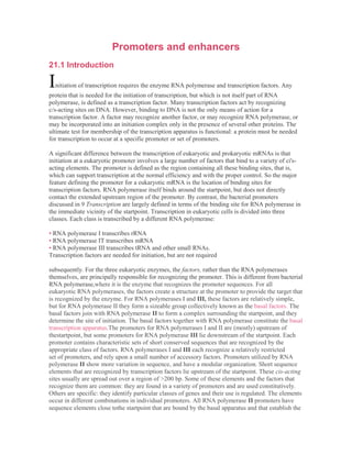
  • 2. site of initiation. The sequences farther upstream determine whether the promoter is expressed in all cell types or is specifically regulated. Promoters that are constitutively expressed (their genes are sometimes called housekeeping genes) have upstream sequence elements that are recognized by ubiquitous activators. No element/factor combination is an essential component of the promoter, which suggests that initiation by RNA polymerase II may be sponsored in many different ways. Promoters that are expressed only in certain times or places have sequence elements that require activators that are available only at those times or places. Sequence components of the promoter are defined operationally by the demand that they must be located in the general vicinity of the startpointand are required for initiation. The enhancer is another type of site involved in initiation. It is identified by sequences that stimulate initiation, but that are located a considerable distance from the startpoint. Enhancer elements are often targets for tissue-specific or temporal regulation. Figure 21.1 illustrates the general properties of promoters and enhancers. The components of an enhancer resemble those of the promoter; they consist of a variety of modular elements. However, the elementsare organized in a closely packed array. The elements in an enhancerfunction like those in the promoter, but the enhancer does not need to benear the startpoint. However, proteins bound at enhancer elements interactwith proteins bound at promoter elements. The distinction betweenpromoters and enhancers is operational, rather than implying a fundamental difference in mechanism. This view is strengthened by the fact that some types of element are found in both promoters and enhancers. Eukaryotic transcription is most often under positive regulation: a transcription factor is provided under tissue-specific control to activate a promoter or set of promoters that contain a common target sequence. Regulation by specific repression of a target promoter is less common. Figure 21.1 A typical gene transcribed by RNA polymerase II has a promoter that extends upstream from the site where transcription is initiated. The promoter contains several short (<10 bp) sequence elements that bind transcription factors, dispersed over >200 bp. An enhancer containing a more closely packed array of elements that also bind transcription factors may be located several kb distant. (DNA may be coiled or otherwise rearranged so that transcription factors at the promoter and at the enhancer interact to form a large protein complex.)
  • 3. 21.2 Eukaryotic RNA polymerases consist of many subunits : Key Concepts • • RNA polymerase I synthesizes rRNA in the nucleolus. : • RNA polymerase II synthesizes mRNA in the nucleoplasm. : • RNA polymerase III synthesizes small RNAs in the nucleoplasm. : • All eukaryotic RNA polymerases have — 12 subunits and are • aggregates of >500 kD. : • Some subunits are common to all three RNA polymerases. : • The largest subunit in RNA polymerase II has a CTD (carboxyi terminal domain) consisting of multiple repeats of a septamer. T he three eukaryotic RNA polymerases have different locations in the nucleus, corresponding with the genes that they transcribe. The most prominent activity is the enzyme RNA polymerase I, which resides in the nucleolus and is responsible for transcribing the genes coding for rRNA. It accounts for most cellular RNA synthesis (in terms of quantity). The other major enzyme is RNA polymerase II, located in the nucleoplasm (the part of the nucleus excluding the nucleolus). It represents most of the remaining cellular activity and is responsible for synthesizing heterogeneous nuclear RNA (hnRNA), the precursor for mRNA. RNA polymerase III is a minor enzyme activity. This nucleoplasmicenzyme synthesizes tRNAs and other small RNAs. All eukaryotic RNA polymerases are large proteins, appearing as aggregates of >500 kD. They typically have ~12 subunits. The purified enzyme can undertake template-dependent transcription of RNA, but is not able to initiate selectively at promoters. The general constitution of a eukaryotic RNA polymerase II enzyme as typified in S. cerevisiaeis illustrated in Figure 21.2. The two largest subunits are homologous to the β and $' subunits of bacterial RNA polymerase. Three of the remaining subunits are common to all the RNA polymerases, that is, they are also components of RNA polymerases I and III. The largest subunit in RNA polymerase II has a carboxy-terminaldomain (CTD), which consists of multiple repeats of a consensussequence of 7 amino acids. The sequence is unique to RNA polymeraseII. There are ~26 repeats in yeast and ~50 in mammals. The number ofrepeats is important, because deletions that remove (typically) more thanhalf of the repeats are lethal (in yeast). The CTD can be highly phosphorylatedon serine or threonine residues; this is involved in the initiationreaction (see 21.11 Initiation is followed by promoter clearance). The RNA polymerases of mitochondria and chloroplasts are smaller, and resemble bacterial RNA polymerase rather than any of the nuclearenzymes. Of course, the organelle genomes are much smaller, the resident polymerase needs to transcribe relatively few genes, and the control of transcription is likely to be very much simpler (if existing at all). So these enzymes are analogous to the phage enzymes that do not need the ability to respond to a more complex environment. A major practical distinction between the eukaryotic enzymes is drawn from their response to the bicyclic octapeptideα-amanitin. In basically all eukaryotic cells the activity of RNA polymerase II is rapidly inhibited by low concentrations of α- amanitin. RNA polymerase I is not inhibited. The response of RNA polymerase III to α-amanitin is less well conserved; in animal cells it is inhibited by high levels, but in yeast and insects it is not inhibited.
  • 4. 21.3 Promoter elements are defined by mutations and footprinting Key Concepts • Promoters are defined by their ability to cause transcription of an attached sequence in an appropriate test system in vitro or in vivo. T he first step in characterizing a promoter is to define the overall length of DNA that contains all the necessary sequence elements. To do this, we need a test system in which the promoter is responsible for the production of an easily assayed product. Historically, several types of systems have been used: • In the oocyte system, a DNA template is injected into the nucleus of the X. laevisoocyte. The RNA transcript can be recovered and analyzed. The main limitation of this system is that it is restricted to the conditions that prevail in the oocyte. It allows characterization of DNA sequences, but not of the factors that normally bind them. • Transfection systems allow exogenous DNA to be introduced into a cultured cell and expressed. (The procedure is discussed in 18.17 Transfectionintroduces exogenous DNA into cells.) The system is genuinely
  • 5. in vivo in the sense that transcription is accomplished by the same apparatus responsible for expressing the cell's own genome. However, it differs from the natural situation because the template consists of a gene that would not usually be transcribed in the host cell. The usefulness of the system may be extended by using a variety of host cells. • Transgenic systems involve the addition of a gene to the germline of an animal. Expression of the transgene can be followed in any or all of the tissues of the animal. Some common limitations apply to transgenic systems and to transfection: the additional gene often is present in multiple copies, and is integrated at a different location from the endogenous gene. Discrepancies between the expression of a gene in vitro and its expression as a transgene can yield important information about the role of the genomic context of the gene. • The in vitro system takes the classic approach of purifying all the components and manipulating conditions until faithful initiation is seen. "Faithful" initiation is defined as production of an RNA starting at the site corresponding to the 5' end of mRNA (or rRNA or tRNA precursors). Ultimately this allows us to characterize the individual sequence elements in the promoter and the transcription factors that bind to them. When a promoter is analyzed, it is important that only the promoter sequence changes. Figure 21.3 shows that the same long upstream sequence is always placed next to the promoter to ensure that it is always inthe same context. Because termination does not occur properlyin the in vitro systems, the template is cut at some distancefrom the promoter (usually ~500 bp downstream), to ensure that all polymerases "run off" at the same point, generating an identifiable transcript. We start with a particular fragment of DNA that can initiate transcription in one of these systems. Then theboundaries of the sequence constituting the promoter can be determined by reducing the length of the fragment from either end, until at some point it ceases to be active, as illustrated in Figure 21.4. The boundary upstream can be identified by progressively removing material from this end until promoter function is lost. To test the boundary downstream, it is necessary to reconnect the shortened promoter to the sequence to be transcribed (since otherwise there is no product to assay). Once the boundaries of the promoter have been defined, the importance of particular bases within it can be determined by introducing point mutations or other rearrangements in the sequence. As with bacterial RNA polymerase, these can be characterized as up or down mutations. Some of these rearrangements affect only the rate of initiation; others influence the site at which initiation occurs, as seen in a change of the startpoint. To be sure that we are dealing with comparable products, in each case it is necessary to characterize the 5' end of the RNA. We can apply several criteria in identifying the sequence components of a promoter (or any other site in DNA): • Mutations in the site prevent function in vitro or in vivo. (Many techniques now exist for introducing point mutations at particular base pairs, and in principle every position in a promoter can be mutated, and the mutant sequence tested in vitro or in vivo.) • Proteins that act by binding to a site may be footprinted on it. There should be a correlation between the ability of mutations to prevent promoter function and to prevent binding of the factor. • When a site recognized by a particular factor is present at multiple promoters, it should be possible to derive a consensus sequence that is bound by the factor. A new promoter should become responsive to this factor when an appropriate copy of the element is introduced.
  • 6. Figure 21.3 A promoter is tested by modifying the sequence that is connected to a constant upstream sequence and a constant downstream transcription unit. Figure 21.4 Promoter boundaries can be determined by making deletions that progressively remove more material from one side. When one deletion fails to prevent RNA synthesis but the next stops transcription, the boundary of the promoter must lie between them.
  • 7. 21.4 RNA polymerase I has a bipartite promoter Key Concepts • The RNA polymerase I promoter consists of a core promoter and an upstream control element. • The factor UBF1 binds to both regions and enables the factor SL1 to bind. • SL1 includes the factor TBP that is involved in initiation by all threeRNA polymerases. • RNA polymerase binds to the UBF1-SL1 complex at the core promoter. R NA polymerase I transcribes only the genes for ribosomal RNA, from a single type of promoter. The transcript includes the sequencesof both large and small rRNAs, which are later released by cleavages and processing. There are many copies of the transcription unit, alternating with nontranscribedspacers, and organized in a cluster as discussed in 4.8Genes for rRNAform tandem repeats. The organizationof the promoter, and the events involved in initiation, are illustrated in Figure 21.5. The promoter consists of two separate regions.The core promoter surrounds the startpoint, extendingfrom -45 to +20, and is sufficient for transcription to initiate. It is generally G-C-rich (unusual for a promoter) except for the only conserved sequence element, a short A-T-rich sequence around the startpointcalled the Inr. However, its efficiency is very much increased by the upstream promoter element (UPE), another G-C-rich sequence, related to the core promoter sequence, which extends from -180 to -107. This type of organization is common to pol I promoters in many species, although the actual sequences vary widely. RNA polymerase I requires two ancillary factors. The factor that binds to the core promoter consists of 4 proteins. (It is called SL1, TIF-IB, Ribl in different species). One of its components, called TBP, is a factor that is required also for initiation by RNA polymerases II and III (see 21.8TBP is a universal factor). TBP does not bind directly to G-C-rich DNA, so DNA-binding is probably the responsibility of the other components of the core-binding factor. It is likely that TBP interacts with RNA polymerase, possibly with a common subunit or a feature that has been conserved among polymerases. Core-binding factor enables RNA polymerase I to initiate from the promoter at a low basal frequency. The core-binding factor has primary responsibility for ensuring that the RNA polymerase is properly localized at the startpoint. We see shortly that a comparable function is provided for RNA polymerases II and III by a factor that consists of TBP associated with other proteins. So a common feature in initiation by all three polymerases is a reliance on a "positioning" factor that consists of TBP associated with proteins that are specific for each type of promoter. For high frequency initiation, the factor UBF is required. This is a single polypeptide that binds to a G-C-rich element in the upstream promoter element. One indication of how UBF interacts with the corebindingfactor is given by the importance of the spacing between the upstream promoter element and the core promoter. This can be changed by distances involving integral numbers of turns of DNA, but not by distances that introduce half turns. This implies that UBF and corebindingfactor need to be bound on the same face of DNA in order to interact. In the presence of UBF, core- binding factor binds more efficiently to the core promoter.
  • 8. Figure 21.5 Transcription units for RNA polymerase I have a core promoter separated by —70 bp from the upstream promoter element. UBF binding to the UPE increases the ability of core-binding factor to bind to the core promoter. Core-binding factor positions RNA polymerase I at the startpoint. 21.5 RNA polymerase III uses both downstream and upstream promoters Key Concepts • RNA polymerase III has two types of promoters. • Internal promoters have short consensus sequences located within the transcription unit and cause initiation to occur a fixed distance upstream. • Upstream promoters contain three short consensus sequences upstream of the startpoint that are bound by transcription factors. R ecognition of promoters by RNA polymerase III strikingly illustrates the relative roles of transcription factors and the polymerase enzyme. The promoters fall into two general classes that are recognized in different ways by different groups of factors. The promoters for 5S and tRNA genes are internal; they lie downstream of thestartpoint. The promoters for snRNA (small nuclear
  • 9. RNA) genes lie upstream of the startpoint in the more conventional manner of other promoters. In both cases, the individual elements that are necessary for promoter function consist exclusively of sequences recognized by transcription factors, which in turn direct the binding of RNA polymerase. Before the promoter of 5S RNA genes was identified inX. laevis, all attempts to identify promoter sequences assumed that they would lie upstream of the startpoint. But deletion analysis showed that the 5S RNA product continues to be synthesized when the entire sequence upstream of the gene is removed! When the deletions continue into the gene, a product very similar in size to the usual 5S RNA continues to be synthesized so long as the deletion ends before base +55. Figure 21.6 shows that the first part of the RNA product corresponds to plasmid DNA; the second part represents the segment remaining of the usual 5S RNA sequence. But when the deletion extends past +55, transcription does not occur. So the promoter lies downstream of position +55, but causes RNA polymerase III to initiate transcription a more or less fixed distance upstream. When deletions extend into the gene from its distal end, transcription is unaffected so long as the first 80 bp remain intact. Once the deletion cuts into this region, transcription ceases. This places the downstream boundary position of the promoter at about position +80. So the promoter for 5S RNA transcription lies between positions+55 and +80 within the gene. A fragment containing this region can sponsor initiation of any DNA in which it is placed, from a startpoint~55 bp farther upstream. (The wild- type startpoint is unique; in deletions that lack it, transcription initiates at the purine base nearest to the position 55 bp upstream of the promoter.) The structures of three types of promoters for RNA polymerase III are summarized in Figure 21.7. There are two types of internal promoter. Each contains a bipartite structure, in which two short sequence elements are separated by a variable sequence. Type 1 consists of a boxA sequence separated from a boxC sequence, and type 2 consists of a boxA sequence separated from a boxB sequence. The distance between boxA and boxB in a type 2 promoter can vary quite extensively, but the boxes usually cannot be brought too close together without abolishing function. Type 3 promoters have three sequence elements all located upstream of the startpoint.
  • 10. 21.6 TF|||B is the commitment factor for pol III promoters Key Concepts • TF|MA and TFmC bind to the consensus sequences and enable TFM|B to bind at the startpoint. • TFmB has TBP as one subunit and enables RNA polymerase to bind. T he detailed interactions are different at the two types of internal promoter, but the principle is the same. TFniC binds downstream of the startpoint, either independently (type 2 promoters) or in conjunction with TFIUA (type 1 promoters). The presence of TFITICenables the positioning factor TFmBto bind at the startpoint. Then RNA polymerase is recruited.Figure 21.8 summarizes the stages of reaction at type 2 internal promoters. TF[][C binds to both boxA and boxB. This enables TFmB to bind at the startpoint. Then RNA polymerase III can bind. The difference at type 1 internal promoters is that TFmA must bind at boxA to enable TFmC to bind at boxC. Figure 21.9 shows that, once TFU1C has bound, events follow the same course as at type 2 promoters, with TFJJJB binding at the startpoint, and RNA polymerase III joining the complex. Type 1 promoters are found only in the genes for 5S rRNA. TFmA and TFmC are assembly factors, whose sole role is to assist the binding of TFIHB at the right location. Once TFmB has bound, TFUIA and TFTljC can be removed from the promoter (by high salt concentration in vitro) without affecting the initiation reaction. TFn]Bremains bound in the vicinity of the startpoint and its presence is sufficientto allow RNA polymerase 111 to bind at the startpoint. So TFniB is the only true initiation factor required by RNA polymerase III. This sequence of events explains how the promoter boxes downstream can cause RNA polymerase to bind at the startpoint, farther upstream. Although the ability to transcribe these genes is conferred by the internal promoter, changes in the region immediately upstream of the startpointcan alter the efficiency of transcription. TFmC is a large protein complex (>500 kD), comparable in size to RNA polymerase itself, and containing 6 subunits. TFinA is a member of an interesting class of proteins containing a nucleic acid-binding motif called a zinc finger (see 22.9 A zinc finger motif is a DNA-binding domain). The positioning factor, TFmIB, consists of three subunits. It includes the same protein, TBP, that is present in the core-binding factor for pol I promoters, and also in the corresponding transcription factor (TFnD) for RNA polymerase II. It also contains Brf, which is related to the factor TFnB that is used by RNA polymerase II. The third subunit is called B"; it is dispensable if the DNA duplex is partially melted, which suggests that its function is to initiate the transcription bubble. The role of B " may be comparable to the role played by sigma factor in bacterial RNA polymerase (see 9.16 Substitution of sigma factors maycontrol initiation). The upstream region has a conventional role in the third class of polymerase III promoters. In the example shown in Figure 21.7, there are three upstream elements. These elements are also found in promoters for snRNA genes that are transcribed by RNA polymerase II. (Genes for some snRNAs are transcribed by RNA polymerase II, while others are transcribed by RNA polymerase III.) The upstream elements function in a similar manner in promoters for both polymerases II and III. Initiation at an upstream promoter for RNA polymerase III can occur on a short region that immediately precedes the startpoint and contains only the TATA element. However, efficiency of transcription is much increased by the presence of the PSE and OCT elements. The factors that bind at these elements interact cooperatively. (The PSE element may be essential at promoters used by RNA polymerase II, whereas it is stimulatory in promoters used by RNA polymerase III; its
  • 11. name stands for proximal sequence element.) The TATA element confers specificity for ftie type of polymerase (II or III) that is recognized by ansnRNA promoter. It is bound by a factor that includes the TBP, which actually recognizes the sequence in DIVA. The TBP is associated with other proteins, which are specific for the type of promoter. The function of TBP and its associated proteins is to position the RNA polymerase correctly at the startpoint. We discuss thisin more detail for RNA polymerase II (see 21.8 TBP is a universal factor). The factors work in the same way for both types of promoters forRNA polymerase III. The factors bind at the promoter before RNA polymerase itself can bind. They form a preinitiation complex that directs binding of the RNA polymerase. RNA polymerase III does not itself recognizes the promoter sequence, but binds adjacent to factors that are themselves bound just upstream of the startpoint. For the type 1 and type 2 internal promoters, the assembly factors ensure that TFmB (which includes TBP) is bound just upstream of the startpoint, to provide the positioning information. For the upstream promoters, TFmB binds directly to the region including the TATA box. So irrespective of the location of the promoter sequences, factor(s) are bound close to the startpoint in order to direct binding of RNA polymerase III.
  • 12. 21.7 The startpoint for RNA polymerase II Key Concepts • RNA polymerase II requires general transcription factors (calledTF||X) to initiate transcription. • RNA polymerase II promoters have a short conserved sequence Py2CAPy5 (the initiator InR) at the startpoint. • The TATA box is a common component of RNA polymerase II promoters and consists of an A-T-rich octamer located —25 bp upstream of the startpoint. • The DPE is a common component of RNA polymerase II promoters that do not contain a TATA box. • A core promoter for RNA polymerase II includes the InR and either a TATA box or a DPE. T he basic organization of the apparatus for transcribing proteincodinggenes was revealed by the discovery that purified RNA polymerase II can catalyze synthesis of mRNA, but cannot initiate transcription unless an additional extract is added. The purification of this extract led to the definition of the general transcription factors—a group of proteins that are needed for initiation by RNA polymerase II at all promoters. RNA polymerase II in conjunction with these factors constitutes the basal transcription apparatus that is needed to transcribe any promoter. The general factors are described as TFj|X, where "X" is a letter that identifies the individual factor. The subunits of RNA polymerase II and the general transcription factors are conserved among eukaryotes. Our starting point for considering promoter organization is to define the core promoter as the shortest sequence at which RNA polymerase II can initiate transcription. A core promoter can in principle be expressed in any cell. It comprises the minimum sequence that enables the general transcription factors to assemble at the startpoint. They are involved in the mechanics of binding to DNA and enable RNA polymerase II to initiate transcription. A core promoter functions at only a low efficiency. Other proteins, called activators, are required for a proper level of function (see 21.13 Short sequence elements bind activators). The activators are not described systematically, but have casual names reflecting their histories of identification. We may expect any sequence components involved in the binding of RNA polymerase and general transcription factors to be conserved at most or all promoters. As with bacterial promoters, when promotersfor RNA polymerase II are compared, homologies in the regions near the startpoint are restricted to rather short sequences. These elements correspond with the sequences implicated in promoter function by mutation. Figure 21.10 shows the construction of a typical pol II core promoter. At the startpoint, there is no extensive homology of sequence, but there is a tendency for the first base of mRNA to be A, flanked on either side by pyrimidines. (This description is also valid for the CAT start sequence of bacterial promoters.) This region is called the initiator(Inr), and may be described in the general form Py2CAPy5. The Inr is contained between positions -3 and +5. Many promoters have a sequence called the TATA box, usually located -25 bp upstream of the startpoint. It constitutes the only upstream promoter element that has a relatively fixed location with respect to the startpoint. The core sequence is TATAA, usually followed by three more A-T base pairs. The TATA box tends to be surrounded by G-C-rich sequences, which could be a factor in its function. It is almost identical with the -10 sequence found in bacterial promoters; in fact, it could pass for one except for the difference in its location at -25 instead of-10. Single base substitutions in trie TATA box act as strong down mutations. Some mutations reverse the orientation of an A-T pair, so base composition alone is not sufficient for its function. So the TATA box comprises an element whose behavior is analogous to our concept of the bacterial promoter: a short, well-defined sequence just upstream of the startpoint, which is necessary for transcription. Promoters that do not
  • 13. contain a TATA element are called TATA-lesspromoters. Surveys of promoter sequences suggest that 50% or more of promoters may be TATA-less. When a promoter does not contain a TATA box, it usually contains another element, the DPE (downstream promoter element) which is located at +28 - +32. A core promoter can consist either of a TATA box plus InR or of an InR plus DPE. 21.8 TBP is a universal factor Key Concepts • TBP is a component of the positioning factor that is required for each type of RNA polymerase to bind its promoter. • The factor for RNA polymerase II is TFMD, which consists of TBP and 11 TAFs, with a total mass - 8 0 0 kD. T he first step in complex formation at a promoter containing a TATA box is binding of the factor TF|,Dto a region that extends upstream from the TATA sequence. TFnD contains two types of component. Recognition of the TATA box is conferred by the TATA-bindingprotein (TBP), a small protein of ~30 kD. The other subunits are called TAFs (for TBP-associated factors). Some TAFs are stoichiometric with TBP; others are present in lesser amounts. TFnDs containing different TAFs could recognize different promoters. Some (substoichiometric) TAFs are tissue-specific. The total mass of TFnD typically is ~800 kD, containing TBP and 11 TAFs, varying in mass from 30- 250 kD. The TAFs in TFnD are named in the form TAFn00, where "00" gives the molecular mass of the subunit. Positioning factors that consist of TBP associated with a set of TAFs are responsible for identifying all classes of promoters. TFmB (for pol III promoters) and SL1 (for pol I promoters) may both be viewed asconsisting of TBP associated with a particular group of proteins that substitute for the
  • 14. TAFs that are found in TFnD. TBP is the key component,and is incorporated at each type of promoter by a different mechanism. In the case of promoters for RNA polymerase II, the key feature in positioning is the fixed distance of the TATA box from the startpoint. Figure 21.11 shows that the positioning factor recognizes the promoter in a different way in each case. At promoters for RNA polymerase III, TFinB binds adjacent to TFIIIC. At promoters for RNA polymerase I, SL1 binds in conjunction with UBF. TFnD is solely responsible for recognizing promoters for RNA polymerase II. At a promoter that has a TATA element, TBP binds specifically to DNA, but at other promoters it may be incorporated by association with other proteins that bind to DNA. Whatever its means of entry into the initiation complex, it has the common purpose of interaction with the RNA polymerase. TFnD is ubiquitous, but not unique. All multicellular eukaryotes also express an alternative complex, which has TLF (TBP like factor) instead of TBP. A TLF is typically -60% similar to TBP. It probably initiates complex formation by the usual set of TFU factors. However, TLF does not bind to the TATA box, and we do not yet know how it works. Drosophila also has a third factor, TRF1, which behaves in the same way as TBP and binds its own set of TAFs, to form a complex that functions as an alternative to TFnD at a specific set of promoters.
  • 15. T BP has the unusual property of binding to DNA in the minor groove. (Virtually all known DNA-binding proteins bind in the major groove.) The crystal structure of TBP suggests a detailed model for its binding to DNA. Figure 21.12 shows that it surrounds one face of DNA, forming a "saddle" around the double helix. In effect, the inner surface of TBP binds to DNA, and the larger outer surface is available to extend contacts to other proteins. The DNA-binding site consists of a C-terminal domain that is conserved between species, while the variable N-terminal tail is exposed to interact with other proteins. It is a measure of the conservation of mechanism in transcriptional initiation that the DNA-binding sequence of TBP is 80% conserved between yeast and Man. Binding of TBP may be inconsistent with the presence of nucleosomes. Because nucleosomes form preferentially by placing A-T-rich sequences with the minor grooves facing inward, they could prevent binding of TBP. This may explain why the presence of nucleosomes prevents initiation of transcription. TBP first binds to the minor groove, and then bends the DNA by ~80°, as illustrated in Figure 21.13. The TATA box bends towards the major groove, widening the minor groove. The distortion is restricted to the 8 bp of the TATA box; at each end of the sequence, the minor groove has its usual width of ~5 A, but at the center of the sequence the minor groove is >9 A. This is a deformation of the structure, but does not actually separate the strands of DNA, because base pairing is maintained.
  • 16. This structure has several functional implications. By changing the spatial organization of DNA on either side of the TATA box, it allows the transcription factors and RNA polymerase to form a closer association than would be possible on linear DNA. The bending at the TATA box corresponds to unwinding of about 1/3 of a turn of DNA, and is compensated by a positive writhe. We do not know yet how this relates to the initiation of strand separation. The presence of TBP in the minor groove, combined with other proteins binding in the major groove, creates a high density of protein- DNA contacts in this region. Binding of purified TBP to DNA invitro protects ~1 turn of the double helix at the TATA box, typically extending from -37 to - 25; but binding of the TFnD complex in the initiation reaction regularly protects the region from -45 to -10, and also extends farther upstream beyond the startpoint. TBP is the only general transcription factor that makes sequence-specific contacts with DNA. Within TFJJD as a free protein complex, the factor TAFn230 binds to TBP, where it occupies the concave DNA-binding surface. In fact, the structure of the binding site, which lies in the N-terminal domain of TAFu230, mimics the surface of the minor groove in DNA. This molecular mimicry allows TAFn230 to control the ability of TBP to bind to DNA; the N-terminal domain of TAFn230 must be displaced from the DNA-binding surface of TBP in order for TFnD to bind to DNA.Some TAFs resemble histones; in particular TAFn42 and TAFn62appear to be (distant) homologues of histones H3 and H4, and they form a heterodimer using the same motif (the histone fold) that histones use for the interaction. (Histones H3 and H4 form the kernel of the histone octamer—the basic complex that binds DNA in eukaryotic chromatin; see 20.8 Organization of the histone octamer.) Together with other TAFs, TAFn42 and TAFn62 may form the basis for a structure resembling a histone octamer; such a structure may be responsible for the nonsequence- specific interactions of TFUD with DNA. Histone folds are also used in pairwise interactions between other TAFns. Some of the TAFns may be found in other complexes as well as in TFHD. In particular, the histone-like TAFns are found also in protein complexes that modify the structure of chromatin prior to transcription (see 23.7 Acetylases are associated with activators). 21.10 The basal apparatus assembles at the promoter Key Concepts • Binding of TFMD to the TATA box is the first step in initiation. • Other transcription factors bind to the complex in a defined order, extending the length of the protected region on DNA. • When RNA polymerase II binds to the complex, it initiates transcription. I nitiation requires the transcription factors to act in a defined order to build a complex that is joined by RNA polymerase. The series of events can be followed by the increasing size of the protein complex associated with DNA. Footprinting of the DNA regions protected by each complex suggests the model summarized inFigure 21.14. As each TF1I factor joins the complex, an increasing length of DNA is covered. RNA polymerase is incorporated at a late stage. Commitment to a promoter is initiated when TFnD binds the TATA box. (TFnD also recognizes the InR sequence at the startpoint.) When TFj[A joins the complex, TFnD becomes able to protect a region extending farther upstream. TFnA may activate TBP by relieving the repression that is caused by the TAFu230.
  • 17. Addition of TFnB gives partial protection of the region of the templatestrand in the vicinity of the startpoint, from —10 to +10. This suggests that TFnB is bound downstream of the TATA box, perhaps loosely associated with DNA and asymmetrically oriented with regard to the two DNA strands. The crystal structure shown in Figure 21.15 extends this model. TFnB binds adjacent to TBP, extending contacts along one face of DNA. It makes contacts in the minor groove downstream of the TATA box, and contacts the major groove upstream of the TATA box, in a region called the BRE. In archaea, the homologue of TFnB actually makes sequence-specific contacts with the promoter in the BRE region. TFUB may provide the surface that is in turn recognized by RNA polymerase, so that it is responsible for the directionality of the binding of the enzyme. The factor TFnF is a heterotetramer consisting of two types of subunit. The larger subunit (RAP74) has an ATP-dependent DNA helicase activity that could be involved in melting the DNA at initiation. The smaller subunit (RAP38) has some homology to the regions of bacterial sigma factor that contact the core polymerase; it binds tightly to RNA polymerase II. TFnF may bring RNA polymerase II to the assembling transcription complex and provide the means by which it binds. The complex of TBP and TAFs may interact with the CTD tail of RNA polymerase, and interaction with TFnB may also be important when TFIIF/polymerase joins the complex. Polymerase binding extends the sites that are protected downstream to +15 on the template strand and +20 on the nontemplate strand. The enzyme extends the full length of the complex, since additional protection is seen at the upstream boundary. What happens at TATA-less promoters? The same general transcription factors, including TFnD, are needed. The Inr provides the positioning element; TFnD binds to it via an ability of one or more of the TAFs to recognize the Inr directly. Other TAFs in TFnD also recognize the DPE element downstream from the startpoint. The function of TBP at these promoters is more like that at promoters for RNA polymerase I and at internal promoters for RNA polymerase III. Assembly of the RNA polymerase II initiation complex provides an interesting contrast with prokaryotic transcription. Bacterial RNA polymerase is essentially a coherent aggregate with intrinsic ability to bind DNA; the sigma factor, needed for initiation but not for elongation, becomes part of the enzyme before DNA is bound, although it is later released. But RNA polymerase II can bind to the promoter only after separate transcription factors have bound. The factors play a role analogous to that of bacterial sigma factor—to allow the basic polymerase to recognize DNA specifically at promoter sequences—but have evolved more independence. Indeed, the factors are primarily responsible for the specificity of promoter recognition. Only some of the factors participate in protein-DNA contacts (and only TBP makes sequence-specific contacts); thus protein-protein interactions are important in the assembly of the complex. When a TATA box is present, it determines the location of the startpoint. Its deletion causes the site of initiation to become erratic, although any overall reduction in transcription is relatively small. Indeed, some TATA-less promoters lack unique startpoints; initiationoccurs instead at any one of a cluster of startpoints. The TATA box aligns the RNA polymerase (via the interaction with TFUD and other factors) so that it initiates at the proper site. This explains why its location is fixed with respect to the startpoint. Binding of TBP to TATA is the predominant feature in recognition of the promoter, but two large TAFs (TAFn250 andTAFn150) also contact DNA in the vicinity of the startpoint and influence the efficiency of the reaction. Although assembly can take place just at the core promoter in vitro,this reaction is not sufficient for transcription in vivo, where interactions with activators that recognize the more upstream elements are required. The activators interact with the basal apparatus at various stages during its assembly (see 22.5 Activators interact with the basal
  • 18. apparatus). 21.11 Initiation is followed by promoter clearance Key Concepts • TFME and TFMH are required to melt DNA to allow polymerase movement. • Phosphorylation of the CTD may be required for elongation to begin. • Further phosphorylation of the CTD is required at some promoters to end abortive initiation. • The CTD may coordinate processing of RNA with transcription. M ost of the transcription factors are required solely to bind RNA polymerase to the promoter, but some act at a later stage. Binding of TFnE causes the boundary of the region protected downstream to be extended by another turn of the double helix, to +30. Two further factors, TFnH and TFnJ, join the complex after TFnE. They do not change the pattern of binding to DNA. TFUH is the only general transcription factor that has independent enzymatic activities. Its several activities include an ATPase, helicases of both polarities, and a kinase activity that can phosphorylate the CTD tail of RNA polymerase II. TFHH is an exceptional factor that may play a role also in elongation. Its interaction with DNA downstream of the startpoint is required for RNA polymerase to escape from the promoter. TFUH is also involved in repair of damage to DNA (see next section).
  • 19. The initiation reaction, as defined by formation of the first phosphodiesterbond, occurs once RNA polymerase has bound. Figure 21.16proposes a model in which phosphorylation of the tail is needed to release RNA polymerase II from the transcription factors so that it can make the transition to the elongating form. Most of the transcription factors are released from the promoter at this stage. On a linear template, ATP hydrolysis, TFnE, and the helicase activity of TFnH (provided by the XPB subunit) are required for polymerasemovement. This requirement is bypassed with a supercoiled template. This suggests that TFUE and TFUH are required to melt DNA to allow polymerase movement to begin. The helicase activity of the XPB subunit of TFnH is responsible for the actual melting of DNA. RNA polymerase II stutters at some genes when it starts transcription. (The result is not dissimilar to the abortive initiation of bacterial RNA polymerase discussed in 9.11 Sigma factor controls binding to DNA, although the mechanism is different.) At many genes, RNA polymerase IIterminates after a short distance. The short RNA product is degraded rapidly. To extend elongation into the gene, a kinase called P-TEFb is required. This kinase is a member of the cdk family that controls the cell cycle (see 29 Cell cycle and growth regulation). P-TEFb acts on the CTD, to phosphorylate it further. We do not yet understand why this effect is required at some promoters but not others or how it is regulated. The CTD may also be involved, directly or indirectly, in processing RNA after it has been synthesized by RNA polymerase II. Figure 21.17 summarizes processing reactions in which the CTD may be involved. The capping enzyme (guanylyltransferase), which adds the G residue to the 5' end of newly synthesized mRNA, binds to the phosphorylated CTD: this may be important in enabling it to modify the 5' end as soon as it is synthesized. A set of proteins called SCAFs bind to the CTD, and they may in turn bind to splicing factors. This may be a means of coordinating transcription and splicing. Some components of the cleavage/polyadenylationapparatus also bind to the CTD. Oddly enough, they do so at the time of initiation, so that RNA polymerase is all ready for the 3' end processing reactions as soon as it sets out! All of this suggests that the CTD may be a general focus for connecting other processes with transcription. In the cases of capping and splicing, the CTD functions indirectly to promote formation of the protein complexes that undertake the reactions. In the case of 3' end generation, it may participate directly in the reaction. The general process of initiation is similar to that catalyzed by bacterial RNA polymerase. Binding of RNA polymerase generates a closed complex, which is converted at a later stage to an open complex in which the DNA strands have been separated. In the bacterial reaction, formation of the open complex completes the necessary structural change to DNA; a difference in the eukaryotic reaction is that further unwinding of the template is needed after this stage.
  • 20.
  • 21. 21.12 A connection between transcription and repair Key Concepts • Transcribed genes are preferentially repaired when DNA damage occurs. • TFMH provides the link to a complex of repair enzymes. • Mutations in the XPD component of TFMH cause three types of human diseases. I n both bacteria and eukaryotes, there is a direct link from RNA polymerase to the activation of repair. The basic phenomenon was first observed because transcribed genes are preferentially repaired. Then it was discovered that it is only the template strand of DNA that is the target—the nontemplate strand is repaired at the same rate as bulk DNA. In bacteria, the repair activity is provided by the uvrexcision-repair system (see 15.21 Excision repair systems in E. coli). Preferential repair is abolished by mutations in the gene mfd, whose product provides the link from RNA polymerase to the Uvr enzymes. Figure 21.18 shows a model for the link between transcription and repair. When RNA polymerase encounters DNA damage in the template strand, it stalls because it cannot use the damaged sequences as a template to direct complementary base pairing. This explains the specificity of the effect for the template strand (damage in the nontemplate strand does not impede progress of the RNA polymerase). The Mfd protein has two roles. First, it displaces the ternary complex of RNA polymerase from DNA. Second, it causes the UvrABCenzyme to bind to the damaged DNA. This leads to repair of DNA by the excision-repair mechanism (see Figure 15.40). After the DNA has been repaired, the next RNA polymerase to traverse the gene is able to produce a normal transcript. A similar mechanism, although relying on different components, is used in eukaryotes. The template strand of a transcribed gene is preferentially repaired following UV-induced damage. The general transcription factor TFnH is involved. TFnH is found in alternative forms, which consist of a core associated with other subunits. TFnH has a common function in both initiating transcription and repairing damage. The same helicase subunit (XPD) creates the initial transcription bubble and melts DNA at a damaged site. Its other functions differ between transcription and repair, as provided by the appropriate form of the complex. Figure 21.19 shows that the basic factor involved in transcription consists of a core (of 5 subunits) associated with other subunits that have a kinase activity. The alternative complex consists of the core associated with a large group of proteins that are coded by repair genes. (The basic model for repair is shown in Figure 15.53.) The repair proteins include a subunit (XPC) that recognizes damaged DNA, which provides the coupling function that enables a template strand to be preferentially repaired when RNA polymerase becomes stalled at damaged DNA. Other proteins associated with the complex include endonucleases (XPG, XPF,ERCC1). Homologous proteins are found in the complexes in yeast (where they are often identified by rad mutations that are defective in repair) and in Man (where they are identified by mutations that cause diseases resulting from deficiencies in repairing damaged DNA). (Subunits with the name XP are coded by genes in which mutations cause the disease xerodermapigmentosum (see 15.28 Eukaryotic cells have conserved repair systems). The kinase complex and the repair complex can associate and dissociatereversibly from the core TFnH. This suggests a model in which thefirst form of TFnH is required for initiation, but may be replaced by theother form (perhaps in response to encountering DNA damage). TFnH dissociates from RNA polymerase at an early stage of elongation (after transcription of ~50 bp); its reassociation at a site of damaged DNA may require additional coupling components.
  • 22. The repair function may require modification or degradation of RNA polymerase. The large subunit of RNA polymerase is degradedwhen the enzyme stalls at sites of UV damage. We do not yet understand the connection between the transcription/repair apparatus as such and the degradation of RNA polymerase. It is possible that removal of the polymerase is necessary once it has become stalled. This degradation of RNA polymerase is deficient in cells from patients with Cockayne's syndrome (a repair disorder). Cockayne's syndrome is caused by mutations in either of two genes (CSA and CSB), both of whose products appear to be part of or bound to TFnH. Cockayne'ssyndrome is also occasionally caused by mutations in XPD. XPD is a pleiotropic protein, in which different mutations can affect different functions. In fact, XPD is required for the stability of the TFj|Hcomplex during transcription, but the helicase activity as such is not needed. Mutations that prevent XPD from stabilizing the complex cause trichothiodystrophy. The helicase activity is required for the repair function. Mutations that affect the helicase activity cause the repair deficiency that results in XP or Cockayne's syndrome.
  • 23. 21.13 Short sequence elements bind activators Key Concepts • Short conserved sequence elements are dispersed in the region preceding the startpoint. • The upstream elements increase the frequency of initiation. • The factors that bind to them to stimulate transcription are called activators. A promoter for RNA polymerase II consists of two types of region.The startpoint itself is identified by the Inr and/or by the TATA box close by. In conjunction with the general transcription factors, RNA polymerase II forms an initiation complex surrounding the startpoint, as we have just described. The efficiency and specificity with which a promoter is recognized, however, depend upon short sequences, farther upstream, which are recognized by a different group of factors, usually called activators. Usually the target sequences are ~100 bp upstream of the startpoint, but sometimes they are more distant. Binding of activators at these sites may influence the formation of the initiation complex at (probably) any one of several stages. An analysis of a typical promoter is summarized in Figure 21.20.Individual base substitutions were introduced at almost every position in the 100 bp upstream of the β-globin startpoint. The striking result is that most mutations do not affect the ability of the promoter to initiate transcription.Down mutations occur in three locations, corresponding to three short discrete elements. The two upstream elements have a greater effect on the level of transcription than the element closest to the startpoint. Up mutations occur in only one of the elements. We conclude that the three short sequences centered at -30, -75, and -90 constitute the promoter. Each of them corresponds to the consensus sequence for a common type of promoter element. The TATA box (centered at —30) is the least effective component of the promoter as measured by the reduction in transcription that is caused by mutations. But although initiation is not prevented when a TATA box is mutated, the startpoint varies from its usual precise location. This confirms the role of the TATA box as a crucial positioning component of the core promoter. The basal elements and the elements upstream of them have different types of functions. The basal elements (the TATA box and Inr) primarily determine the location of the startpoint, but can sponsor initiation only ata rather low level. They identify the location at which the general transcriptionfactors assemble to form the basal complex. The sequence elementsfarther upstream influence the frequency of initiation, most likelyby acting directly on the general transcription factors to enhance the efficiencyof assembly into an initiation complex (see 22.5 Activators interactwith the basal apparatus). The sequence at -75 is the CAAT box. Named for its consensus sequence, it was one of the first common elements to be described. It is often located close to -80, but it can function at distances that vary considerably from the startpoint. It functions in either orientation. Susceptibility to mutations suggests that the CAAT box plays a strong role in determining the efficiency of the promoter, but does not influence its specificity. The GC box at -90 contains the sequence GGGCGG. Often multiple copies are present in the promoter, and they occur in either orientation. It too is a relatively common promoter component.
  • 24. Figure 21.20 Saturation mutagenesis of the upstream region of the β-globin promoter identifies three short regions (centered at -30, -75, and -90) that are needed to initiate transcription. These correspond to the TATA, CAAT, and GC boxes. 21.14 Promoter construction is flexible but context can be important Key Concepts • No individual upstream element is essential for promoter function, although one or more elements must be present for efficient initiation. • Some elements are recognized by multiple factors, and the factor that is used at any particular promoter may be determined by the context of the other factors that are bound. P romoters are organized on a principle of "mix and match." A variety of elements can contribute to promoter function, but none is essential for all promoters. Some examples are summarized in Figure 21.21. Four types of elements are found altogether in these promoters: TATA, GC boxes, CAAT boxes, and theoctamer (an 8 bp element). The elements found in any individual promoter differ in number, location, and orientation. No element is common to all of the promoters. Although the promoter conveys directional information (transcription proceeds only in the downstream direction), the GC and CAAT boxes seem to be able to function in either orientation. This implies that the elements function solely as DNA-binding sites to bring transcription factors into the vicinity of the startpoint; the structure of a factor must be flexible enough to allow it to make protein-protein contacts with the basal apparatus irrespective of the way in which its DNA-binding domain is oriented and its exact distance from the startpoint.
  • 25. Activators that are more or less ubiquitous are assumed to be availableto any promoter that has a copy of the element that they recognize. Common elements recognized by ubiquitous activators include the CAAT box, GC box, and the octamer. All promoters probably require one or more of these elements in order to function efficiently. An activator typically has a consensus sequence of < 10 bp, but actually covers a length of -20 bp of DNA. Given the sizes of the activators, and the length of DNA each covers, we expect that the various proteins will together cover the entire region upstream of the startpoint in which the elements reside. Usually a particular consensus sequence is recognized by a corresponding activator (or by a member of a family of factors). However, sometimes a particular promoter sequence can be recognized by one of several activators. A ubiquitous activator, Oct-1, binds to the octamer to activate the histone H2B (and presumably also other) genes. Oct-1 is the only octamer-binding factor in nonlymphoid cells. But in lymphoid cells, a differentactivator, Oct-2, binds to the octamer to activate the immunoglobulinK light gene. So Oct-2 is a tissue-specific activator, while Oct-1 is ubiquitous. The exact details of recognition are not so important to know as the fact that a variety of activators recognize CAAT boxes. The use of the same octamer in the ubiquitously expressed H2B gene and the lymphoid-specific immunoglobulin genes poses a paradox.Why does the ubiquitous Oct-1 fail to activate the immunoglobulin genes in nonlymphoid tissues? The context must be important: Oct-2 rather than Oct-1 may be needed to interact with other proteins that bind at the promoter. These results mean that we cannot predict whether a gene will be activated by a particular activator simply on the basis of the presence of particular elements in its promoter. 21.15 Enhancers contain bidirectional elements that assist initiation
  • 26. Key Concepts • An enhancer activates the nearest promoter to it, and can be any distance either upstream or downstream of the promoter. • A UAS (upstream activator sequence) in yeast behaves like an enhancer but works only upstream of the promoter. • Similar sequence elements are found in enhancers and promoters. • Enhancers form complexes of activators that interact directly or indirectly with the promoter. W e have considered the promoter so far as an isolated region responsible for binding RNA polymerase. But eukaryotic promoters do not necessarily function alone. In at least some cases, the activity of a promoter is enormously increased by the presence of an enhancer,which consists of another group of elements, but located at a variable distance from those regarded as comprising part of the promoter itself. The concept that the enhancer is distinct from the promoter reflects two characteristics. The position of the enhancer relative to the promoter need not be fixed, but can vary substantially. Figure 21.22 shows that it can be either upstream or downstream. And it can function in either orientation (that is, it can be inverted) relative to the promoter. Manipulations of DNA show that an enhancer can stimulate any promoter placed in its vicinity. In natural genomes, enhancers can be located within genes (that is, just downstream of the promoter) or tens of kilobases away in either direction. For operational purposes, it is sometimes useful to define the promoter as a sequence or sequences of DNAthat must be in a (relatively) fixed location with regard tothe startpoint. By this definition, the TATA box and other upstream elements are included, but the enhancer is excluded. This is, however, a working definition rather than a rigid classification. Elements analogous to enhancers, called upstream activator sequences (UAS), are found in yeast. They can function in either orientation, at variable distances upstream of the promoter, but cannot function when located downstream. They have a regulatory role: in several cases the UAS is bound by the regulatory protein(s) that activates the genes downstream.Reconstruction experiments in which the enhancer sequence isremoved from the DNA and then is inserted elsewhere show that normaltranscription can be sustained so long as it is present anywhere on theDNA molecule. If a β-globin gene is placed on a DNA molecule thatcontains an enhancer, its transcription is increased in vivo more than200-fold, even when the enhancer is several kb upstream or downstreamof the startpoint, in either orientation. We have yet to discover at whatdistance the enhancer fails to work.
  • 27. Figure 21.22 An enhancer can activate a promoter from upstream or downstream locations, and its sequence can be inverted relative to the promoter. 21.16 Enhancerscontainthesameelements that are found at promoters Key Concepts • Enhancers are made of the same short sequence elements that are found in promoters. • The density of sequence components is greater in the enhancer than in the promoter. A difference between the enhancer and a typical promoter is presented by the density of regulatory elements. Figure 21.23 summarizes the susceptibility of the SV40 enhancer to damage by mutation; and we see that a much greater proportion of its sites directly influences its function than is the case with the promoter analyzed in the same way in Figure 21.20. There is a corresponding increase in the density of protein-binding sites. Many of these sites are common elements in promoters; for example, API and the octamer. The specificity of transcription may be controlled by either a promoter or an enhancer. A promoter may be specifically regulated, and a nearby enhancer used to increase the efficiency of initiation; or a promoter may lack specific regulation, but become active only when a nearby enhancer is specifically activated. An example is provided by immunoglobulin genes, which carry enhancers within the transcription unit. The immunoglobulin enhancers appear to be active only in the B lymphocytes in which the immunoglobulin genes are expressed. Such enhancers provide part of the regulatory network by which gene expression is controlled. A difference between enhancers and promoters may be that an enhancer shows greater cooperativity between the binding of factors. A complex that assembles at the enhancer that responds to IFN (interferon) y assembles cooperatively to form a functional structure called the enhanceosome. Binding of the nonhistone protein HMGI(Y) bends the DNA into a structure that then binds several activators (NFKB, IRF, ATF-Jun). In contrast with the "mix and match" constructionof promoters, all of these components are required to create an active structure at the enhancer. These components do not themselves directly bind to RNA polymerase, but they create a surface that binds a coactivatingcomplex. The complex helps the pre-initiation complex of basal transcription factors that is assembling at the promoter to recruit RNA polymerase. We discuss the function ofcoactivators in more detail in 22.5 Activators interact with the basal apparatus.
  • 28. Figure 21.23 An enhancer contains several structural motifs. The histogram plots the effect of all mutations that reduce enhancer function to <75% of wild type. Binding sites for proteins are indicatedbelowthehistogram. 21.17 Enhancers work by increasing the concentration of activators near the promoter Key Concepts • Enhancers usually work only in cisconfiguration with a target promoter. • Enhancers can be made to work in transconfiguration by linking the DNA that contains the target promoter to the DNA that contains the enhancer via a protein bridge or by catenating the two molecules. • The principle is that an enhancer works in any situation in which it is constrained to be in the same proximity as the promoter. H ow can an enhancer stimulate initiation at a promoter that can be located any distance away on either side of it? When enhancers were first discovered, several possibilities were considered for their action as elements distinctly different from promoters: • An enhancer could change the overall structure of the template—for example, by influencing the density of supercoiling. • It could be responsible for locating the template at a particular place within the cell—for example, attaching it to the nuclear matrix. • An enhancer could provide an "entry site," a point at which RNA polymerase (or some other essential protein) initially associates with chromatin. Now we take the view that enhancer function involves the same sort of interaction with the basal apparatus as the interactions sponsored by upstream promoter elements. Enhancers are modular, like promoters. Some elements are found in both enhancers and promoters. Some individual elements found in promoters share with enhancers the ability to function at variable distance and in either orientation. So the distinction between enhancers and promoters is blurred: enhancers might be viewed as containing promoter elements that are grouped closely together, with the ability to function at increased distances from the startpoint. The essential role of the enhancer may be to increase the concentration of activator in the vicinity of the promoter (vicinity in this sense being a relative term). Two types of experiment illustrated in Figure 21.24 suggest that this is the case.
  • 29. A fragment of DNA that contains an enhancer at one end and a promoter at the other is not effectively transcribed, but the enhancer can stimulate transcription from the promoter when they are connected by a protein bridge. Since structural effects, such as changes in supercoiling, could not be transmitted across such a bridge, this suggests that the critical feature is bringing the enhancer and promoter into close proximity.A bacterial enhancer provides a binding site for the regulator NtrC,which acts upon RNA polymerase using promoters recognized by When the enhancer is placed upon a circle of DNA that iscatenated (interlocked) with a circle that contains the promoter, initiationis almost as effective as when the enhancer and promoter areon the same circular molecule. But there is no initiation when theenhancer and promoter are on separated circles. Again this suggeststhat the critical feature is localization of the protein bound at the enhancer, to increase its chance of contacting a protein bound at the promoter. If proteins bound at an enhancer several kb distant from a promoter interact directly with proteins bound in the vicinity of the startpoint, the organization of DNA must be flexible enough to allow the enhancer and promoter to be closely located. This requires the intervening DNA to be extruded as a large "loop." Such loops have been directly observed in the case of the bacterial enhancer. There is an interesting exception to the rule that enhancers are exacting in natural situations. This is seen in the phenomenon of transvection. Pairing of somatic chromosomes allows an enhancer on one chromosome to activate a promoter on the partner chromosome. This reinforces the view that enhancers work by proximity. What limits the activity of an enhancer? Typically it works upon the nearest promoter. There are situations in which an enhancer is located between two promoters, but activates only one of them on the basis of specific protein-protein contacts between the complexes bound at the two elements. The action of an enhancer may be limited by an insulator—an element in DNA that prevents it from acting on promoters beyond (see 21.20 Insulators block the actions of enhancers andheterochromatin). The generality of enhancement is not yet clear. We do not know what proportion of cellular promoters require an enhancer to achieve their usual level of expression. Nor do we know how often an enhancer provides a target for regulation. Some enhancers are activated only in the tissues in which their genes function, but others could be active in all cells.
  • 30. Figure 21.24 An enhancer may function by bringing proteins into the vicinity of the promoter. An enhancer does not act on a promoter at the opposite end of a long linear DNA, but becomes effective when the DNA is joined into a circle by a protein bridge. An enhancer and promoter on separate circular DNAs do not interact, but can interact when the two molecules are catenated. 21.18 Gene expression is associated with demethylation Key Concepts • Demethylation at the 5' end of the gene is necessary for transcription. M ethylation of DNA is one of the parameters that controls transcription. Methylation in the vicinity of the promoter is associated with the absence of transcription. This is one of several regulatory events that influence the activity of a promoter; like the other regulatory events, typically this will apply to both (allelic) copies of the gene. However, methylation also occurs as an epigenetic event that can distinguish alleles whose sequences are identical. This can result in differences in the expression of the paternal and maternal alleles (see 23.20 DNAmethylation is responsible for imprinting). In this chapter we are concerned with the means by which methylation influences transcription. The distribution of methyl groups can be examined by taking advantage of restriction enzymes that cleave target sites containing the CG doublet. Two types of restriction activity are compared in Figure 21.25.These isoschizomersare enzymes that cleave the same target sequencein DNA, but have a different response to its state of methylation.The enzyme Hpall cleaves the sequence CCGG (writing thesequence of only one strand of DNA). But if the second C is methylated,the enzyme can no longer recognize the site. However, the enzyme Mspl cleaves the same target site irrespective of the state of methylationat this C. So Mspl can be used to identify all the CCGG sequences; and Hpall can be used to determine whether or not they are methylated.
  • 31. With a substrate of nonmethylated DNA, the two enzymes would generate the same restriction bands. But in methylated DNA, the modified positions are not cleaved by Hpall. For every such position, one larger Hpall fragment replaces two Mspl fragments. Figure 21.26 gives an example.Many genes show a pattern in which the state of methylation is constant at most sites, but varies at others. Some of the sites are methylated in all tissues examined; some sites are unmethylated in all tissues. Aminority of sites are methylated in tissues in which the gene is notexpressed, but are not methylated in tissues in which the gene is active.So an active gene may be described as undermethylated.Experiments with the drug 5- azacytidine produce indirect evidence that demethylation can result in gene expression. The drug is incorporated into DNA in place of cytidine, and cannot be methylated, because the 5' position is blocked. This leads to the appearance of demethylatedsites in DNA as the consequence of replication (following the scheme on the right of Figure 14.35). The phenotypic effects of 5-azacytidine include the induction of changes in the state of cellular differentiation; for example, muscle cells are induced to develop from nonmuscle cell precursors. The drug also activates genes on a silent X chromosome, which raises the possibility that the state of methylation could be connected with chromosomal inactivity. As well as examining the state of methylation of resident genes, we can compare the results of introducing methylated or nonmethylated DNA into new host cells. Such experiments show a clear correlation: the methylated gene is inactive, but the nonmethylated gene is active. What is the extent of the undermethylated region? In the chicken α-globin gene cluster in adult erythroid cells, the undermethylation is confined to sites that extend from - 500 bp upstream of the first of the
  • 32. two adult a genes to ~500 bp downstream of the second. Sites of undermethylation are present in the entire region, including the spacer between the genes. The region of undermethylation coincides with the region of maximum sensitivity to DNAase I. This argues that undermethylation is a feature of a domain that contains a transcribed gene or genes. As with other changes in chromatin, it seems likely that the absence of methyl groups is associated with the ability to be transcribed rather than with the act of transcription itself. Our problem in interpreting the general association between undermethylation and gene activation is that only a minority (sometimes a small minority) of the methylated sites are involved. It is likely that the state of methylation is critical at specific sites or in a restricted region. It is also possible that a reduction in the level of methylation (or even the complete removal of methyl groups from some stretch of DNA) is part of some structural change needed to permit transcription to proceed. In particular, demethylation at the promoter may be necessary to make it available for the initiation of transcription. In the -y-globin gene, for example, the presence of methyl groups in the region around thestartpoint, between -200 and +90, suppresses transcription. Removal of the 3 methyl groups located upstream of the startpoint orof the 3 methyl groups located downstream does not relieve the suppression. But removal of all methyl groups allows the promoter to function. Transcription may therefore require a methyl-free region at the promoter (see next section). There are exceptions to this general relationship. Some genes can be expressed even when they are extensively methylated. Any connection between methylation and expression thus is not universal in an organism, but the general rule is that methylation prevents gene expression and demethylation is required for expression. 21.19 CpG islands are regulatory targets Key Concepts * CpG islands surround the promoters of constitutively expressed genes where they are unmethylated. * They are also found at the promoters of some tissue-regulated genes. * There are —29,000 CpG islands in the human genome.
  • 33. * Methylation of a CpG island prevents activation of a promoter within it. * Repression is caused by proteins that bind to methylated CpG doublets. T he presence of CpG islands in the 5' regions of some genes is connected with the effect of methylation on gene expression. These islands are detected by the presence of an increased density of the dinucleotide sequence, CpG. The CpG doublet occurs in vertebrate DNA at only -20% of the frequency that would be expected from the proportion of G-C base pairs. (This may be because CpG doublets are methylated on C, and spontaneous deamination of methyl-C converts it to T, introducing a mutation that removes the doublet.) In certain regions, however, the density of CpG doublets reaches the predicted value; in fact, it is increased by 10X relative to the rest of the genome. The CpG doublets in these regions are unmethylated. These CpG-rich islands have an average G-C content of ~60%, compared with the 40% average in bulk DNA. They take the form of stretches of DNA typically 1-2 kb long. There are ~45,000 such islands altogether in the human genome. Some of the islands are present in repeatedAlu elements, and may just be the consequence of their high G-C-content. The human genome sequence confirms that, excluding these, there are - 29,000 islands. There are fewer in the mouse genome, -15,500. About 10,000 of the predicted islands in both species appear to reside in a context of sequences that are conserved between the species, suggesting that these may be the islands with regulatory significance. The structure of chromatin in these regions has changes associated with gene expression (see 23.11 Promoter activation involves an ordered series of events); there is a reduced content of histone HI (which probably means that the structure is less compact), the other histones are extensively acetylated (a feature that tends to be associated with gene expression), and there are hypersensitive sites (as would be expected of active promoters). In several cases, CpG-rich islands begin just upstream of a promoter and extend downstream into the transcribed region before petering out. Figure 21.27 compares the density of CpG doublets in a "general"region of the genome with a CpG island identified from the DNA sequence. The CpGisland surrounds the 5' region of the APRT gene, which is constitutively expressed. All of the "housekeeping" genes that are constitutively expressed haveCpG islands; this accounts for about half of the islands altogether. The other half of the islands occur at the promoters of tissue-regulated genes; only a minority (<40%) of these genes have islands. In these cases, the islands are unmethylated irrespective of the state of expression of the gene. The presence of unmethylatedCpG-rich islands may be necessary, but therefore is not sufficient, for transcription. So the presence of unmethylatedCpG islands may be taken as an indication that a gene is potentially active, rather than inevitably transcribed. Many islands that are nonmethylated in the animal become methylated in cell lines in tissue culture, and this could be connected with the inability of these lines to express all of the functions typical of the tissue from which they were derived. Methylation of a CpG island can affect transcription. Two mechanisms can be involved: • Methylation of a binding site for some factor may prevent it from binding. This happens in a case of binding to a regulatory site other than the promoter (see 23.21 Oppositely imprinted genes can be controlled by a single center). • Or methylation may cause specific repressors to bind to the DNA. Repression is caused by either of two types of protein that bind to methylatedCpG sequences. The protein MeCPl requires the presence of several methyl groups to bind to DNA, while MeCP2 and a family of
  • 34. related proteins can bind to a single methylated CpG base pair. This explains why a methylation- free zone is required for initiation of transcription. Binding of proteins of either type prevents transcription in vitro by a nuclear extract. MeCP2, which directly represses transcription by interacting with complexes at the promoter, is bound also to the Sin3 repressor complex, which contains histone deacetylase activities (see Figure 23.15). This observation provides a direct connection between two types of repressive modifications: methylation of DNA and acetylation of histones. The absence of methyl groups is associated with gene expression. However, there are some difficulties in supposing that the state of methylation provides a general means for controlling gene expression. In the case of D. melanogaster (and other Dipteran insects), there is very little methylation of DNA (although there is gene potentially coding amethyltransferase), and in the nematode C. elegans there is no methylation of DNA. The other differences between inactive and active chromatin appear to be the same as in species that display methylation. So in these organisms, any role that methylation has in vertebrates is replaced by some other mechanism.
  • 35. 21.20 Insulators block the actions of enhancers and heterochromatin Key Concepts • Insulators are able to block passage of any activating or inactivating effects from enhancers, silencers, or LCRs. • Insulators may provide barriers against the spread of heterochromatin. E lements that prevent the passage of activating or inactivating effects are called insulators. They have either or both of two key properties: • When an insulator is placed between an enhancer and a promoter, it prevents the enhancer from activating the promoter. The blocking effect is shown in Figure 21.28. This may explain how the action of an enhancer is limited to a particular promoter. • When an insulator is placed between an active gene and heterochromatin, it provides a barrier that protects the gene against the inactivating effect that spreads from the heterochromatin. (Heterochromatin is a region of chromatin that is inactive as the result of its higher order structure; see 23.13 Heterochromatin propagates from a nucleation event.) The barrier effect is shown in Figure 21.29. Some insulators possess both these properties, but others have only one, or the blocking and barrier functions can be separated. Although both actions are likely to be mediated by changing chromatin structure, they may involve different effects. In either case, however, the insulator defines a limit for long-range effects. What is the purpose of an insulator? A major function may be to counteract the indiscriminate actions of enhancers on promoters. Most enhancers will work with any promoter in the vicinity. An insulator can restrict an enhancer by blocking the effects from passing beyond a certain point, so that it can act only on a specific promoter. Similarly, when a gene is located near heterochromatin, an insulator can prevent it from being inadvertently inactivated by the spread of the heterochromatin. Insulators therefore function as elements for increasing the precision of generegulation.
  • 36. 21.21 Insulators can define a domain Key Concepts • Insulators are specialized chromatin structures that have hypersensitive sites. Two insulators can protect the region between them from all external effects. I nsulators were discovered during the analysis of the region of the D. melanogaster genome summarized in Figure 21.30. Two genes for the protein Hsp70 lie within an 18 kb region that constitutes band 87A7. Special structures, called scsand scs' (specialized chromatin structures), are found at the ends of the band. Each consists of a region that is highly resistant to degradation by DNAase I, flanked on either side by hypersensitive sites, spaced at about 100 bp. The cleavage pattern at these sites is altered when the genes are turned on by heat shock. The scselements insulate the hsp 70 genes from the effects of surrounding regions. If we take scsunits and place them on either side of a whitegene, the gene can function anywhere it is placed in the genome, even in sites where it would normally be repressed by context, for example, in heterochromatic regions. The scsand scs' units do not seem to play either positive or negative roles in controlling gene expression, but just restrict effects from passing from one region to the next. If adjacent regions have repressive effects, however, the scselements might be needed to block the spread of such effects, and therefore could be essential for gene expression. In this case, deletion of such elements could eliminate the expression of theadjacent gene(s). scsand scs' have different structures and each appears to have a different basis for its insulator activity. The key sequence in the scselement is a stretch of 24 bp that binds the product of the zw5 gene. The insulator property of scs' resides in a series of CGATA repeats. The repeats bind a group of related proteins called BEAF-32. The protein shows discrete localization within the nucleus, but the most remarkable data derive from its localization on polytene chromosomes. Figure 21.31 shows that an anti-BEAF-32 antibody stains -50% of the interbands of the polytene chromosomes. This suggests that there are many insulators in the genome, and that BEAF-32 is a common part of the insulating apparatus. It would imply that the band is a functional unit, and that interbands often have insulators that block the propagation of activating or inactivating effects. Another example of an insulator that defines a domain is found in the chickβ-globin LCR (the group of hypersensitive sites that controls expression of all β-globin genes; see 20.16 An LCR may control a domain). The leftmost hypersensitive site of the chick β-globin LCR (HS4) is an insulator that marks the 5' end of the functional domain. This restricts the LCR to acting only on the globin genes in the domain. A gene that is surrounded by insulators is usually protected against the propagation of inactivating effects from the surrounding regions. The test is to inserted DNA into a genome at random locations by transfection. The expression of a gene in the inserted sequence is often erratic; in some instances it is properly expressed, but in others it is extinguished (see18.18 Genes can be injected into animal eggs). However, when insulators that have a barrier function are placed on either side of the gene in the inserted DNA, its expression typically is uniform in every case.
  • 37. Figure 21.31 A protein that binds to the insulatorscs' is localized at interbands in Drosophila polytene chromosomes. Red staining identifies the DNA (the bands) on both the upper and lower samples; green staining identifies BEAF32 (often at interbands) on the upper sample. Yellow shows coincidence of the two labels (meaning that BEAF32 is in a band). Photograph kindly provided by UliLaemmli.
  • 38. 21.22 Insulators may act in one direction Key Concepts • Some insulators have directionality, and may stop passage of effects in one direction but not the other. I nsulators may have directional properties. Insertions of the transposon gypsyinto the yellow (y) locus of D. melanogaster cause loss of gene function in some tissues, but not in others. The reason is that the y locus is regulated by four enhancers, as shown in Figure 21.32. Wherever gypsyis inserted, it blocks expression of all enhancers that it separates from the promoter, but not those that lie on the other side. The sequence responsible for this effect is an insulator that lies at one end of the transposon. The insulator works irrespective of its orientation of insertion. Some of the enhancers are upstream of the promoter and others are downstream, so the effect cannot depend on position with regard to the promoter, nor can it require transcription to occur through the insulator. This is difficult to explain in terms of looping models for enhancer-promoter interaction, which essentially predict the irrelevance of the intervening DNA. The obvious model to invoke is a tracking mechanism, in which some component must move unidirectionally from the enhancer to the promoter, but this is difficult to reconcile with previous characterizations of the independence of enhancers from such effects. Proteins that act upon the insulator have been identified through the existence of two other loci that affect insulator function in a transacting manner. Mutations in su(Hw) abolish insulation: y is expressed in all tissues in spite of the presence of the insulator. This suggests thatsu(Hw) codes for a protein that recognizes the insulator and is necessary for its action. Su(Hw) has a zinc finger DNA- motif; mapping to polytene chromosomes shows that it is bound at a large number of sites. The insulator contains 12 copies of a 26 bp sequence that is bound by Su(Hw). Manipulations show that the strength of the insulator is determined by the number of copies of the binding sequence. The second locus is mod(mdg4), in which mutations have the opposite effect. This is observed by the loss of directionality. These mutations increase the effectiveness of the insulator by extending its effects so that it blocks utilization of enhancers on both sides. su(Hw) is epistatic tomod(mdg4); this means that in a double mutant we see only the effect of su(Hw). This implies that mod(mdg4) acts through su(Hw). The basic role of the wild-type protein from the mod(mdg4) locus is therefore to impose directionality on the ability of su(Hw) to insulate promoters from the boundary. Binding of su(Hw) to DNA, followed by binding of mod(mdg4) to su(Hw), therefore creates a unidirectional block to activation of a promoter. This suggests that the insulator bound by su(Hw) can spread inactivity in both directions, but mod(mdg4) stops the effect from spreading in one direction. Perhaps there is some intrinsic directionality to chromatin, which results ultimately in the incorporation of su(Hw), mod(mdg4), or some other component in one orientation, presumably by virtue of an interaction with some component of chromatin that is itself preferentially oriented. Any such directionality would need to reverse at the promoter. It is likely that insulators act by making changes in chromatin structure. One model is prompted by the observation that Su(Hw) and mod(mdg4) binding sites are present at >500 locations in the Drosophila genome. But visualization of the sites where the proteins are bound in the nucleus shows that they are colocalized at ~25 discrete sites around the nuclear periphery. This suggests the model of Figure 21.33 in which Su(Hw) proteins bound at different sites on DNA are brought together by binding to mod(mdg4). The Su(Hw)/mod(mdg4) complex is localized at the nuclear periphery. The DNA bound to it is organized into loops. An average complex might have ~20 such loops. Enhancer-promoter actions can occur only within a loop, and cannot propagatebetweenthem.
  • 39. 21.23 Insulators can vary in strength Key Concepts • Insulators can differ in how effectively they block passage of an activatingsignal. S ometimes elements with different cw-acting properties are combined to generate regions with complex regulatory effects. The Fab-7 region is defined by deletions in the bithoraxlocus of Drosophila, This locus contains a series of cw-acting regulatory elements that control the activities of three transcription units (see Figure 31.36). The relevant part of the locus is drawn in Figure 21.34. The regulatory elementsiab-6 and iab-7 control expression of the adjacent geneAbd-B in successive regions of the embryo (segments A6 and A7). A deletion of Fab-7 causes A6 to develop like A7, instead of in the usual way. This is a dominant effect, which suggests that iab-7 has taken over control from iab-6. We can interpret this in molecular terms by supposing that Fab-7 provides a boundary that prevents iab-7 from acting when iab-6 is usually active. Like other boundary elements, Fab-7 contains a distinctive chromatin structure that is marked by a series of hypersensitive sites. The region can be divided into two types of elements by smaller deletions and by testing fragments for their ability to provide a boundary. A sequence of ~3.3 kb behaves as an insulator when it is placed in other constructs. A sequence of ~0.8 kb behaves as a repressor that acts on iab-7. The presence of these two elements explains the complicated genetic behavior of Fab-7 (which we have not described in detail). An insight into the action of the boundary element is provided by the effects of substituting other insulators for Fab-7. The effect of Fab- 7 is simply to prevent interaction between iab-6 and iab-7. But when Fab-7 is replaced by a different insulator [in fact a binding site for the protein Su(Hw)], a stronger effect is seen: iab-5 takes over from iab-7. And when anscselement is used, the effect
  • 40. extends to iab-4. This suggests a scheme in which stronger elements can block the actions of regulatory sequences that lie farther away. This conclusion introduces a difficulty for explaining the action of boundary elements. They cannot be functioning in this instance simply by preventing the transmission of effects past the boundary. This argues against models based on simple tracking or inhibiting the linear propagation of structural effects. It suggests that there may be some sort of competitive effect, in which the strength of the element determines how far its effect can stretch. The situation is further complicated by the existence of anti-insulator elements, which allow an enhancer to overcome the blocking effects of an insulator. This again suggests that these effects are mediated by some sort of control over local chromatin structure.
  • 41. 21.24 What constitutes a regulatory domain? Key Concepts • A domain has an insulator, an LCR, a matrix attachment site, and transcription unit(s). I f we now put together the various types of structures that have been found in different systems, we can think about the possible nature of a chromosomal domain. The basic feature of a regulatory domain is that regulatory elements can act only on transcription units within the same domain. A domain might contain more than one transcription unit and/or enhancer. Figure 21.35 summarizes the structures that might be involved in defining a domain. An insulator stops activating or repressing effects from passing. In its simplest form, an insulator blocks either type of effect from passing across it, but there can be more complex relationships in which the insulator blocks only one type of effect and/or acts directionally. We assume that insulators act by affecting higher order chromatin structure, but we do not know the details and varieties of such effects. A matrix attachment site (MAR) may be responsible for attaching chromatin to a site on the nuclear periphery (see 19.6 Specific sequences attach DNA to an interphase matrix). These are likely to be responsible for creating physical domains of DNA that take the form of loops extending out from the attachment sites. This looks like one model for insulator action. In fact, some MAR elements behave as insulators in assays in vitro, but it seems that the their ability to attach DNA to the matrix can be separated from the insulator function, so there is not a simple cause and effect. It would not be surprising if insulator and MAR elements were associated to maintain a relationship between regulatory effects and physical structure. An LCR functions at a distance and may be required for any and all genes in a domain to be expressed (see 20.16 An LCR may control a domain). When a domain has an LCR, its function is essential for all genes in the domain, but LCRs do not seem to be common. Several types of cw- acting structures could be required for function. As defined originally, the property of the LCR rests with an enhancer-like hypersensitive site that is needed for the full activity of promoter(s) within the domain. The organization of domains may help to explain the large size of the genome. A certain amount of space could be required for such a structure to operate, for example, to allow chromatin to become decondensed and to become accessible. Although the exact sequences of much of the unit might be irrelevant, there might be selection for the overall amount of DNA within it, or at least selection might prevent the various transcription units from becoming too closely spaced.
  • 42. 21.25 Summary O f the three eukaryotic RNA polymerases, RNA polymerase I transcribesrDNA and accounts for the majority of activity, RNA polymerase II transcribes structural genes for mRNA and has the greatest diversity of products, and RNA polymerase III transcribes small RNAs. The enzymes have similar structures, with two large subunits and many smaller subunits; there are some common subunits among the enzymes. None of the three RNA polymerases recognize their promoters directly. A unifying principle is that transcription factors have primary responsibility for recognizing the characteristic sequence elements of any particular promoter, and they serve in turn to bind the RNA polymerase and to position it correctly at the startpoint.At each type of promoter, the initiation complex is assembled by a series of reactions in which individual factors join (or leave) the complex. The factor TBP is required for initiation by all three RNA polymerases. In each case it provides one subunit of a transcription factor that binds in the vicinity of the startpoint. A promoter consists of a number of short sequence elements in the region upstream of the startpoint. Each element is bound by a transcription factor. The basal apparatus, which consists of the TF M factors, assembles at the startpoint and enables RNA polymerase to bind. The TATA box (if there is one) near the startpoint, and the initiator region immediately at the startpoint, are responsible for selection of the exact startpoint at promoters for RNA polymerase II. TBP binds directly to the TATA box when there is one; in TATA-less promoters it is located near the startpoint by binding to the DPE down- stream. After binding of TFMD, the other general transcription factors for RNA polymerase II assemble the basal transcription apparatus at the promoter. Other elements in the promoter, located upstream of the TATA box, bind activators that interact with the basal apparatus. The activators and basal factors are released when RNA polymerase begins elongation. The CTD of RNA polymerase II is phosphorylated during the initiation reaction. TFMD and SRB proteins both may interact with the CTD. It may also provide a point of contact for proteins that modify the RNA transcript, including the 5' capping enzyme, splicing factors, and the 3' processing complex. Promoters may be stimulated by enhancers, sequences that can act at great distances and in either orientation on either side of a gene. Enhancers also consist of sets of elements, although they are more compactly organized. Some elements are found in both promoters and enhancers. Enhancers probably function by assembling a protein complex that interacts with the proteins bound at the promoter, requiring that DNA between is "looped out." An insulator blocks the transmission of activating or inactivating effects in chromatin. An insulator that is located between an enhancer and a promoter prevents the enhancer from activating the promoter. Two insulators define the region between them as a regulatory domain; regulatory interactions within the domain are limited to it, and the domain is insulated from outside effects. Most insulators block regulatory effects from passing in either direction, but some are directional. Insulators usually can block both activating effects (enhancer-promoter interactions) and inactivating effects (mediated by spread of heterochromatin), but some are limited to one or the other. Insulators are thought to act via changing higher order chromatin structure, but the details are not certain. CpG islands contain concentrations of CpG doublets and often surround the promoters of constitutively expressed genes, although they are also found at the promoters of regulated genes. The island including a promoter must be unmethylated for that promoter to be able to initiate transcription. A specific protein binds to the methylated CpG doublets and prevents initiation of transcription.