VIP Call Girls Mumbai Arpita 9910780858 Independent Escort Service Mumbai
Muscles of the lower limb
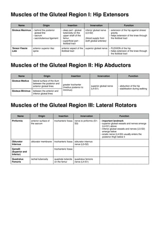
1. Muscles of the Gluteal Region I: Hip Extensors
Name
Origin
Insertion
Innervation
Function
Gluteus Maximus - behind the posterior
gluteal line
- sacrum
- sacrotuberous ligament
- deep part - gluteal
tuberosity on the
upper shaft of the
femur
- superficial part iliotibial tract
inferior gluteal nerve
(L5-S2)
Tensor Fascia
Lata
anterior aspect of the
iliotibial tract
superior gluteal nerve - FLEXION of the hip
- helps extension of the knee through
the iliotibial tract
anterior superior iliac
spine
(blood supply from
both gluteal arteries)
- extension of the hip against stress/
gravity
- helps extension of the knee through
the iliotibial tract
Muscles of the Gluteal Region II: Hip Abductors
Name
Gluteus Medius
Gluteus Minimus
Origin
Insertion
lateral surface of the ilium
between the posterior and
anterior gluteal lines
between the anterior and
inferior gluteal lines
greater trochanter
(medius posterior to
minimus)
Innervation
superior gluteal nerve
(L4-S1)
Function
- abduction of the hip
- stabilisation during walking
Muscles of the Gluteal Region III: Lateral Rotators
Name
Origin
Insertion
Innervation
Piriformis
anterior surface of
the sacrum
trochanteric fossa nerve to piriformis (S1S3)
Obturator
Internus
obturator membrane
trochanteric fossa obturator internus
nerve (L5-S2)
Gemelli
(Superior and
Inferior)
Quadratus
Femoris
trochanteric fossa
ischial tuberosity
quadrate tubercle
on the femur
quadratus femoris
nerve (L4-S1)
Function
- important landmark
- superior gluteal vessels and nerves emerge
(L4-S1) above
- inferior gluteal vessels and nerves (L5-S2)
emerge below
- sciatic nerve (L4-S3) usually enters the
posterior thigh below it
2. Posterior Compartment of the Thigh
Name
Origin
Insertion
Semitendinosus
Semimembranosus
medial surface of the
proximal shaft of the
tibia
ischial tuberosity
short head of biceps
originates from the
lateral shaft of the femur
Biceps Femoris
- medial tibial condyle
- recurrent slip of tendon
across the back of the
knee joint to the
oblique popliteal
ligament
Innervation
Function
tibial nerve (L5-S1)
short head of
biceps supplied by
common fibular
(peroneal) nerve
- extensors of the thigh at the hip
joint
- flexors of the knee
- small degree of rotation
head of the fibula
Anterior Compartment of the Thigh
Name
Origin
Insertion
Innervation
Function
Iliopsoas
- psoas major - L1-L5
lesser trochanter of the
(vertebrae, intercalated femur
discs, transverse
processes)
- iliacus - iliac fossa
- psoas major direct from L1-L3
- iliacus - femoral
nerve (L2-L3)
- flexes the thigh
- when fixed, flexes the trunk
Sartorius
anterior superior iliac
spine
medial aspect shaft of
the tibia
femoral nerve (L2L3)
- flexion of hip
- lateral rotation and abduction of hip
- flexion of the knee
- crossed leg position
Pectineus
pecten of the pubis
spiral line of the femur
femoral nerve (L2L3) and/or
obturator nerve
(L2-L4)
- adducts the hip
tibial tuberosity
femoral nerve (L2L3)
- main extensor of the knee
Quadriceps Femoris - rectus femoris anterior inferior iliac
spine
- vastus medialis medial side of the linea
aspera
- vastus lateralis - lateral
side of the linea aspera
- vastus intermedius front of the femoral
shaft
patella is found in the
common (quadriceps)
tendon, after which is
called the
ligamentum patellae
Medial Compartment of the Thigh
Name
Gracilis
Origin
pubic bone
Insertion
Innervation
medial shaft of the tibia
Function
adducts the hip
shared with sartorius and
semitendinosus
Obturator Externus
obturator membrane
+ surrounding bone
trochanteric fossa
Adductor Longus
linea aspera?
Adductor Brevis
Adductor Magnus
pubic bone
- adductor part - entire
length of linea aspera
- hamstring part - adductor
tubercle on the medial
epichondyle of the tibia
obturator nerve (L2-L4)
eternally rotates the hip
hamstring portion of
adductor magnus tibial
nerve (L5-S1)
adducts the hip
3. Anterior Compartment of the Leg
Name
Origin
Insertion
Tibialis Anterior
lateral surface of tibia
medial side of foot (medial
cuneiform and base of 1st
metatarsal)
Extensor Digitorum
Longus
- interosseous
membrane
- fibula (EDL superior to
the EHL)
distal phalanx of the 2nd-5th
digits
Innervation
Extensor Hallucis
Longus
Function
- dorsiflexes the foot
- inverts the foot (elevation
of the medial side of the
foot)
deep fibular nerve
distal phalanx of the 1st digit
(big toe)
extends 2nd-4th digits
extends 1st digit (big toe)
Lateral Compartment of the Leg
Name
Fibularis (prev.
Peroneus) Longus
Fibularis (prev.
Peroneus) Brevis
Origin
lateral surface of the
fibula (longus, upper
part; brevis, lower part)
Insertion
Innervation
Function
medial cuneiform and base
of 1st metatarsal
styloid process of the 5th
metatarsal
superficial fibular nerve everts the foot
Posterior Compartment of the Leg
Name
Origin
Insertion
Gastrocnemius
below the knee joint
Plantaris
lateral epicondyle of the
femur
Tibialis Posterior
interosseous membrane
tuberosity of the navicular
bone (medial side of the
sole of the foot)
Flexor Digitorum
Longus
tibia
distal phalanx of the 2nd-5th
digits
Flexor Hallucis
Longus
fibula
distal phalanx of the 1st digit
Function
above the knee joint
Soleus
Innervation
(superficial)
(deep)
tendocalcaneus
tibial nerve
plantar flexion of the foot