Western_Chemi_Tech_Note
•
0 likes•382 views

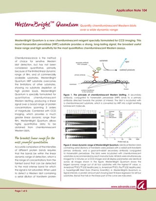
This document describes the performance of WesternBright Quantum, a new chemiluminescent reagent for Western blot detection. It provides a strong, long-lasting signal over a wide dynamic range of protein concentrations spanning 3 orders of magnitude. This allows both low and high abundance proteins to be accurately quantified in a single experiment. WesternBright Quantum shows superior sensitivity, signal duration, and linear dynamic range compared to other commercial chemiluminescent substrates. It is well-suited for quantitative Western blotting using CCD imaging.

Recommended
Recommended
More Related Content
More from WISBIOMED
More from WISBIOMED (18)
Recently uploaded
Recently uploaded (20)
Western_Chemi_Tech_Note
- 1. Application Note 104 WesternBrightTM Quantum Quantify chemiluminescent Western blots over a wide dynamic range WesternBright Quantum is a new chemiluminescent reagent specially formulated for CCD imaging. This novel Horseradish peroxidase (HRP) substrate provides a strong, long-lasting signal, the broadest useful linear range and high sensitivity for the most quantitative chemiluminescent Western assays. Chemiluminescence is the method of choice for sensitive Western blot detection, but has not been considered quantitative, primarily because of the limited linear dynamic range of film, and of commercially available substrates. WesternBright Quantum HRP substrate overcomes the limitations of other substrates, showing no substrate depletion at high protein loads. WesternBright Quantum is specially formulated for Figure 1. The principle of chemiluminescent Western blotting. A secondary quantitative chemiluminescent antibody, conjugated to horseradish peroxidase (HRP) binds to a primary Western blotting, producing a linear antibody directed towards the protein of interest. The blot is incubated with a chemiluminescent substrate, which is converted by HRP into a light emitting signal over a broad range of protein luminescent molecule. concentrations spanning 3 orders of magnitude. Combined with CCD 72.9 pg 36.4 pg 18.2 pg 9.33 ng 4.66 ng 2.33 ng 1.16 ng 0.58 ng 0.29 ng 0.14 ng 9.1 pg 4.6 pg 2.3 pg 1.1 pg 0.6 pg 0.3 pg e imaging, which provides a much a greater linear dynamic range than film, WesternBright Quantum allows b highly quantitative data to be obtained from chemiluminescent c Western blots. d The broadest linear range for the most powerful quantitation Figure 2. Linear dynamic range of WesternBright Quantum. Identical Western blots Accurate comparison of the intensities containing serial dilutions of transferrin were probed with a rabbit-anti-transferrin of different protein bands requires primary antibody, and a goat-anti-rabbit secondary antibody conjugated that the bands be within the linear to Horseradish peroxidase. The blots were incubated with chemiluminescent dynamic range of detection, which is substrates as recommended by each manufacturer. All blots were simultaneously imaged for 2 minutes on a CCD imager and all display parameters are identical the range of concentrations from the across all images shown in the figure. WesternBright Quantum shows the faintest band that can be detected largest dynamic range out of all four substrates with the highest R2 value. a. to the most intense band for which Amersham™ ECL™ (GE Healthcare); b. Amersham™ ECL Plus™ (GE Healthcare); the signal is not saturated. When used c. SuperSignal® West Dura (Thermo Scientific); d. WesternBrights Quantum; e. to detect a Western blot containing Signal intensity vs protein amount plot showing best fit linear regression for all four a serial dilution of transferrin protein substrates. Bands that fall on the linear part of the curve are indicated. www.advansta.com Page 1 of 5
- 2. Application Note 104 (Figure 2), WesternBright Quantum provides the broadest Substrate Linear Dynamic Range linear dynamic range when compared to several other WesternBright Quantum 4.6 pg – 4.7 ng commercially available chemiluminescent substrates SuperSignal West Dura 9.1 pg – 2.3 ng (Figure 2e, Table 1). Notably, no substrate depletion is seen at any protein loads with WesternBright Quantum, ECL Plus 9.1 pg – 0.29 ng while substrate depletion interferes with detection ECL 73 pg – 1.2 ng of high amounts of protein by both ECL Plus and Table 1. Linear dynamic range for four chemiluminescent SuperSignal West Dura (seen as reverse intensity bands substrates. The linear dynamic range includes data points in Figures 2b and 2c). The ability to detect high amounts that produce the best possible linear regression fit (Figure 2e). of protein without substrate depletion contributes to the Analysis of the experiment depicted in Figure 2. increased dynamic range provided by WesternBright Quantum. a For determining linear range, best fit data analysis was performed using linear regression. Data points corresponding to high protein amounts were excluded from datasets one by one as outliers to obtain the broadest range with R2 value above 0.98. Using this method, WesternBright Quantum produced the broadest linear range with the highest R2 value. Long lasting signal CCD exposure times with WesternBright Quantum are as quick as film exposures with other substrates. In addition, WesternBright Quantum provides greater signal stability than the competition, allowing long-term CCD camera exposures to be conducted, if desired, b 5 min c 60 min d 10 hr to detect faint bands and low-abundance proteins. The long-lasting signal also means there is no need to rush to image a blot, since the signal will remain strong for several hours. To follow signal strength over time, GAPDH was detected on quadruplicate Western blots Figure 3. Signal duration of WesternBright Quantum. Blots containing serial dilutions of HeLa cell lysate (Figure 3). detected using WesternBright Quantum and three other A band which was determined to be within the linear chemiluminescent substrates were imaged simultaneously range of all four chemiluminescent substrates tested at several times over a 10 hour period. Each exposure was 2 min long for all blots and all substrates. Intensities of bands was quantified at several time points over the next containing the same amount of protein are plotted for each 10 hours. WesternBright Quantum maintained signal substrate in 3a. Images of this band on the WesternBright strength, with signal declining only approximately 30% Quantum blot obtained after 5 min (3b), 60 min (3c) and 10 over a 60 minute span, while the signal from ECL, ECL hr (3d) are shown. Plus and SuperSignal West Dura each decayed by almost 90%, to barely detectable levels, over the same period (Figure 3a). In fact, the WesternBright Quantum signal is still detectable in a 2 minute exposure 10 hours later (inset, Figure 3a and 3d). www.advansta.com Page 2 of 5
- 3. Application Note 104 Low background for high sensitivity a b c d WesternBright Quantum produces an exceptionally strong signal with little to no background, for high signal to noise ratio and excellent sensitivity. The low background is especially apparent when WesternBright Quantum is is used with film detection. To demonstrate the relative lack of background with WesternBright Quantum compared to other chemiluminescent substrates, quadruplicate Western blots were detected Figure 4. Extremely low background with WesternBright using various HRP substrates as described in Methods. Quantum. Duplicate Western blots were developed using The four blots were simultaneously exposed to a single (a) ECL, (b) ECL Plus, (c) SuperSignal West Dura, or (d) film. After a 20 minute exposure, the blot detected using WesternBright Quantum as substrates. After a 20 minute exposure to film, WesternBright Quantum displays the best WesternBright Quantum remains clear of background combination of sensitivity and signal with low background. All (Figure 4d) compared to ECL Plus (Figure 4b) or display parameters are identical across all images shown in SuperSignal West Dura (Figure 4c). this figure. Figure 5 demonstrates the superior sensitivity of WesternBright Quantum on film; duplicate blots were a b c detected using WesternBright Quantum or SuperSignal West Pico. In a short exposure, WesternBright Quantum (Figure 5a) detects a band of a truncated protein not visible with the other reagent (Figure 5b) unless a longer exposure is used (Figure 5c). WesternBright SuperSignal SuperSignal Accurately quantify low and high abundance Quantum West Pico West Pico proteins 45 sec exposure 45 sec exposure 15 min exposure Figure 5. Superior sensitivity of WesternBright Quantum with WesternBright Quantum's high sensitivity, low film detection. WesternBright Quantum (a) or SuperSignal background and broad linear range combine to West Pico (Thermo Scientific) (b, c) were used to detect allow the accurate quantitation of low and high duplicate Western blots. A band (arrow in panel a) is detected abundance proteins in a single experiment. Low by WesternBright Quantum in a brief exposure, while a much abundance proteins can be detected due to the longer exposure is needed to detect the same band with SuperSignal West Pico (c). excellent sensitivity, and the broad linear dynamic range allows high abundance proteins to be detected with the same exposure, without saturation of signal. Figure 6 demonstrates the superior performance of WesternBright Quantum when probing for either a high abundance (actin) or low abundance (STAT-1) protein. Serial dilutions of extracts from A431 cells were assayed by replicate Western blots, probed for actin and STAT- 1, and visualized using WesternBright Quantum or other commercially available chemiluminescent substrates as described in Methods. For quantitative detection on the linear part of the intensity vs protein curve, WesternBright Quantum proved to be 8-times more www.advansta.com Page 3 of 5
- 4. Application Note 104 sensitive than ECL Plus in detecting STAT-1 (Figure 6g), actin STAT-1 5.00 µg 2.50 µg 1.25 µg 5.00 µg 2.50 µg 1.25 µg and twice as sensitive as ECL in detecting actin (Figure 625 ng 313 ng 156 ng 625 ng 313 ng 156 ng 78 ng 78 ng 6c). a d Simply the best choice for CCD imaging ECL e WesternBright Quantum outperforms other enhanced chemiluminescent reagents, providing superior ECL Plus sensitivity, signal duration, and linear dynamic range. b f Specially formulated for CCD imaging, WesternBright Quantum maintains a detectable signal in a two-minute WesternBright Quantum exposure for at least 10 hours, long after the signals from c g other substrates have decayed to levels undetectable without exposures many times longer. Methods Gel electrophoresis Proteins were separated on self-made 12 % polyacrylamide gels using a Laemmli buffer system. Figure 6. Detect high and low abundance proteins with Gels were 10 cm wide and 0.8 mm thick. WesternBright Quantum. Identical blots containing serial Transfer dilutions of cell A431 lysates were probed with a mixture or Proteins were transferred to PVDF membrane using a primary antibodies to actin and STAT-1. The blots were then detected with WesternBright Quantum (panels b and f) or tank (Idea Scientific) and buffers developed by Bolt one of two other chemiluminescent substrates and imaged and Mahoney (1). Transfers were conducted for 25 min simultaneously with a 2 minute exposure using a CCD imager. at 24 V. The intensities of the bands were plotted against total protein loaded (panels c and g), demonstrating that WesternBright Blocking, Primary and Secondary Antibodies, and Quantum provided the highest sensitivity and the broadest Washing linear dynamic range for both the high and low abundance Membranes were blocked with 2% non-fat dry milk proteins. in AdvanWash buffer for 1 hour at room temperature (RT). Primary and secondary antibodies were diluted as Experiment Primary Dilution Secondary Dilution described in Table 2 in blocking buffer, and applied to antibody antibody membranes for 1 hour at RT. Washing was conducted Linear Rabbit anti- 1:10,000 HRP-Goat- 1:20,000 dynamic transferrin anti-rabbit with AdvanWash buffer according to the WesternBright range (Abcam, (Advansta, cat. user manual. cat. no. no. R-05072-500) ab122301) Substrate incubation Signal Mouse 1:2,000 HRP-Goat- 1:2,500 duration anti-GAPDH anti-mouse After washing, blots were incubated with (Millipore, (Advansta, cat. chemiluminescent substrates as recommended by cat. no. no. R-05071-500) MAB374) manufacturers. Incubation with WesternBright Quantum Cell lysates Mouse 1:1,000 HRP-Goat- 1:2,500 substrate was done for 2 minutes. anti-actin anti-mouse (Sigma, cat. (Advansta, cat. Imaging no. A4700) no. R-05071-500) After incubation, blots were placed on a plastic Mouse 1:250 HRP-Goat- 1:2,500 sheet, covered with Saran wrap, and imaged using a anti-STAT 1 anti-mouse (BD, cat. no. (Advansta, cat. FluorChem Q (Cell Biosciences). Images were analyzed 10116) no. R-05071-500) using AlphaView® software (Cell Biosciences). Table 2. Antibodies and dilutions used. www.advansta.com Page 4 of 5
- 5. Application Note 104 References Copyright © 2010 Advansta. All rights reserved. The Advansta logo is a registered trademarks of the Company. WesternBright™, AdvanBlock™, AdvanWash™ and LucentBlue™ are trademarks of the Company. All other trademarks, service 1. Bolt M.W. and Mahoney P.A. 1997. Anal Biochem. marks and tradenames appearing in this brochure are the property of their respective owners. 247. 185-192 Catalog Number Product Size K-12042-C20 WesternBright™ Quantum Western Blotting HRP Substrate Trial kit size 20 ml (200 cm2) K-12042-D10 WesternBright™ Quantum Western Blotting HRP Substrate (for 1000 cm2 membrane) 100 ml (1000 cm2) K-12042-D20 WesternBright™ Quantum Western Blotting HRP Substrate 200 ml (2000 cm2) (for 2000 cm2 membrane) L-07014-100 LucentBlue™ X-ray film 100 sheets R-03024-D50 AdvanWash™ 10x washing solution 500 ml R-05072-500 Goat-anti-rabbit HRP-conjugated secondary antibody 500 μl R-05071-500 Goat-anti-mouse HRP-conjugated secondary antibody 500 μl L-08001-010 Low Fluorescence Western Membrane (PVDF) 7x9 cm 10 sheets L-08002-010 Nitrocellulose Transfer Membrane 0.45 μm 7x9 cm 10 sheets L-08003-010 Nitrocellulose Transfer Membrane 0.22 μm 7x9 cm 10 sheets Advansta Corporation 1455 Adams Drive, Ste. 1160 | Menlo Park, CA 94025 Tel: 650.325.1980 | Fax: 650.325.1904 | Email: sales@advansta.com D-08104-023 www.advansta.com Page 5 of 5