SlideShare une entreprise Scribd logo
1  sur  16
Télécharger pour lire hors ligne
CASE OF THE WEEK
                   PROFESSOR YASSER METWALLY
CLINICAL PICTURE

CLINICAL PICTURE

30 Years old male patient presented clinically with bilateral pyramidal tract signs, pseudo-bulbar palsy, with oro-
genital ulcerations. The patient also gave a history of nonspecific relapsing headache. Examination of sensation did
not show any abnormality.

RADIOLOGICAL FINDINGS

RADIOLOGICAL FINDINGS  


Figure 1. Precontrast MRI T1 images showing focal signal hypointensity in the left basis pontis, left crus cerebri
(cerebral peduncle) and the posterior limb of the internal capsule. The focal hypointense lesions has no or very mild
mass effect and they are apparently continuos cephalocaudally. Contralaterally, the pontine basis is showing ? a
very small hypointense lesion in the right side. The lesions are located in the presumed anatomical area of the
corticospinal (pyramidal) tract.
Figure 2. Postcontrast MRI T1 images showing enhancement of the hypointense lesions shown in figure 1. The
lesion in the right anterior pontine zone is more definite now and is better delineated by contrast enhancement. All
enhanced areas are surrounded by a hypointense edema area with mild mass effect. Also notice contrast
enhancement of the meningeal covering of the pons and midbrain which indicates the existence of a meningitic
element.


Figure 3. Postcontrast coronal MRI T1 images showing that the pontine, midbrain and capsular lesions described in
figure 1,2 are continuous cephalocaudally as a linear long lesion, the lesion is surrounded by a hypointense edema
area and is located in the presumed anatomical area of the corticospinal (pyramidal) tract. The corticospinal tract
was actually mapped by contrast enhancement. The contralateral side is involved to a lessor degree. Notice
meningeal enhancement which reflects a meningitic element. Contrast enhancement of the corticospinal tract in this
case is due to break down of the blood brain barrier along the course of the corticospinal tract and it is a good sign
of vasogenic edema which is mainly due to break down of the blood brain barrier (70). Notice middle line crossing
of the lesions suggesting its venous rather that arterial origin. Also notice the mild mass effect induced by the
pyramidal tract lesion in the left side which could be due to edema.
Figure 4. Postcontrast sagittal MRI T1 images showing that the pontine, midbrain and capsular lesions described in
figure 1,2,3 are continuous cephalocaudally as a linear long lesion in the anterior parts of the pons and midbrain,
the lesion is located in the presumed anatomical area of the corticospinal (pyramidal) tract and is seen surrounded
by a hypointense edema area. The corticospinal tract was actually mapped by contrast enhancement. Also notice
meningeal enhancement. Thrombosis of the superficial and deep venous systems is also seen.


                                      Figure 5. MRI T2 image showing the capsular lesion (in the posterior limb of
                                      the internal capsule), The lesion is mainly hyperintense with some
                                      hypointense zones probably representing a hemorrhagic element.


In general the observed pathology in this case represents demyelination and edema along the cortico-spinal tract
fibers (in fact the pyramidal tract is actually anatomically drawn by the MRI signal changes of vasogenic edema and
contrast enhancement) and is found to be located mainly in the midbrain/thalamus (which probably constitutes the
primary site of involvement) with upward extension to the posterior limb of the internal capsule and downward
extension to the basis pontis. The upward and downward extension of the primary midbrain/thalamic lesions might
be explained by wallerian degeneration and this was noted in some pathologic studies as well (20, 43, 44).

DIAGNOSIS:

DIAGNOSIS: NEURO-BEHCET SYNDROME

DISCUSSION

DISCUSSION

Behcet's disease (BD) is an uncommon, relapsing and remitting, multisystem inflammatory disorder characterized
by the triad of oral ulceration, genital ulceration, and uveitis. 61 A number of additional features are commonly
present, including arthritis, retinal and cutaneous vasculitis, thrombophlebitis, and gastroenteric disorders. The
essential lesion is a focus of chronic inflammation, typically in the vicinity of a small blood vessel. Lesions in the
CNS resemble those in other organ systems. Characteristically there are multiple small foci of softening that may
eventually become confluent. These correspond to myelin loss and, to a lesser degree, drop out of neural elements
with replacement by foamy macrophages. The lesions are not fundamentally demyelinative and more closely
resemble minute ischemic foci. As in the periphery, lesions tend to occur near blood vessels, but vasculitis is
uncommon. Pathologic findings are most extensive in the midbrain, pons (especially the basis pontis) and the
medulla, and there is typically a lesser degree of involvement of the spinal cord, internal capsule, globus pallidus,
optic nerves, and hypothalamus. In some cases, the cerebral white matter, cortex, hippocampus, basal ganglia, and
thalamus are involved. The cerebellum is usually spared. The meninges are typically thickened and adherent to the
brain surface, and acutely there may be considerable meningeal inflammation; hence the common reference to
neuro-Behcet's disease as a meningoencephalitis. (The x ray room) (Print this case report)

In general the most common presentation of parenchymal CNS involvement is a subacute brainstem syndrome with
cranial nerve findings, dysarthria, and cerebellar or corticospinal tract signs. More infrequent presentations include
a strokelike presentation (with the acute onset of unilateral neurologic findings and signs of cortical involvement
including seizures) and psychiatric features, such as psychosis. Sinus venous thrombosis may evolve relatively
slowly and results in intracranial hypertension, resulting headache, vomiting, and bilateral papilledema. Impaired
memory (long-term verbal and nonverbal) and visuospatial skills occur frequently in patients with active disease
who are taking large doses of steroids. Pure spinal cord or PNS involvement is rarely reported. Impairment of
sensation is uncommon. (70)

Between 5% and 20% of patients have neurologic disease. (70) The most common neurologic manifestation of
Behcet's disease is a relapsing and remitting, but often progressive, focal meningoencephalitis that most often affects
the brain stem. (70) Patients present with a host of cranial nerve and long tract signs and eventually develop spastic
quadriparesis and a bulbar or pseudobulbar palsy, frequently with rather dramatic emotional incontinence. (70)
Cochlear and vestibular dysfunctions are common. Often there are additional features of impairment in frontal lobe
and memory function characteristic of a subcortical dementia. A number of cases of nearly pure subcortical
dementia have been reported, but typically long tract signs are prominent, and bulbar involvement eventually
develops. Patients may also present initially with features of transverse myelitis, but bulbar signs characteristically
appear as the disease progresses. Cerebral cortical involvement is unusual, but seizures and aphasia have been
reported. The optic nerves are commonly involved, but symptoms are indiscriminable from those of uveitis, which is
usually present in patients with neurologic features. CNS vasculitis is rare, but stroke caused by large artery
involvement has been reported. Pseudotumor cerebri is relatively common and almost always caused by venous
sinus thrombosis. Cerebral venous thrombosis may affect up to 10% of all patients with Behcet's disease and one
third of those with neurologic involvement. CNS involvement generally manifests several years after the onset of
systemic Behcet's disease, but occasionally it may be an initial feature and even precede other disease
manifestations. As a rule, flares of neurologic disease parallel flares of systemic disease.

Neuro-Behçet's syndrome can be classified as an acute type and as a chronic progressive type (70). It should be
pointed out that the two types of neuro-Behçet's syndrome are currently considered to represent different stages
rather than independent clinical entities. Acute neuro-Behçet's syndrome is characterized by acute
meningoencephalitis with a focal midbrain pyramidal tract lesions, the lesions are anteriorly located in the cerebral
peduncles, bilateral, asymmetrical, extending up and down within the brain stem as a longitudinal linear lesion, and
presenting high-intensity areas in the MRI T2-weighted images or the fluid attenuated inversion recovery (FLAIR)
images (70). Acute neuro-Behçet's syndrome responds to steroid therapy, and is usually self-limiting.

By contrast, the chronic progressive type of neuro-Behçet's syndrome is characterized by intractable, slowly
progressive dementia, ataxia and dysarthria, with persistent elevation of cerebrospinal fluid IL-6 activity (> 20
pg/ml) (70). Most patients with the chronic progressive type of neuro-Behçet's syndrome were HLA-B51-positive,
and they had history of attacks of acute type neuro-Behçet's syndrome prior to the development of progressive
neurological symptoms (70).

The neurological involvement in Behçet's disease is either caused by primary neural parenchymal lesions (neuro-
Behçet's syndrome, or brain stem meningoencephalitis) or is secondary to major vascular involvement. The latter
type is rarely complicated with the parenchymal lesions and should be called vascular-Behçet's disease. This
vasculo-Behçet's disease type generally has a better prognosis compared with the parenchymal type. (70) Both
vascular and parenchymal types can coexist, as in this case, but this is not common.

      The primary parenchymal neuro-Behçet type
   It is primary parenchymal neural disease, a focal brain stem meningoencephalitis affecting the midbrain
              primarily with selective involvement of the corticospinal tract bilaterally. The most common clinical
              findings are pyramidal signs. Sensory symptoms or signs are much less frequent in neuro-Behcet
              disease. (73,74) The corticospinal tract lesions extend cephalocaudally in a linear fashion from the
              mesencephalon down to the pons and less frequently to the medulla and up to the posterior limb of the
              internal capsule. The cephalocaudal extension of the midbrain corticospinal tract lesions could be either
              due to wallerian degeneration or extension of vasogenic edema along the myelinated fibers of the
              corticospinal tract. (70,73,74)

             The corticospinal tract pathology is manifested by MR imaging as bilateral and asymmetrical
              longitudinal linear lesions extending from the posterior limb of the internal capsule down to the
              midbrain, pons and less frequently the medulla. The midbrain is more severely affected. The
              corticospinal tract linear lesions are hyperintense on the MRI T2 images and hypointense on the MRI
              T1 images and show contrast enhancement in the acute stage. Contrast enhancement is due to break
              down of the blood brain barrier and this is in favour of the corticospinal tract MRI signal changes being
              due vasogenic edema. (70,73,74)

      Vascular-Behçet's type

             Involvement of veins and arteries in Behçet's disease is usually called vasculo-Behçet's disease. Venous
              thrombosis appeared to be the major vascular involvement in 7–33% of patients with Behçet's disease,
              and represents 85–93% of vascular-Behçet's disease (70). The brain parenchymal involvement seen in
              the primary neuro-Behçet type is rarely seen in the vascular type of Behçet disease. Parenchymal
              involvement in the vascular type of Behçet disease is more commonly secondary to venous sinus
              thrombosis. Deep vein thrombosis or cerebral sinus thrombosis are significantly associated with the
              male gender and a positive pathergy test (70). Venous thrombosis is a reflection of thrombophilia in
              vascular Behçet's disease. (70,73,74). To the best of the author's information, cerebral sinovenous
              thrombosis in the vascular-Behçet type is indistinguishable from sinovenous thrombosis due to any
              other cause and is only distinct from the view point that it is commonly associated clinically with oro-
              genital ulcerations. Probably vascular-Behçet syndrome must be put in the differential diagnosis of cases
              presented with cerebral sinovenous thrombosis of undetermined aetiology.

             The clinical picture of vascular Behçet is different from that of primary neuro-Behçet. The clinical
              picture of neuro-Behçet is dominated by a mainly motor clinical picture with bilateral pyramidal tract
              signs, pseudo-bulbar palsy, subcortical dementia, etc. and sensory symptoms are uncommon, while the
              clinical picture of vascular Behçet is that of acute and chronic cerebral sinovenous thrombosis and its
              parenchymal complications (headache, seizures, focal sensory and motor deficits, bilateral papilledema,
              pseudotumor cerebri).

             A number of studies have explored the pathogenesis of thrombophilia in Behçet's disease. Neither
              deficiency in protein C, in protein S, in factor V Leiden and in antithrombin III nor resistance to
              activated protein C and anticardiolipin antibody levels seemed to be correlated with vascular
              thrombosis in Behçet's disease (75,76). There were increased thrombin generation, fibrinolysis, and
              thrombomodulin in Behçet's disease, but these abnormalities were not related to thrombosis (76). These
              results therefore suggest that thrombophilia in Behçet's disease might be related more to inflammation
              than to clotting disorder.

             Arterial involvement, although rare, does occur in Behçet's disease. The arterial manifestations in
              Behçet's disease resemble those of Takayasu's arteritis, including arterial occlusion and aneurysm
              formation. Histopathological studies revealed that the number of vasa vasorum with infiltration of
              neutrophils and lymphocytes was significantly increased in vascular-Behçet's disease compared with in
              Takayasu's arteritis and other inflammatory aneurysms (70). It was therefore suggested that arterial
              involvement in vascular-Behçet's (vasculo-Behçet) disease might be caused by a neutrophilic vasculitis
              targeting the vasa vasorum, leading to degeneration of the arterial wall. (70,73,74)

Table 1. Lesion characteristics in the primary parenchymal neuro-Behçet type
    The lesion in the primary parenchymal neuro-Behçet type is a focal brain stem meningoencephalitis
     affecting the midbrain primarily with selective involvement of the corticospinal tract bilaterally.

      The most common clinical findings are pyramidal signs. Sensory symptoms or signs are much less frequent
in neuro-Behcet disease.

       The corticospinal tract lesions extend cephalocaudally in a linear fashion from the mesencephalon down to
        the pons and less frequently to the medulla and up to the posterior limb of the internal capsule. The
        cephalocaudal extension of the midbrain corticospinal tract lesions could be either due to wallerian
        degeneration or extension of vasogenic edema along the myelinated fibers of the corticospinal tract.

       The corticospinal tract pathology is manifested by MR imaging as bilateral and asymmetrical longitudinal
        linear lesions extending from the posterior limb of the internal capsule down to the midbrain, pons and less
        frequently the medulla. The midbrain is more severely affected. The corticospinal tract linear lesions are
        hyperintense on the MRI T2 images and hypointense on the MRI T1 images and show contrast enhancement
        in the acute stage. Contrast enhancement is due to break down of the blood brain barrier and this is in
        favour of the corticospinal tract MRI signal changes being due vasogenic edema. (70,73,74)

       The corticospinal tract linear lesions are sometimes surrounded by a hypointense edema area with positive
        mild mass effect.

Table 2. The differences between the primary parenchymal neuro-Behçet type and the vascular-Behçet's type

Parameter        The primary parenchymal neuro-Behçet type                       Vascular-Behçet's type
                                                                                 The clinical picture of vascular
                                                                                 Behçet is that of acute and chronic
                 The clinical picture of neuro-Behçet is dominated by a mainly   cerebral sinovenous thrombosis
Clinical         motor clinical picture with bilateral pyramidal tract signs,    and its parenchymal complications
picture          pseudo-bulbar palsy, subcortical dementia etc. Sensory          (headache, seizures, focal sensory
                 symptoms are uncommon                                           and motor deficits, bilateral
                                                                                 papilledema,          pseudotumor
                                                                                 cerebri, etc.).
Incidence    More common (70-93%)                                                Less common (7-33%)
             It is primary parenchymal neural disease, a focal brain stem        Venous thrombosis is a reflection
             meningoencephalitis affecting the midbrain primarily with           of thrombophilia in Behçet's
Pathology & upward and downward extension (along the pyramidal tract             disease. Thrombophilia in Behçet's
pathogenesis fibers to the posterior limb of the internal capsule, the basis     disease might be related more to
             pontis and the medulla) and with selective involvement of the       inflammation than to clotting
             corticospinal tract bilaterally.                                    disorder.
             The corticospinal tract pathology is manifested by MR imaging
             as bilateral and asymmetrical longitudinal linear lesions
             extending from the posterior limb of the internal capsule down
             to the midbrain, pons and less frequently the medulla. The
             midbrain is more severely affected. The corticospinal tract         The MRI picture is that of
Radiological
             linear lesions are hyperintense on the MRI T2 images and            sinovenous thrombosis and its
findings
             hypointense on the MRI T1 images and show contrast                  parenchymal complications (70)
             enhancement in the acute stage. Contrast enhancement is due
             to break down of the blood brain barrier and this is in favour
             of the corticospinal tract MRI signal changes being due
             vasogenic edema. (70,73,74)
             Bad, higher incidence of being transformed into the progressive
Prognosis                                                                        Better
             type with subcortical dementia, etc.
                                                                                 Anticoagulants, steroid may play a
Management Steroid responsive
                                                                                 role

Currently, the most widely used diagnostic criteria of Behcet's disease is the International Study Group's
classification, which requires recurrent oral ulcerations plus two of the following in order to establish a definite
diagnosis: recurrent genital ulcerations, skin or eye lesions, or a positive pathergy test (13). The epidemiology of
disease shows geographic variation, encountered more commonly along the Silk Road, which extends from the
Mediterranean region to Japan (15). This is coupled with a similar variation in HLA B51 (human leukocyte
antigen), which has been reported to be strongly associated with the disease in the high prevalence areas (16–19).
Despite broadened clinical understanding of this disease, the etiologic factors remain obscured and speculative:
viral agents, immunologic factors, genetic causes, bacterial factors, and fibrinolytic defects have all been implicated
(3, 20–25). Vessel wall and perivascular mononuclear cell infiltration, which is consistent with vasculitis involving
both arterial and venous systems, has been shown in histopathologic studies (20, 21). It has been postulated that
genetic susceptibility together with a possible trigger by an extrinsic factor, such as an infectious agent, is
responsible for the observed vasculitis (24, 26).

                   Table 3. The International Study Group criteria for the diagnosis of Behcet disease
       At least 3 episodes of oral ulceration must occur in a 12-month period. They must be observed by a physician
        or the patient and may be herpetiform or aphthous in nature.

       At least 2 of the following must occur:
             Recurrent, painful genital ulcers that heal with scarring.
             Ophthalmic lesions, including anterior or posterior uveitis, hypopyon, or retinal vasculitis.
             Skin lesions, including erythema nodosum, pseudofolliculitis, or papulopustular lesions (may also
              include atypical acne).
             Pathergy: defined as a sterile erythematous papule larger than 2 mm in size appearing 48 hours after
              skin pricks with a sharp, sterile needle (a dull needle may be used as a control).

Neurologic involvement in Behcet's disease, was first reported in 1941 by Knapp (27), and the term neuro-Behçet
syndrome was introduced by Cavara and D'Ermo in 1954 (28). The reported rate of development of neurologic
involvement among Behcet's disease patients ranges from 4% to 49% (9, 29). This rate was found to be 6.7% in our
large nonselective series of patients referred from the Behçet's Disease Research Center (30).

Neuro-Behçet syndrome may present as an acute focal or multifocal CNS dysfunction, and the clinical picture of
neuro-Behçet syndrome may resemble multiple sclerosis (MS) (7, 31–35). It has been observed that a substantial
number of patients with neuro-Behçet syndrome will have a relapsing-remitting course while others may develop a
secondary-progressive course; some neuro-Behçet syndrome patients have an insidious onset, with primary-
progressive CNS dysfunction, and others may display symptoms attributable to intracranial hypertension
associated with dural venous sinus thrombosis (12, 36, 37). Although non-neurologic involvement generally precedes
neurologic findings, the non-neurologic involvement may go unrecognized in some cases or it may appear late in the
patient's course, thus posing diagnostic difficulties (38–40). Peripheral nerve involvement, although reported in
neuro-Behçet syndrome, is relatively uncommon (41).

The most common imaging lesion seen in the acute or subacute stage of neuro-Behçet syndrome is an asymmetric
mesodiencephalic junction lesions (7–9, 33). These lesions extend along long fiber tracts and spare the red nucleus,
suggesting that this downward extension is due to edema. The reversibility of the extension, leaving small residual
lesions at the center, as observed on follow-up MR studies, further supports their edematous nature. This feature
was also noted in earlier publications (9, 11, 42). Accordingly, pontobulbar involvement was an extension of lesions
located at other sites, particularly the mesodiencephalic junction. The distribution and intensity of changes of the
residual lesions closely corresponded to pathologic descriptions of secondary demyelination (20, 21, 43, 44). In the
chronic cases, Changes may extend to the cervical cord, along the corticospinal tract, and this might be explained by
wallerian degeneration. This was noted in some pathologic studies as well (20, 43, 44).

Taken together, the current case (case version number 1.4) and the previously reported case in version 1.3 of this
publication emphasized and highlighted the following common clinical-radiological findings in neuro-Behçet
syndrome:

  1. In general the observed pathology in these cases represents demyelination and edema along the cortico-spinal
     tract fibers (in fact the pyramidal tract is actually anatomically drawn by the MRI signal changes of vasogenic
     edema and contrast enhancement) and is found to be located mainly in the midbrain/thalamus (which
     probably constitutes the primary site of involvement) with upward extension to the posterior limb of the
     internal capsule and downward extension to the basis pontis. The upward and downward extension of the
     primary midbrain/thalamic lesions might be explained by wallerian degeneration or vasogenic edema (which
     characteristically spreads along the myelinated fibers of long tracts) and this was noted in some pathologic
     studies as well (20, 43, 44)

  2. The selective involvement of the corticospinal tract would explain the clinical picture of these patients which is
     dominated by pyramidal tract involvement and pseudo-bulbar palsy with absence of sensory changes. In fact
     the clinical picture and the MRI findings in neuro-Behçet syndrome closely resemble those of motor neuron
     disease with pseudo-bulbar palsy (they both shared common topographic distribution and spatial extension of
the corticospinal tract lesions), however the corticospinal tract lesion in motor neuron disease does not show
     contrast enhancement. (70). The corticospinal tract linear lesion appeared larger in size in acute neuro-Behçet
     syndrome compared with that of motor neuron disease probably due to the contribution of edema in neuro-
     Behçet syndrome. The corticospinal tract lesion is reversible in the acute neuro-Behçet syndrome, however it
     is irreversible in motor neuron disease. In general the lesion in acute neuro-Behçet syndrome has an acute
     onset and a regressive course, while that of motor neuron disease has a chronic progressive course. The MRI
     signal changes along the course of the corticospinal tract are mainly due to reversible vasogenic edema in
     acute neuro-Behçet syndrome, while the MRI signal changes along the course of the corticospinal tract in
     motor neuron disease are mainly due to wallerian degeneration. See table 4

  3. Both cases (this case and the previously reported case in version 1.3 of this publication) shared common
     clinical (purely motor clinical presentation) and common MRI findings (selective pyramidal tract
     involvement). However sinus thrombosis was demonstrated only in the current case. Which simply means that
     the parenchymal lesion demonstrated in neuro-Behçet syndrome is a primary neuronal lesion and not
     secondary to sinus thrombosis (Behcet's disease is a relapsing and remitting, but often progressive, focal
     meningoencephalitis that most often affects the brain stem). Sinus thrombosis is probably a product of the
     primary pathology. In the current patient the presence of the midbrain lesions characteristic of the primary
     neural type of Behçet disease associated with cerebral sinus thrombosis simply means that both the primary
     parenchymal neural type and the vascular type of Behçet disease can coexist in a single patient, but this
     uncommon. Vascular Behçet is rarely associated with neuro-Behçet. The predominant histopathological
     features in the inflamed tissues in Behcet's disease are infiltration of lymphocytes and monocytes, and
     sometimes polymorph nuclear leukocytes, through small veins without microscopic changes in the vessel walls.
     Thrombophilia or thrombophlebitis involving small and large veins is also common, whereas arteritis is rare.
     In these regards, Behçet's disease is unique compared with other vasculitides.

  4. The existence of meningeal enhancement at the midbrain and pontine level in this case (version number 1.4) is
     simply a reflection of a meningitic process at the pontine and midbrain levels. The meninges are typically
     thickened and adherent to the brain surface in neuro-Behçet syndrome, and acutely there may be
     considerable meningeal inflammation; hence the common reference to neuro-Behcet's disease as a
     meningoencephalitis. (70) Meningeal enhancement could not be appreciated radiologically in the previously
     reported case (number 1.3).

Table 4. Differences between motor neuron disease and acute neuro-Behçet syndrome, with regard to the
corticospinal tract affection

The corticospinal tract lesion in neuro-Behçet          The corticospinal tract lesion in motor neuron disease
syndrome
The corticospinal tract linear lesion appeared larger   Smaller in size
in size probably due to the contribution of edema
Reversible                                              Irreversible
Show contrast enhancement                               Does not show contrast enhancement
Has an acute onset and a regressive course              Has a chronic progressive course
MRI signal changes are mainly due to reversible         MRI signal changes are mainly due to wallerian
vasogenic edema                                         degeneration

In presented patient, an asymmetric subcortical and deep periventricular white matter involvement (involving
mainly the posterior limb of the internal capsule) without cortical involvement was observed. The patient also had
concomitant mesodiencephalic junction lesions. In the majority of reported cases with hemispheric involvement, the
lesions were located subcortically, particularly within temporal and occipital regions (7–11, 34, 39, 45, 46). In the
acute stage the periventricular white matter changes are due to edema, while in the chronic stage these white matter
changes are due to wallerian degeneration, gliosis, and demyelination (4, 20, 21, 43, 44, 47, 48).

Vasculitis is regarded as the key feature in neuro-Behçet syndrome (3, 26), as biopsy specimens from mucous and
cutaneous lesions show those changes (54, 55). Arterial and venous large vessel involvement, such as narrowing,
occlusion, and aneurysmal formation, has been reported in up to 27% to 35% of cases, with 12% arterial and 88%
venous (52). An even greater proportion of patients with neuro-Behçet syndrome may have small vessel vasculitis,
and recently this has been validated as the pathologic basis of various histologic changes observed in different organ
systems (26).
Lesions of arterial origin may be observed in some patients, in whom these lesions, for example, may lie within the
pons without crossing the midline, consistent with involvement of the penetrating arteries. Although such lesions,
resulting from small or medium-sized intracranial arteries, have been reported either microscopically or
radiologically in neuro-Behçet syndrome, they are not as common as the arterial lesions observed with the
involvement of other systems (20, 21, 40, 43, 47, 48, 50–52). There are also a few publications concerning the
involvement of large intracranial arteries (40, 51–53). Hemorrhagic lesions seen in some patients most likely
resulted from quot;diapedesis of red cells around veins,quot; as already reported, and are not of arterial origin (21).

Autopsy studies and biopsy specimens of the CNS lesions are consistent with vasculitis as well, and they show a clear
venous predominance (20, 21, 49). Radiologic studies support this finding that lesions seen in neuro-Behçet
syndrome are not compatible with arterial territories. Furthermore, significant perilesional edema with a tendency
to disappear or to leave disproportionally small residua on follow-up studies has been reported. This feature is
consistent with venous infarction, since not all signal intensity changes seen in venous occlusive disease necessarily
represent infarction, but rather an accumulation of water within interstitial spaces (56, 57). This information,
together with our observations, supports the probable inflammatory-venous pathogenesis for the CNS lesions seen
in Behcet's disease.

Table 5. Factors that support the probable inflammatory-venous pathogenesis for the CNS lesions seen in neuro-
Behçet syndrome
Arterial lesions are unilateral and do not cross the middle line, while lesions observed in neuro-Behcet syndrome
are frequently bilateral with frequent midline crossing.
Autopsy studies and biopsy specimens of the CNS lesions are consistent with vasculitis with a clear venous
predominance (20, 21, 49).
Significant perilesional edema with a tendency to disappear or to leave disproportionally small residua on follow-
up studies has been reported. This feature is consistent with venous infarction, since not all signal intensity
changes seen in venous occlusive disease necessarily represent infarction, but rather an accumulation of water
within interstitial spaces (56, 57).

If one considers the possible venous territories in which brain stem lesions have occurred in neuro-Behçet
syndrome, it is clear that the affected venous structures are indeed small intraaxial veins of the brain stem (58).
That particular predilection of occurrence raises the question of why these veins are affected or why occlusion of
them causes symptoms. The literature on autopsy studies states no apparent tendency toward these venous
channels, and brain stem veins and cerebral hemispherical veins are equally affected, although there is a clear-cut
predominance of lesions in the brain stem. The question can also be addressed according to regional hemodynamic
properties. It is well known that cerebral hemispherical structures are drained by superficial and deep venous
systems, both of which anastomose via medullary veins. They interconnect superficial pial veins to the internal
cerebral vein and the basal vein of Rosenthal, the former being more common than the latter (59), whereas in the
brain stem, intraparenchymal radial and longitudinal anastomotic channels are nearly absent (58). In the spinal
cord, intraaxial anastomoses are claimed to be prominent at the thoracic level (60). The particular arrangement of
veins in the cerebral hemisphere permits them to flow in both directions via medullary veins, as seen with certain
disorders, such as deep arteriovenous malformations, Sturge-Weber disease, and the developmental venous
anomalies (60), possibly explaining the small diameter and unimportance of parenchymal lesions when such a vein
is thrombosed. At the mesencephalic, diencephalic, and pontine levels, thrombosis of small veins might be
accompanied by a very large, sometimes hemorrhagic lesion, since there is nearly no collateral venous pathway. The
same anatomic arrangement might also explain the vulnerability of the cervical spinal cord (7, 44). It appears,
therefore, that the variability of venous anatomic arrangements at different levels of the CNS might explain the
predilection of lesions for different regions.

Vasculitis, like inflammation in other tissues, is caused by many different agents and pathogenic mechanisms;
however, these different causes produce only a limited number of histologic expressions of injury. The major type of
injury to nervous tissue in vasculitis is ischemia. Therefore, the same clinical manifestations can result from
etiologically and pathogenetically different vasculitic diseases. In vasculitic processes, location, extension, and
distribution of vascular involvement might point to a specific diagnosis, such as Takayasu or temporal arteritis (26,
61–65). In neuro-Behçet syndrome, lesions therefore appear secondary to the small vessel venous vasculitis, and the
anatomy of those intraaxial venous structures explains the dominant involvement of the upper brain stem and
diencephalic structures.

Pathologically proved small vessel arteritis, either alone or with venous inflammation, has also been reported in
conjunction with neuro-Behçet syndrome, and it is probable that some of the cerebral hemisphere and midbrain
lesions might result from small vessel arteritis (62, 63). In this case, there appeared to be at least one lesion that
occurred within a probable pontine arterial territory, and the hemispheric lesions seen in neuro-Behçet syndrome
were not significantly different from the CNS lesions of other vasculopathies of accepted arteritic origin.

It is possible to explain the selective involvement of the corticospinal fibers observed in this patient. If the primary
pathology in Behcet disease is inflammatory vascular process with secondary demyelination, then the myelinated
fibers in the long tracts will suffer more. The posterior part of the brain stem is mainly reticular while the anterior
part is compact and is occupied mainly by the pyramid in the cerebral peduncle, crus cerebral and basis pontis. It is
quite illogical to see inflammatory demyelination in the reticular posterior parts of the brain stem. The
inflammatory demyelinating process will affect the anteriorly located long myelinated fibers in the corticospinal
tract.

Vasogenic edema is the most common type of edema associated with brain tumors, venous congestion and other
causes and results from local disruption of the blood brain barrier. This leads to extravasation of protein-rich
filtrate of plasma into the interstitial space, with subsequent accumulation of vascular fluid. This disruption results
from loosening of the tight junctions between endothelial cells, and the neoformation of pinocytic vesicles. Once the
barrier is breached, hydrostatic and osmotic forces work together to extravasate intravascular fluid. Once
extravasated, fluid is retained outside the vasculature, mostly in the white matter of the brain, and within the
bundles of myelinated axons of long tracts and commissural fibers. This is because axons run in parallel bundles of
fibres with loose extracellular space (that offer low resistance and facilitates the extension of vasogenic edema along
myelinated axons which are spreaded apart by the edema) as opposed to gray matter, which has high cell density
and is enmeshed in an interwoven network of connecting fibres that offer high resistance to the formation and
spread of edema. By definition, this type of edema is confined to the extracellular space. (70) In neuro-Behcet case
reports of diffusion weighted imaging (DWI) of the T2 hyperintense lesions revealed elevated ADC values compared
to normal subjects, indicating restricted diffusion and the presence of vasogenic edema (71,72). Contrast
enhancement of the corticospinal tract in this case is due to break down of the blood brain barrier along the course
of the corticospinal tract and it is a good sign of vasogenic edema which is mainly due to break down of the blood
brain barrier. (70)

Anther explanation for the selective involvement of the corticospinal tract in neuro-Behcet is that the signal changes
observed in the MRI study is due to vasogenic edema secondary to venous congestion, vasogenic interstitial edema
spreads in the white matter along myelinated fibers of long tracts and commissural fibers (This is because axons of
long tracts run in parallel bundles of fibres with loose extracellular space that offer low resistance to the formation
and spread of edema as opposed to grey matter). The long tracts are anteriorly located within the brain stem in the
crus cerebra, cerebral peduncle, and basis pontis. Subsequently brain stem interstitial or vasogenic edema is much
more likely to be concentrated anteriorly within the corticospinal tract as vasogenic edema spreads the myelinated
fibers apart and extends along them. In the presented case the MRI signal changes were maximum in the upper
midbrain and internal capsule, present to a lesser extent in the pons and to a much less extent in the medulla.
Anatomically the corticospinal tract remains abundant, compact and in a single bundle as it transverses in the
posterior limb of the internal capsule down to the cerebral peduncle (crus cerebri) in the midbrain. The
corticospinal and corticobulbar fibers gradually decreases in number as they descend down to the pons, and
through the medulla to the spinal cord. Also starting from the level of pons and downward the corticospinal tract
starts to fragment into scattered, irregular and isolated bundles. The corticospinal fibers, being more abundant and
more compact at the internal capsule and midbrain levels, compared with corticospinal fibers at the level of pons
and medulla, would explain why MRI signal changes were maximum in the internal capsule and upper midbrain.

However selective involvement of the corticospinal tract in Behcet disease still needs further explanation. Taken
together, this case and the case reported in version 1.3 of this publication clearly demonstrate the selective
pyramidal tract involvement in neuro-Behcet.

Concerning the differential diagnosis, hemispheric white matter lesions are not common in neuro-Behçet syndrome,
and when they are present, they are more likely to be subcortical than periventricular. Furthermore, these are
generally associated with brain stem–diencephalic lesions. That combination is unlikely in systemic lupus
erythematosus (SLE) and non-Behçet vasculitides. CNS involvement due to SLE and other systemic vasculitis tends
to involve arterial territories, and as a result, cortical involvement is frequently observed (34, 66, 67). We have not
observed cortical involvement in neuro-Behçet syndrome, despite pathologic studies in which such involvement has
been reported (21). These changes, however, are minor in neuro-Behçet syndrome, which may explain the
radiologic-pathologic discrepancy. Periventricular and ovoid lesions suggestive of MS are not expected to be seen in
neuro-Behçet syndrome.

Extensive confluent periventricular changes that are seen in MS and occasionally in sarcoidosis were not observed
in this patient with neuro-Behçet syndrome. Posterior fossa lesions, particularly those located around the fourth
ventricle with or without the associated supratentorial lesions seen in MS, are not similar to the neuro-Behçet
syndrome lesions described above. Brain stem lesions in MS are usually small, even in the acute stage, and
prominent brain stem and/or cerebellar atrophy without cerebral volume loss, which is observed in the chronic
phase of neuro-Behçet syndrome, is unusual in MS (7, 34). When one considers cervical involvement, this rarely
extends more than a few vertebral segments in MS (68), unlike the more extensive lesions we observed in neuro-
Behçet syndrome. Leptomeningeal contrast enhancement is a typical finding of sarcoidosis (69). We did not
encounter this finding in patients with neuro-Behçet syndrome. Devlin et al (38) reported abnormal leptomeningeal
enhancement in two of their patients with neuro-Behçet syndrome. Abnormal meningeal enhancement secondary to
dural venous occlusion or to lumber puncture should be excluded before attributing it to the disease itself (70).

Table 6. Differences between neuro-Behçet syndrome. and other similar diseases

Pathology Comment
CNS
lupus
          CNS involvement due to SLE and other systemic vasculitis tends to involve arterial territories, and as a
versus
          result, cortical involvement is frequently observed (34, 66, 67).
neuro-
Behcet
          Periventricular and ovoid lesions suggestive of MS are not expected to be seen in neuro-Behçet
          syndrome.
          Extensive confluent periventricular changes that are seen in MS and occasionally in sarcoidosis are not
          observed in neuro-Behçet syndrome.
          Posterior fossa lesions, particularly those located around the fourth ventricle with or without the
MS versus associated supratentorial lesions seen in MS, are not similar to the neuro-Behçet syndrome lesions.
neuro-
Behcet    When one considers cervical involvement, this rarely extends more than a few vertebral segments in
          MS (68), unlike the more extensive lesions observed in neuro-Behçet syndrome.
          Brain stem lesions in MS are usually small, even in the acute stage, and prominent brain stem and/or
          cerebellar atrophy without cerebral volume loss, which is observed in the chronic phase of neuro-
          Behçet syndrome, is unusual in MS (7, 34)
          Abnormal meningeal enhancement is unusual in MS and reported in neuro-Behçet syndrome. (38)

Inflammatory demyelinating diseases, such as MS, and inflammatory vascular disorders (vasculitides), such as
neuro-Behçet syndrome and SLE, can affect the CNS primarily or secondarily, and onset tends to occur in young
adulthood. Although the clinical presentation of these diseases may be similar, the radiologic findings of neuro-
Behçet syndrome are quite distinct, which may help differentiate it from other disorders, even in the absence of
overt systemic involvement.

The diagnosis of Behcet's disease is clinical. Patients with Behcet's disease and venous thrombosis are often
heterozygotes or homozygotes for the factor V Leiden allele. The CSF in patients with neurologic involvement is
typically characterized by a pleocytosis (usually less than 100/mm3) that is equally likely to be neutrophil or
lymphocyte predominant. CSF protein is only slightly elevated. There have been reports of intrathecal synthesis of
oligoclonal IgA and IgM. An immunofixation electrophoretic pattern of oligoclonal bands has been reported in 16%
of patients. Magnetic resonance imaging in patients with CNS disease typically reveals multiple 3-10 mm in
diameter sharply marginated, irregular, and often confluent lesions in the spinal cord, brain stem, thalamus, basal
ganglia, and deep cerebral white matter. The lesions may be quite extensive. In some cases, they may be difficult to
distinguish from those of multiple sclerosis, but Dawson's fingers are not seen. (70)

High-dose corticosteroids (prednisone 60 mg/day) are effective in suppressing skin, mucosal, and arthritic
manifestations of Behcet's disease. Pulse corticosteroids (methylprednisolone I gm/day for 3 days) are often
employed effectively for acute flares of neurologic disease, but the effect of corticosteroid treatment on chronic or
recurrent neurologic disease is less satisfactory. Corticosteroids are clearly ineffective in halting the progression of
uveitis and chlorambucil; 0.1-0.2 mg/kk/day has been used with considerable success in treating this disabling
manifestation. (70) O'Duffy, (3) also induced remission in eight of nine patients with meningoencephalitis using
chlorambucil. Azathioprine (Immuran), cyclosporine A, and colchicine have been shown in prospective, randomized
studies to be highly effective in halting or preventing the ocular manifestations of Behcet's disease; however,
cyclosporine A is relatively contraindicated in the presence of neurologic disease. Acute neurologic symptoms are
remarkably reversible and justify aggressive treatment during flares. Patients with cerebral venous thrombosis
respond well to chronic anticoagulation without significant risk of intracranial hemorrhage. (70)
Thalidomide (Thalomid) is highly effective in suppressing oral and genital manifestations of Behcet's disease;
however, between 6% and 50% of patients develop an axonal sensorimotor polyneuropathy that may be dose
related. (70)


SUMMARY


The parenchymal distribution of lesions in neuro-Behçet syndrome seems to support the hypothesis of small vessel
vasculitis, mainly venular involvement. The known anatomic arrangement of CNS intraaxial veins explains the
predominant involvement of the brain stem structures observed in these patients. This pattern of lesion distribution
might help to differentiate neuro-Behçet syndrome from other vasculitides as well as from the inflammatory-
demyelinating diseases of the CNS, such as MS. Our experience with neuro-Behçet syndrome has caused us to
consider neuro-Behçet syndrome in the differential diagnosis of patients who have brain stem and/or diencephalic
lesions that extend along the long tracts and have a tendency to resolve on subsequent imaging studies, whether or
not they are associated with periventricular and subcortical lesions. CNS involvement generally manifests several
years after the onset of systemic Behcet's disease, but occasionally it may be an initial feature and even precede
other disease manifestations. According to the diagnostic criteria of Behcet's disease of the International Study
Group's classification (13), it is very difficult to diagnose Behçet disease without oro-genital ulcerations. However
and in the author experience, primary parenchymal neuro-Behçet disease with selective involvement of the
corticospinal tract (and with a purely motor clinical presentation) taken together with the characteristic MR
imaging findings of selective pyramidal tract involvement are highly suggestive of Behçet disease even if CNS
involvement precedes other disease manifestations.


      Addendum

            A new version of this PDF file (with a new case) is uploaded in my web site every week (every Saturday and
             remains available till Friday.)

            To download the current version follow the link quot;http://pdf.yassermetwally.com/case.pdfquot;.
            You can also download the current version from my web site at quot;http://yassermetwally.comquot;.
            To download the software version of the publication (crow.exe) follow the link:
             http://neurology.yassermetwally.com/crow.zip
            The case is also presented as a short case in PDF format, to download the short case follow the link:
             http://pdf.yassermetwally.com/short.pdf
            At the end of each year, all the publications are compiled on a single CD-ROM, please contact the author to
             know more details.
            Screen resolution is better set at 1024*768 pixel screen area for optimum display.
            For an archive of the previously reported cases go to www.yassermetwally.net, then under pages in the right
             panel, scroll down and click on the text entry quot;downloadable case records in PDF formatquot;


REFERENCES

References

  1. Behçet H. Uber residivierende, aphtöse, durch ein virus verursachte Geschwüre am Mund, am Auge und an
     den Genitalien. Derm Woschenscr 1937;105:1152-1157

  2. Yazici H, Yurdakul S, Hamuryudan V. Behçet's syndrome. In: Klippel J, Dieppe P, eds. Rheumatology.
     London: Gower Medical; 1997;7.26:1–6
3. O'Duffy JD. Vasculitis in Behçet's disease. Rheum Dis Clin North Am 1990;16:423-43

 4. Lakhanpal S, Tani K, Lie JT, Katoh K, Ishigatsuba Y, Ohokubo T. Pathological features of Behçet's
    syndrome: a review of Japanese autopsy registry data. Hum Pathol 1985;16:790-795

 5. Inaba G. Clinical features of neuro-Behçet syndrome. In: Lehner T, Barnes CG, eds. Recent Advances in
    Behçet's Disease. International Congress and Symposium Series Number 103. London: Royal Society of
    Medicine Services; 1986:235–246

 6. Siva A, Necdet V, Yurdakul S, Yardim M, Denkta F, Yazici H. Neuro-radiologic findings in neuro-Behçet
    syndrome. In: O'DuffyI D, Kokmen E, eds. Behçet's Disease: Basic and Clinical Aspects. New York: Dekker;
    1991;323–329

 7. Morrisey SP, Miller DH, Hermaszewski R, et al. Magnetic resonance imaging of the central nervous system in
    Behçet's disease. Eur Neurol 1993;33:287-293

 8. Wechsler B, Dell'sola B, Vidailhet M, et al. MRI in 31 patients with Behçet's disease and neurological
    involvement: prospective study with clinical correlation. J Neurol Neurosurg Psychiatry 1993;56:783-789

 9. Al Kawi MZ, Bohlega S, Banna M. MRI findings in neuro-Behçet's disease. Neurology 1991;41:405-408

10. Banna M, El-Ramahi K. Neurologic involvement in Behçet disease: imaging findings in 16 patients. AJNR Am
    J Neuroradiol 1991;12:791-796

11. Fukuyama H, Kameyama M, Nebatame H, et al. Magnetic resonance images of neuro-Behçet syndrome show
    precise brain stem lesions: report of a case. Acta Neurol Scand 1987;57:70-73

12. Wechsler B, Vidailhet N, Piette JC, et al. Cerebral venous thrombosis in Behçet's disease: clinical study and
    long-term follow-up of 25 cases. Neurology 1992;42:614-618

13. The International Study Group for Behçet's Disease. Evaluation of diagnostic (quot;classificationquot;) criteria in
    Behçet's disease: towards internationally agreed criteria. Br J Rheumatol 1992;31:299-308

14. Osborne AG. Diagnostic Neuroradiology.. St Louis: Mosby; 1994:154–197

15. Ohno S. Behçet's disease in the world. In: Lehner T, Barnes CG, eds. Recent Advances in Behçet's Disease.
    London: Royal Society of Medicine Service; 1986:181–186

16. Yazici H, Akhan G, Yalçin B, Müftüo lu A. The high prevalence of HLA-B5 in Behçet's disease. Clin Exp
    Immunol 1977;30:259-261Yurdakul S, Günaydin I, Tüzün H, et al. The prevalence of Behçet's syndrome in a
    rural area in northern Turkey. J Rheumatol 1988;15:820-822

17. Ohno S, Ohguchi M, Hirose S, Matsuda H, Wakisaka A, Aizava M. Close association of HLA-BW51 with
    Behçet's disease. Arch Opthalmol 1982;100:1455-1458

18. Mizuki N, Inoko H, Ohno S. Molecular genetics (HLA) of Behçet's disease. Yonsei Med J 1997;38:423-427

19. McMenemey WH, Lawrence BJ. Encephalomyelopathy in Behçet's disease: report of necropsy findings in two
    cases. Lancet 1957;24:353-358

20. Kawakita H, Nishimura M, Satoh Y, Shibata N. Neurological aspects of Behçet's disease: a case report and
    clinico-pathological review of the literature in Japan. J Neurol Sci 1967;5:417-438

21. Gamble CN, Wiesner KB, Shapiro RF, Boyer WJ. The immune complex pathogenesis of glomerulonephritis
    and pulmonary vasculitis in Behçet's disease. Am J Med 1979;66:1031-1039

22. Hirohata S, Tekeuchi A, Miyamoto T. Association of cerebrospinal fluid IgM index with central nervous
    system involvement in Behçet's disease. Arthritis Rheum 1986;29:793-796

23. Kansu E. Endothelial cell dysfunction in Behçet's disease. In: Ansell BM, Bacon PA, Lie JT, Yazici H, eds.
Vasculitides. London: Chapman & Hall; 1996:207–221

24. Yazici H. The place of Behçet's syndrome among the autoimmune diseases. Int Rev Immunol 1997;14:1-10

25. Lie JT. Primary (granulomatous) angiitis of the central nervous system: a clinicopathologic analysis of 15 new
    cases and a review of the literature. Hum Pathol 1992;23:164-171

26. Knapp P. Beitrag zur Symptomatologie und Therapie der rezidiverenden Hypopyoniritis und der
    begleitenden apthösen Schleimhauterkrankungen. Shweiz Med Wochenschr 1941;71:1288-1290

27. Cavara V, D'Ermo E. A case of neuro-Behçet's syndrome. Acta XVII Concili Ophtalmologici 1954;3:1489

28. Serdaroglu P. Behçet's disease and the nervous system. J Neurol 1998;245:197-205

29. Siva A, Saip S, Kantarci O, et al. Neuro-Behçet syndrome (NBS): clinical and imaging correlates. J Neurol
    1997;244(Suppl 3):545

30. O'Duffy JD, Goldstein NP. Neurologic involvement in seven patients with Behçet's disease. Am J Med
    1976;61:170-178

31. Motomura S, Tabira T, Koroiwa Y. A clinical comparative study of multiple sclerosis and neuro-Behçet's
    syndrome. J Neurol Neurosurg Psychiatry 1980;43:210-213

32. Herskovits S, Lipton R, Lantos G. Neuro-Behçet's disease: CT and clinical correlates. Neurology
    1988;38:1714-1720

33. Miller DH, Ormerod IEC, Gibson A, DuBoulay EPGH, Rudge P, McDonald WI. MRI brain scanning in
    patients with vasculitis: differentiation from multiple sclerosis. Neuroradiology 1987;29:226-231

34. Siva A, Saip S, Kantarci O, et al. Neuro-Behçet syndrome (NBS): clinical and imaging features (abstr).
    Neurology 1997;48(Suppl 1):A362

35. Saip S, Siva A, Basibüyük N, et al. Neuro-Behçet syndrome (NBS): clinical and MRI features. Neurology
    1992;42(Suppl 3):339

36. Harper CM, O'Neill BP, O'Duffy JD, Forbes GS. Intracranial hypertension in Behçet's disease:
    demonstration of sinus occlusion with use of digital subtraction angiography. Mayo Clin Proc 1985;60:419-
    422

37. Devlin T, Gray L, Allen NB, Friedman AH, Tien R, Morgenlander JC. Neuro-Behçet's disease: factors
    hampering proper diagnosis. Neurology 1995;45:1754-1757

38. Kozin F, Haughton V, Bernhard GC. Neuro-Behçet disease: two cases and neuroradiologic findings.
    Neurology 1977;27:1148-1152

39. Iragui VJ, Maravi E. Behçet syndrome presenting as cerebrovascular disease. J Neurol Neurosurg Psychiatry
    1986;49:838-840

40. Baso P, Kara I, Eker E, et al. Clinical, electrophysiological, immunological and electronmicroscopic
    investigation of Behçet's disease. In: Dilsen N, Koniçe M, Ovul C, eds. Behçet's Disease. Amsterdam: Excerpta
    Medica International Congress Series; 1979;469:178–182

41. Nüssel F, Wegmüller H, Laseyras F, Posse S, Herschkowitz N, Huber P. Neuro-Behçet: acute and sequential
    aspects by MRI and MRIS. Eur Neurol 1991;31:399-402

42. Totsuka S, Midorikawa T. Some clinical and pathological problems in neuro-Behçet's syndrome. Folia
    Psychiatry Neurol Jpn 1972;26:275-284

43. Ishino H, Higashi H, Otsuki S. Neuro-Behçet's syndrome: case report with pathological findings. Folia
    Psychiatry Neurol Jpn 1971;25:27-36
44. Patel DV, Neuman MJ, Hier DB. Reversibility of CT and MRI findings in neuro-Behçet disease. J Comput
    Assist Tomogr 1989;13:669-673

45. Wildhagen K, Meyer GJ, Stoppe G, Heintz P, Deicher H, Hundeshagen H. PET and MRI imaging in a neuro-
    Behçet syndrome. Eur Neurol 1991;31:399-402

46. Sugihara H, Mutoh Y, Tsuchiyama H. Neuro-Behçet's syndrome: report of two autopsy cases. Acta Pathol
    Jpn 1969;19:95-101

47. Adams JH, Duchen LW. Virus diseases. In: Esiri MM, Kennedy PGE, eds. Greenfield's Neuropathology. 5th
    ed. London: Edward Arnold; 1992:335–385

48. Akar Z, Hanci M, Kaynar YM, Uzan M, Kuday C. Neuro-Behçet's disease: progressively enlarging temporal
    mass. J Ankara Med Sch 1992;14:295-300

49. Kansu T, Kansu E, Zileli T, Kirkali P. Neuro-ophthalmologic manifestations of Behçet's disease. Neuro-
    ophthalmology 1991;11:7-11

50. Zelenski JD, Capraro JA, Holden D, Calabrase H. Central nervous system vasculitis in Behçet's syndrome:
    angiographic improvement after therapy with cytotoxic agents. Arthritis Rheum 1986;32:217-220

51. Koç Y, Güllü I, Akpek G, et al. Vascular involvement in Behçet's disease. J Rheumatol 1992;19:402-410

52. Nishimura M, Satah K, Suga M, Oda M. Cerebral angio and neuro-Behçet's syndrome: neuroradiological and
    pathological study of one case. J Neurol Sci 1991;106:19-24

53. Matsumoto T, Uekusa T, Fukuda Y. Vasculo-Behçet's disease: a pathologic study of eight cases. Hum Pathol
    1991;22:45-51

54. Jorizzo JL, Abernethy JL, White WL, et al. Mucocutaneous criteria for the diagnosis of Behçet's disease: an
    analysis of clinicopathologic data from multiple international centers. J Am Acad Dermatol 1995;32:968-976

55. Yuh WTC, Simonson MT, Wang A, et al. Venous sinus occlusive disease: MRI findings. AJNR Am J
    Neuroradiol 1994;15:309-316

56. Dormont D, Anxionnat R, Evrard S, Louallile C, Chiras J, Marsault C. IRM des tromboses veineus
    cerebrales. J Neuroradiol 1994;21:81-99

57. Duvernoy HM. Human Brainstem Vessels.. Berlin: Springer; 1978:2–34

58. Bracard S, Braun M, Meder JF, Velut S. Anatomie et radioanatomie du systeme veineux intracranien.
    Neurochirurgie 1996;42(Suppl 1):11-44

59. Lasjaunias P, Berenstein A. Surgical Neuroangiography.. Berlin: Springer; 1990;3:68–80, 240–245

60. Jennette JC, Falk RJ, Milling DM. Pathogenesis of vasculitis. Semin Neurol 1994;14:291-299

61. Harris KC, Tran DD, Sickels WJ, Cornell SH, Yuh WTC. Diagnosing intracranial vasculitis: the roles of MRI
    and angiography. AJNR Am J Neuroradiol 1994;15:317-330

62. Greenan TJ, Grossman RI, Goldberg HI. Cerebral vasculitis: MRI imaging and angiographic correlation.
    Radiology 1992;182:65-72

63. Hurst RW, Grossman RI. Neuroradiology of central nervous system vasculitis. Semin Neurol 1994;14:4:320-
    340

64. Cohen BA, Biller J. Hemorrhagic stroke due to cerebral vasculitis and the role of immunosuppressive
    therapy. Neurosurg Clin North Am 1992;3:611-624

65. Vermess M, Bernstein RM, Bydder GM, et al. Nuclear magnetic resonance (NMR) imaging of the brain in
systemic lupus erythematosus. J Comput Assist Tomogr 1983;7:461-467

66. Aisen AM, Gabrielsen TO, McCune WJ. MR imaging of systemic lupus erythematosus involving the brain.
    AJR Am J Roentgenol 1985;144:1027-1031

67. Nijeholt GJL, Barkhof F, Scheltens P, et al. MR of the spinal cord in multiple sclerosis: relation to clinical
    subtype and disability. AJNR Am J Neuroradiol 1997;18:1041-1048

68. Sato N, Sze G, Kim JH. Cystic pituitary mass in neurosarcoidosis. AJNR Am J Neuroradiol 1997;18:1182-118

69. River Y, Schwartz A, Gomori JM, Soffer D, Siegal T. Clinical significance of diffuse dural enhancement
    detected by magnetic resonance imaging. J Neurosurg 1996;85:777-783

70. Metwally, MYM. Textbook of neuroimaging, CD-ROM based publication, in Metwally (ed), WEB-CD agency
    for electronic publication, version 8.4a ; 2007

71. Suner NR. Neuro-Behcet's disease: diffusion MR imaging and proton MR spectroscopy. AJNR Am J
    Neuroradiol. 2003 Sep;24(8):1612-4.

72. Baysal T, Dogan M, Karlidag R, Ozisik HI, Baysal O, Bulut T, Sarac K. Diffusion-weighted imaging in
    chronic Behcet patients with and without neurological findings. Neuroradiology. 2005 Jun;47(6):431-7.

73. Serdaroglu P. Behçet's disease and the nervous system. J Neurol. 1998;245:197–205.

74. Kidd D, Steuer A, Denman AM, Rudge P. Neurological complications in Behcet's syndrome. Brain.
    1999;122:2183–2194.

75. Houman MH, Ben Ghorbel I, Khiari Ben Salah I, Lamloum M, Ben Ahmed M, Miled M. Deep vein
    thrombosis in Behcet's disease. Clin Exp Rheumatol. 2001;19(suppl 24):S48–S50.

76. Espinosa G, Font J, Tassies D, Vidaller A, Deulofeu R, Lopez-Soto A, Cervera R, Ordinas A, Ingelmo M,
    Reverter JC. Vascular involvement in Behcet's disease: relation with thrombophilic factors, coagulation
    activation, and thrombomodulin. Am J Med. 2002;112:37–43.

Contenu connexe

Tendances

Alveolar hemorrhage
Alveolar hemorrhageAlveolar hemorrhage
Alveolar hemorrhagePRABHAKAR K
 
Approach To Diffuse Parenchymal Lung Diseases
Approach To  Diffuse Parenchymal Lung DiseasesApproach To  Diffuse Parenchymal Lung Diseases
Approach To Diffuse Parenchymal Lung DiseasesGamal Agmy
 
Connective tissue disease associated ILD
Connective tissue disease associated ILDConnective tissue disease associated ILD
Connective tissue disease associated ILDRMLIMS
 
Connective tissue Disease associated Interstitial Lung Disease
Connective tissue Disease associated Interstitial Lung DiseaseConnective tissue Disease associated Interstitial Lung Disease
Connective tissue Disease associated Interstitial Lung DiseaseOpeyemi Muyiwa
 
Interstitial Lung Disease
Interstitial Lung DiseaseInterstitial Lung Disease
Interstitial Lung Diseasedranimesharya
 
Idiopathic interstitial pneomonia part2
Idiopathic interstitial pneomonia part2Idiopathic interstitial pneomonia part2
Idiopathic interstitial pneomonia part2Vrishit Saraswat
 
Interstitial Lung Diseases & Pathology Of Lung In AIDS
Interstitial Lung Diseases &Pathology Of Lung In AIDSInterstitial Lung Diseases &Pathology Of Lung In AIDS
Interstitial Lung Diseases & Pathology Of Lung In AIDSDr Siddartha
 
interstitial lung diseases
interstitial lung diseasesinterstitial lung diseases
interstitial lung diseasesDrBasith Lateef
 
Diagnostic Imaging of Pulmonary infections
Diagnostic Imaging of Pulmonary infectionsDiagnostic Imaging of Pulmonary infections
Diagnostic Imaging of Pulmonary infectionsMohamed M.A. Zaitoun
 
Respiratory Complication Of Rheumatic Disease
Respiratory Complication Of Rheumatic DiseaseRespiratory Complication Of Rheumatic Disease
Respiratory Complication Of Rheumatic Diseasedrmomusa
 
Approach to ild & update
Approach to ild & updateApproach to ild & update
Approach to ild & updateNahid Sherbini
 
Pulmonary embolism
Pulmonary embolism Pulmonary embolism
Pulmonary embolism Zahra Khan
 
Hemorragia Alveolar Difusa
Hemorragia Alveolar DifusaHemorragia Alveolar Difusa
Hemorragia Alveolar DifusaFlávia Salame
 
Imaging of Pulmonary Vascular Lesions ``
Imaging of Pulmonary Vascular Lesions ``Imaging of Pulmonary Vascular Lesions ``
Imaging of Pulmonary Vascular Lesions ``Gamal Agmy
 

Tendances (20)

Alveolar hemorrhage
Alveolar hemorrhageAlveolar hemorrhage
Alveolar hemorrhage
 
Approach To Diffuse Parenchymal Lung Diseases
Approach To  Diffuse Parenchymal Lung DiseasesApproach To  Diffuse Parenchymal Lung Diseases
Approach To Diffuse Parenchymal Lung Diseases
 
Connective tissue disease associated ILD
Connective tissue disease associated ILDConnective tissue disease associated ILD
Connective tissue disease associated ILD
 
Interstitial lung disease
Interstitial lung diseaseInterstitial lung disease
Interstitial lung disease
 
Connective tissue Disease associated Interstitial Lung Disease
Connective tissue Disease associated Interstitial Lung DiseaseConnective tissue Disease associated Interstitial Lung Disease
Connective tissue Disease associated Interstitial Lung Disease
 
Interstitial Lung Disease
Interstitial Lung DiseaseInterstitial Lung Disease
Interstitial Lung Disease
 
Idiopathic interstitial pneomonia part2
Idiopathic interstitial pneomonia part2Idiopathic interstitial pneomonia part2
Idiopathic interstitial pneomonia part2
 
Pulmonary Renal Syndorme
Pulmonary Renal Syndorme Pulmonary Renal Syndorme
Pulmonary Renal Syndorme
 
Interstitial Lung Diseases & Pathology Of Lung In AIDS
Interstitial Lung Diseases &Pathology Of Lung In AIDSInterstitial Lung Diseases &Pathology Of Lung In AIDS
Interstitial Lung Diseases & Pathology Of Lung In AIDS
 
interstitial lung diseases
interstitial lung diseasesinterstitial lung diseases
interstitial lung diseases
 
Diagnostic Imaging of Pulmonary infections
Diagnostic Imaging of Pulmonary infectionsDiagnostic Imaging of Pulmonary infections
Diagnostic Imaging of Pulmonary infections
 
Bd ppt
Bd pptBd ppt
Bd ppt
 
Respiratory Complication Of Rheumatic Disease
Respiratory Complication Of Rheumatic DiseaseRespiratory Complication Of Rheumatic Disease
Respiratory Complication Of Rheumatic Disease
 
Approach to ild & update
Approach to ild & updateApproach to ild & update
Approach to ild & update
 
ild
ildild
ild
 
Pulmonary manifestation of systemic lupus
Pulmonary  manifestation of systemic lupusPulmonary  manifestation of systemic lupus
Pulmonary manifestation of systemic lupus
 
Idiopathic Pulmonary Fibrosis (IPF)
Idiopathic Pulmonary Fibrosis (IPF)Idiopathic Pulmonary Fibrosis (IPF)
Idiopathic Pulmonary Fibrosis (IPF)
 
Pulmonary embolism
Pulmonary embolism Pulmonary embolism
Pulmonary embolism
 
Hemorragia Alveolar Difusa
Hemorragia Alveolar DifusaHemorragia Alveolar Difusa
Hemorragia Alveolar Difusa
 
Imaging of Pulmonary Vascular Lesions ``
Imaging of Pulmonary Vascular Lesions ``Imaging of Pulmonary Vascular Lesions ``
Imaging of Pulmonary Vascular Lesions ``
 

Similaire à Case record... Behcet disease

Topic of the month: Radiological pathology of behcet disease
Topic of the month: Radiological pathology of behcet diseaseTopic of the month: Radiological pathology of behcet disease
Topic of the month: Radiological pathology of behcet diseaseProfessor Yasser Metwally
 
Neuroradiology case presentation
Neuroradiology case presentationNeuroradiology case presentation
Neuroradiology case presentationVamshi Medico
 
Case record...Multiple dural arteriovenous fistulas
Case record...Multiple dural arteriovenous fistulasCase record...Multiple dural arteriovenous fistulas
Case record...Multiple dural arteriovenous fistulasProfessor Yasser Metwally
 
Radiology of demyelinating diseases
Radiology of demyelinating diseases Radiology of demyelinating diseases
Radiology of demyelinating diseases NeurologyKota
 
lecture for physiothrapy.pdf
lecture for physiothrapy.pdflecture for physiothrapy.pdf
lecture for physiothrapy.pdfeyobkaseye
 
Presentation1.pptx, radiological imaging of intra cranial calcification.
Presentation1.pptx, radiological imaging of intra cranial calcification.Presentation1.pptx, radiological imaging of intra cranial calcification.
Presentation1.pptx, radiological imaging of intra cranial calcification.Abdellah Nazeer
 
Case record...Idiopathic postinfectious transverse myelitis
Case record...Idiopathic postinfectious transverse myelitisCase record...Idiopathic postinfectious transverse myelitis
Case record...Idiopathic postinfectious transverse myelitisProfessor Yasser Metwally
 
Imaging of common viral brain infection in india PPT
Imaging of common  viral brain infection in india PPTImaging of common  viral brain infection in india PPT
Imaging of common viral brain infection in india PPTNaba Kumar Barman
 
Presentation1.pptx,radiological imaging of cord myelopathy.
Presentation1.pptx,radiological imaging of cord myelopathy.Presentation1.pptx,radiological imaging of cord myelopathy.
Presentation1.pptx,radiological imaging of cord myelopathy.Abdellah Nazeer
 
Case record..Multiple sclerosis presented clinically with bilateral trigemina...
Case record..Multiple sclerosis presented clinically with bilateral trigemina...Case record..Multiple sclerosis presented clinically with bilateral trigemina...
Case record..Multiple sclerosis presented clinically with bilateral trigemina...Professor Yasser Metwally
 

Similaire à Case record... Behcet disease (20)

Case record...Neuro-Behcet
Case record...Neuro-BehcetCase record...Neuro-Behcet
Case record...Neuro-Behcet
 
Topic of the month: Radiological pathology of behcet disease
Topic of the month: Radiological pathology of behcet diseaseTopic of the month: Radiological pathology of behcet disease
Topic of the month: Radiological pathology of behcet disease
 
Neuroradiology case presentation
Neuroradiology case presentationNeuroradiology case presentation
Neuroradiology case presentation
 
Caser record... Rasmussen's encephalitis
Caser record... Rasmussen's encephalitisCaser record... Rasmussen's encephalitis
Caser record... Rasmussen's encephalitis
 
DDX Paraplegia
DDX ParaplegiaDDX Paraplegia
DDX Paraplegia
 
Case record...Multiple dural arteriovenous fistulas
Case record...Multiple dural arteriovenous fistulasCase record...Multiple dural arteriovenous fistulas
Case record...Multiple dural arteriovenous fistulas
 
Case record...Postinfectious cerebellitis
Case record...Postinfectious cerebellitisCase record...Postinfectious cerebellitis
Case record...Postinfectious cerebellitis
 
Picture Medicine
Picture MedicinePicture Medicine
Picture Medicine
 
Radiology of demyelinating diseases
Radiology of demyelinating diseases Radiology of demyelinating diseases
Radiology of demyelinating diseases
 
Case record... Behcet disease
Case record... Behcet diseaseCase record... Behcet disease
Case record... Behcet disease
 
lecture for physiothrapy.pdf
lecture for physiothrapy.pdflecture for physiothrapy.pdf
lecture for physiothrapy.pdf
 
Presentation1.pptx, radiological imaging of intra cranial calcification.
Presentation1.pptx, radiological imaging of intra cranial calcification.Presentation1.pptx, radiological imaging of intra cranial calcification.
Presentation1.pptx, radiological imaging of intra cranial calcification.
 
Case record...Idiopathic postinfectious transverse myelitis
Case record...Idiopathic postinfectious transverse myelitisCase record...Idiopathic postinfectious transverse myelitis
Case record...Idiopathic postinfectious transverse myelitis
 
Imaging cns tb
Imaging   cns tbImaging   cns tb
Imaging cns tb
 
Imaging of common viral brain infection in india PPT
Imaging of common  viral brain infection in india PPTImaging of common  viral brain infection in india PPT
Imaging of common viral brain infection in india PPT
 
Presentation1.pptx,radiological imaging of cord myelopathy.
Presentation1.pptx,radiological imaging of cord myelopathy.Presentation1.pptx,radiological imaging of cord myelopathy.
Presentation1.pptx,radiological imaging of cord myelopathy.
 
Case record..Multiple sclerosis presented clinically with bilateral trigemina...
Case record..Multiple sclerosis presented clinically with bilateral trigemina...Case record..Multiple sclerosis presented clinically with bilateral trigemina...
Case record..Multiple sclerosis presented clinically with bilateral trigemina...
 
Progressive multifocal leukoencephalopathy
Progressive multifocal leukoencephalopathyProgressive multifocal leukoencephalopathy
Progressive multifocal leukoencephalopathy
 
Case record...Neuromyelitis optica
Case record...Neuromyelitis opticaCase record...Neuromyelitis optica
Case record...Neuromyelitis optica
 
Dyke david off mason syndrome
Dyke  david off mason syndromeDyke  david off mason syndrome
Dyke david off mason syndrome
 

Plus de Professor Yasser Metwally

The Snake, the Scorpion, the turtle in Egypt
The Snake, the Scorpion, the turtle in EgyptThe Snake, the Scorpion, the turtle in Egypt
The Snake, the Scorpion, the turtle in EgyptProfessor Yasser Metwally
 
Radiological pathology of epileptic disorders
Radiological pathology of epileptic disordersRadiological pathology of epileptic disorders
Radiological pathology of epileptic disordersProfessor Yasser Metwally
 
Radiological pathology of cerebrovascular disorders
Radiological pathology of cerebrovascular disordersRadiological pathology of cerebrovascular disorders
Radiological pathology of cerebrovascular disordersProfessor Yasser Metwally
 
Radiological pathology of spontaneous cerebral hemorrhage
Radiological pathology of spontaneous cerebral hemorrhageRadiological pathology of spontaneous cerebral hemorrhage
Radiological pathology of spontaneous cerebral hemorrhageProfessor Yasser Metwally
 
Radiological pathology of cerebral amyloid angiography
Radiological pathology of cerebral amyloid angiographyRadiological pathology of cerebral amyloid angiography
Radiological pathology of cerebral amyloid angiographyProfessor Yasser Metwally
 
Radiological pathology of cerebral microbleeds
Radiological pathology of cerebral microbleedsRadiological pathology of cerebral microbleeds
Radiological pathology of cerebral microbleedsProfessor Yasser Metwally
 
Issues in radiological pathology: Radiological pathology of watershed infarct...
Issues in radiological pathology: Radiological pathology of watershed infarct...Issues in radiological pathology: Radiological pathology of watershed infarct...
Issues in radiological pathology: Radiological pathology of watershed infarct...Professor Yasser Metwally
 
Radiological pathology of cortical laminar necrosis
Radiological pathology of cortical laminar necrosisRadiological pathology of cortical laminar necrosis
Radiological pathology of cortical laminar necrosisProfessor Yasser Metwally
 

Plus de Professor Yasser Metwally (20)

The Egyptian Zoo in Cairo 2015
The Egyptian Zoo in Cairo 2015The Egyptian Zoo in Cairo 2015
The Egyptian Zoo in Cairo 2015
 
End of the great nile river in Ras Elbar
End of the great nile river in Ras ElbarEnd of the great nile river in Ras Elbar
End of the great nile river in Ras Elbar
 
The Lion and The tiger in Egypt
The Lion and The tiger in EgyptThe Lion and The tiger in Egypt
The Lion and The tiger in Egypt
 
The monkeys in Egypt
The monkeys in EgyptThe monkeys in Egypt
The monkeys in Egypt
 
The Snake, the Scorpion, the turtle in Egypt
The Snake, the Scorpion, the turtle in EgyptThe Snake, the Scorpion, the turtle in Egypt
The Snake, the Scorpion, the turtle in Egypt
 
The Egyptian Parrot
The Egyptian ParrotThe Egyptian Parrot
The Egyptian Parrot
 
The Egyptian Deer
The Egyptian DeerThe Egyptian Deer
The Egyptian Deer
 
The Egyptian Pelican
The Egyptian PelicanThe Egyptian Pelican
The Egyptian Pelican
 
The Flamingo bird in Egypt
The Flamingo bird in EgyptThe Flamingo bird in Egypt
The Flamingo bird in Egypt
 
Egyptian Cats
Egyptian CatsEgyptian Cats
Egyptian Cats
 
Radiological pathology of epileptic disorders
Radiological pathology of epileptic disordersRadiological pathology of epileptic disorders
Radiological pathology of epileptic disorders
 
Radiological pathology of cerebrovascular disorders
Radiological pathology of cerebrovascular disordersRadiological pathology of cerebrovascular disorders
Radiological pathology of cerebrovascular disorders
 
Radiological pathology of spontaneous cerebral hemorrhage
Radiological pathology of spontaneous cerebral hemorrhageRadiological pathology of spontaneous cerebral hemorrhage
Radiological pathology of spontaneous cerebral hemorrhage
 
Radiological pathology of cerebral amyloid angiography
Radiological pathology of cerebral amyloid angiographyRadiological pathology of cerebral amyloid angiography
Radiological pathology of cerebral amyloid angiography
 
Radiological pathology of cerebral microbleeds
Radiological pathology of cerebral microbleedsRadiological pathology of cerebral microbleeds
Radiological pathology of cerebral microbleeds
 
The Egyptian Zoo in Cairo
The Egyptian Zoo in CairoThe Egyptian Zoo in Cairo
The Egyptian Zoo in Cairo
 
Progressive multifocal leukoencephalopathy
Progressive multifocal leukoencephalopathyProgressive multifocal leukoencephalopathy
Progressive multifocal leukoencephalopathy
 
Issues in radiological pathology: Radiological pathology of watershed infarct...
Issues in radiological pathology: Radiological pathology of watershed infarct...Issues in radiological pathology: Radiological pathology of watershed infarct...
Issues in radiological pathology: Radiological pathology of watershed infarct...
 
Radiological pathology of cortical laminar necrosis
Radiological pathology of cortical laminar necrosisRadiological pathology of cortical laminar necrosis
Radiological pathology of cortical laminar necrosis
 
Neurological examination PDF manual
Neurological examination  PDF manualNeurological examination  PDF manual
Neurological examination PDF manual
 

Dernier

Student Profile Sample - We help schools to connect the data they have, with ...
Student Profile Sample - We help schools to connect the data they have, with ...Student Profile Sample - We help schools to connect the data they have, with ...
Student Profile Sample - We help schools to connect the data they have, with ...Seán Kennedy
 
Q-Factor HISPOL Quiz-6th April 2024, Quiz Club NITW
Q-Factor HISPOL Quiz-6th April 2024, Quiz Club NITWQ-Factor HISPOL Quiz-6th April 2024, Quiz Club NITW
Q-Factor HISPOL Quiz-6th April 2024, Quiz Club NITWQuiz Club NITW
 
4.16.24 Poverty and Precarity--Desmond.pptx
4.16.24 Poverty and Precarity--Desmond.pptx4.16.24 Poverty and Precarity--Desmond.pptx
4.16.24 Poverty and Precarity--Desmond.pptxmary850239
 
ICS2208 Lecture6 Notes for SL spaces.pdf
ICS2208 Lecture6 Notes for SL spaces.pdfICS2208 Lecture6 Notes for SL spaces.pdf
ICS2208 Lecture6 Notes for SL spaces.pdfVanessa Camilleri
 
Congestive Cardiac Failure..presentation
Congestive Cardiac Failure..presentationCongestive Cardiac Failure..presentation
Congestive Cardiac Failure..presentationdeepaannamalai16
 
Transaction Management in Database Management System
Transaction Management in Database Management SystemTransaction Management in Database Management System
Transaction Management in Database Management SystemChristalin Nelson
 
Grade 9 Quarter 4 Dll Grade 9 Quarter 4 DLL.pdf
Grade 9 Quarter 4 Dll Grade 9 Quarter 4 DLL.pdfGrade 9 Quarter 4 Dll Grade 9 Quarter 4 DLL.pdf
Grade 9 Quarter 4 Dll Grade 9 Quarter 4 DLL.pdfJemuel Francisco
 
Beauty Amidst the Bytes_ Unearthing Unexpected Advantages of the Digital Wast...
Beauty Amidst the Bytes_ Unearthing Unexpected Advantages of the Digital Wast...Beauty Amidst the Bytes_ Unearthing Unexpected Advantages of the Digital Wast...
Beauty Amidst the Bytes_ Unearthing Unexpected Advantages of the Digital Wast...DhatriParmar
 
4.16.24 21st Century Movements for Black Lives.pptx
4.16.24 21st Century Movements for Black Lives.pptx4.16.24 21st Century Movements for Black Lives.pptx
4.16.24 21st Century Movements for Black Lives.pptxmary850239
 
Textual Evidence in Reading and Writing of SHS
Textual Evidence in Reading and Writing of SHSTextual Evidence in Reading and Writing of SHS
Textual Evidence in Reading and Writing of SHSMae Pangan
 
Grade Three -ELLNA-REVIEWER-ENGLISH.pptx
Grade Three -ELLNA-REVIEWER-ENGLISH.pptxGrade Three -ELLNA-REVIEWER-ENGLISH.pptx
Grade Three -ELLNA-REVIEWER-ENGLISH.pptxkarenfajardo43
 
Unraveling Hypertext_ Analyzing Postmodern Elements in Literature.pptx
Unraveling Hypertext_ Analyzing  Postmodern Elements in  Literature.pptxUnraveling Hypertext_ Analyzing  Postmodern Elements in  Literature.pptx
Unraveling Hypertext_ Analyzing Postmodern Elements in Literature.pptxDhatriParmar
 
week 1 cookery 8 fourth - quarter .pptx
week 1 cookery 8  fourth  -  quarter .pptxweek 1 cookery 8  fourth  -  quarter .pptx
week 1 cookery 8 fourth - quarter .pptxJonalynLegaspi2
 
Team Lead Succeed – Helping you and your team achieve high-performance teamwo...
Team Lead Succeed – Helping you and your team achieve high-performance teamwo...Team Lead Succeed – Helping you and your team achieve high-performance teamwo...
Team Lead Succeed – Helping you and your team achieve high-performance teamwo...Association for Project Management
 
INTRODUCTION TO CATHOLIC CHRISTOLOGY.pptx
INTRODUCTION TO CATHOLIC CHRISTOLOGY.pptxINTRODUCTION TO CATHOLIC CHRISTOLOGY.pptx
INTRODUCTION TO CATHOLIC CHRISTOLOGY.pptxHumphrey A Beña
 
Measures of Position DECILES for ungrouped data
Measures of Position DECILES for ungrouped dataMeasures of Position DECILES for ungrouped data
Measures of Position DECILES for ungrouped dataBabyAnnMotar
 
Decoding the Tweet _ Practical Criticism in the Age of Hashtag.pptx
Decoding the Tweet _ Practical Criticism in the Age of Hashtag.pptxDecoding the Tweet _ Practical Criticism in the Age of Hashtag.pptx
Decoding the Tweet _ Practical Criticism in the Age of Hashtag.pptxDhatriParmar
 
Man or Manufactured_ Redefining Humanity Through Biopunk Narratives.pptx
Man or Manufactured_ Redefining Humanity Through Biopunk Narratives.pptxMan or Manufactured_ Redefining Humanity Through Biopunk Narratives.pptx
Man or Manufactured_ Redefining Humanity Through Biopunk Narratives.pptxDhatriParmar
 
Blowin' in the Wind of Caste_ Bob Dylan's Song as a Catalyst for Social Justi...
Blowin' in the Wind of Caste_ Bob Dylan's Song as a Catalyst for Social Justi...Blowin' in the Wind of Caste_ Bob Dylan's Song as a Catalyst for Social Justi...
Blowin' in the Wind of Caste_ Bob Dylan's Song as a Catalyst for Social Justi...DhatriParmar
 

Dernier (20)

Student Profile Sample - We help schools to connect the data they have, with ...
Student Profile Sample - We help schools to connect the data they have, with ...Student Profile Sample - We help schools to connect the data they have, with ...
Student Profile Sample - We help schools to connect the data they have, with ...
 
Q-Factor HISPOL Quiz-6th April 2024, Quiz Club NITW
Q-Factor HISPOL Quiz-6th April 2024, Quiz Club NITWQ-Factor HISPOL Quiz-6th April 2024, Quiz Club NITW
Q-Factor HISPOL Quiz-6th April 2024, Quiz Club NITW
 
4.16.24 Poverty and Precarity--Desmond.pptx
4.16.24 Poverty and Precarity--Desmond.pptx4.16.24 Poverty and Precarity--Desmond.pptx
4.16.24 Poverty and Precarity--Desmond.pptx
 
ICS2208 Lecture6 Notes for SL spaces.pdf
ICS2208 Lecture6 Notes for SL spaces.pdfICS2208 Lecture6 Notes for SL spaces.pdf
ICS2208 Lecture6 Notes for SL spaces.pdf
 
Congestive Cardiac Failure..presentation
Congestive Cardiac Failure..presentationCongestive Cardiac Failure..presentation
Congestive Cardiac Failure..presentation
 
Transaction Management in Database Management System
Transaction Management in Database Management SystemTransaction Management in Database Management System
Transaction Management in Database Management System
 
Grade 9 Quarter 4 Dll Grade 9 Quarter 4 DLL.pdf
Grade 9 Quarter 4 Dll Grade 9 Quarter 4 DLL.pdfGrade 9 Quarter 4 Dll Grade 9 Quarter 4 DLL.pdf
Grade 9 Quarter 4 Dll Grade 9 Quarter 4 DLL.pdf
 
Beauty Amidst the Bytes_ Unearthing Unexpected Advantages of the Digital Wast...
Beauty Amidst the Bytes_ Unearthing Unexpected Advantages of the Digital Wast...Beauty Amidst the Bytes_ Unearthing Unexpected Advantages of the Digital Wast...
Beauty Amidst the Bytes_ Unearthing Unexpected Advantages of the Digital Wast...
 
4.16.24 21st Century Movements for Black Lives.pptx
4.16.24 21st Century Movements for Black Lives.pptx4.16.24 21st Century Movements for Black Lives.pptx
4.16.24 21st Century Movements for Black Lives.pptx
 
Textual Evidence in Reading and Writing of SHS
Textual Evidence in Reading and Writing of SHSTextual Evidence in Reading and Writing of SHS
Textual Evidence in Reading and Writing of SHS
 
Grade Three -ELLNA-REVIEWER-ENGLISH.pptx
Grade Three -ELLNA-REVIEWER-ENGLISH.pptxGrade Three -ELLNA-REVIEWER-ENGLISH.pptx
Grade Three -ELLNA-REVIEWER-ENGLISH.pptx
 
Unraveling Hypertext_ Analyzing Postmodern Elements in Literature.pptx
Unraveling Hypertext_ Analyzing  Postmodern Elements in  Literature.pptxUnraveling Hypertext_ Analyzing  Postmodern Elements in  Literature.pptx
Unraveling Hypertext_ Analyzing Postmodern Elements in Literature.pptx
 
week 1 cookery 8 fourth - quarter .pptx
week 1 cookery 8  fourth  -  quarter .pptxweek 1 cookery 8  fourth  -  quarter .pptx
week 1 cookery 8 fourth - quarter .pptx
 
Team Lead Succeed – Helping you and your team achieve high-performance teamwo...
Team Lead Succeed – Helping you and your team achieve high-performance teamwo...Team Lead Succeed – Helping you and your team achieve high-performance teamwo...
Team Lead Succeed – Helping you and your team achieve high-performance teamwo...
 
INTRODUCTION TO CATHOLIC CHRISTOLOGY.pptx
INTRODUCTION TO CATHOLIC CHRISTOLOGY.pptxINTRODUCTION TO CATHOLIC CHRISTOLOGY.pptx
INTRODUCTION TO CATHOLIC CHRISTOLOGY.pptx
 
Measures of Position DECILES for ungrouped data
Measures of Position DECILES for ungrouped dataMeasures of Position DECILES for ungrouped data
Measures of Position DECILES for ungrouped data
 
Decoding the Tweet _ Practical Criticism in the Age of Hashtag.pptx
Decoding the Tweet _ Practical Criticism in the Age of Hashtag.pptxDecoding the Tweet _ Practical Criticism in the Age of Hashtag.pptx
Decoding the Tweet _ Practical Criticism in the Age of Hashtag.pptx
 
Man or Manufactured_ Redefining Humanity Through Biopunk Narratives.pptx
Man or Manufactured_ Redefining Humanity Through Biopunk Narratives.pptxMan or Manufactured_ Redefining Humanity Through Biopunk Narratives.pptx
Man or Manufactured_ Redefining Humanity Through Biopunk Narratives.pptx
 
Paradigm shift in nursing research by RS MEHTA
Paradigm shift in nursing research by RS MEHTAParadigm shift in nursing research by RS MEHTA
Paradigm shift in nursing research by RS MEHTA
 
Blowin' in the Wind of Caste_ Bob Dylan's Song as a Catalyst for Social Justi...
Blowin' in the Wind of Caste_ Bob Dylan's Song as a Catalyst for Social Justi...Blowin' in the Wind of Caste_ Bob Dylan's Song as a Catalyst for Social Justi...
Blowin' in the Wind of Caste_ Bob Dylan's Song as a Catalyst for Social Justi...
 

Case record... Behcet disease

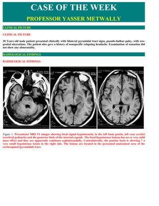
  • 1. CASE OF THE WEEK PROFESSOR YASSER METWALLY CLINICAL PICTURE CLINICAL PICTURE 30 Years old male patient presented clinically with bilateral pyramidal tract signs, pseudo-bulbar palsy, with oro- genital ulcerations. The patient also gave a history of nonspecific relapsing headache. Examination of sensation did not show any abnormality. RADIOLOGICAL FINDINGS RADIOLOGICAL FINDINGS   Figure 1. Precontrast MRI T1 images showing focal signal hypointensity in the left basis pontis, left crus cerebri (cerebral peduncle) and the posterior limb of the internal capsule. The focal hypointense lesions has no or very mild mass effect and they are apparently continuos cephalocaudally. Contralaterally, the pontine basis is showing ? a very small hypointense lesion in the right side. The lesions are located in the presumed anatomical area of the corticospinal (pyramidal) tract.
  • 2. Figure 2. Postcontrast MRI T1 images showing enhancement of the hypointense lesions shown in figure 1. The lesion in the right anterior pontine zone is more definite now and is better delineated by contrast enhancement. All enhanced areas are surrounded by a hypointense edema area with mild mass effect. Also notice contrast enhancement of the meningeal covering of the pons and midbrain which indicates the existence of a meningitic element. Figure 3. Postcontrast coronal MRI T1 images showing that the pontine, midbrain and capsular lesions described in figure 1,2 are continuous cephalocaudally as a linear long lesion, the lesion is surrounded by a hypointense edema area and is located in the presumed anatomical area of the corticospinal (pyramidal) tract. The corticospinal tract was actually mapped by contrast enhancement. The contralateral side is involved to a lessor degree. Notice meningeal enhancement which reflects a meningitic element. Contrast enhancement of the corticospinal tract in this case is due to break down of the blood brain barrier along the course of the corticospinal tract and it is a good sign of vasogenic edema which is mainly due to break down of the blood brain barrier (70). Notice middle line crossing of the lesions suggesting its venous rather that arterial origin. Also notice the mild mass effect induced by the pyramidal tract lesion in the left side which could be due to edema.
  • 3. Figure 4. Postcontrast sagittal MRI T1 images showing that the pontine, midbrain and capsular lesions described in figure 1,2,3 are continuous cephalocaudally as a linear long lesion in the anterior parts of the pons and midbrain, the lesion is located in the presumed anatomical area of the corticospinal (pyramidal) tract and is seen surrounded by a hypointense edema area. The corticospinal tract was actually mapped by contrast enhancement. Also notice meningeal enhancement. Thrombosis of the superficial and deep venous systems is also seen. Figure 5. MRI T2 image showing the capsular lesion (in the posterior limb of the internal capsule), The lesion is mainly hyperintense with some hypointense zones probably representing a hemorrhagic element. In general the observed pathology in this case represents demyelination and edema along the cortico-spinal tract fibers (in fact the pyramidal tract is actually anatomically drawn by the MRI signal changes of vasogenic edema and contrast enhancement) and is found to be located mainly in the midbrain/thalamus (which probably constitutes the primary site of involvement) with upward extension to the posterior limb of the internal capsule and downward extension to the basis pontis. The upward and downward extension of the primary midbrain/thalamic lesions might be explained by wallerian degeneration and this was noted in some pathologic studies as well (20, 43, 44). DIAGNOSIS: DIAGNOSIS: NEURO-BEHCET SYNDROME DISCUSSION DISCUSSION Behcet's disease (BD) is an uncommon, relapsing and remitting, multisystem inflammatory disorder characterized by the triad of oral ulceration, genital ulceration, and uveitis. 61 A number of additional features are commonly
  • 4. present, including arthritis, retinal and cutaneous vasculitis, thrombophlebitis, and gastroenteric disorders. The essential lesion is a focus of chronic inflammation, typically in the vicinity of a small blood vessel. Lesions in the CNS resemble those in other organ systems. Characteristically there are multiple small foci of softening that may eventually become confluent. These correspond to myelin loss and, to a lesser degree, drop out of neural elements with replacement by foamy macrophages. The lesions are not fundamentally demyelinative and more closely resemble minute ischemic foci. As in the periphery, lesions tend to occur near blood vessels, but vasculitis is uncommon. Pathologic findings are most extensive in the midbrain, pons (especially the basis pontis) and the medulla, and there is typically a lesser degree of involvement of the spinal cord, internal capsule, globus pallidus, optic nerves, and hypothalamus. In some cases, the cerebral white matter, cortex, hippocampus, basal ganglia, and thalamus are involved. The cerebellum is usually spared. The meninges are typically thickened and adherent to the brain surface, and acutely there may be considerable meningeal inflammation; hence the common reference to neuro-Behcet's disease as a meningoencephalitis. (The x ray room) (Print this case report) In general the most common presentation of parenchymal CNS involvement is a subacute brainstem syndrome with cranial nerve findings, dysarthria, and cerebellar or corticospinal tract signs. More infrequent presentations include a strokelike presentation (with the acute onset of unilateral neurologic findings and signs of cortical involvement including seizures) and psychiatric features, such as psychosis. Sinus venous thrombosis may evolve relatively slowly and results in intracranial hypertension, resulting headache, vomiting, and bilateral papilledema. Impaired memory (long-term verbal and nonverbal) and visuospatial skills occur frequently in patients with active disease who are taking large doses of steroids. Pure spinal cord or PNS involvement is rarely reported. Impairment of sensation is uncommon. (70) Between 5% and 20% of patients have neurologic disease. (70) The most common neurologic manifestation of Behcet's disease is a relapsing and remitting, but often progressive, focal meningoencephalitis that most often affects the brain stem. (70) Patients present with a host of cranial nerve and long tract signs and eventually develop spastic quadriparesis and a bulbar or pseudobulbar palsy, frequently with rather dramatic emotional incontinence. (70) Cochlear and vestibular dysfunctions are common. Often there are additional features of impairment in frontal lobe and memory function characteristic of a subcortical dementia. A number of cases of nearly pure subcortical dementia have been reported, but typically long tract signs are prominent, and bulbar involvement eventually develops. Patients may also present initially with features of transverse myelitis, but bulbar signs characteristically appear as the disease progresses. Cerebral cortical involvement is unusual, but seizures and aphasia have been reported. The optic nerves are commonly involved, but symptoms are indiscriminable from those of uveitis, which is usually present in patients with neurologic features. CNS vasculitis is rare, but stroke caused by large artery involvement has been reported. Pseudotumor cerebri is relatively common and almost always caused by venous sinus thrombosis. Cerebral venous thrombosis may affect up to 10% of all patients with Behcet's disease and one third of those with neurologic involvement. CNS involvement generally manifests several years after the onset of systemic Behcet's disease, but occasionally it may be an initial feature and even precede other disease manifestations. As a rule, flares of neurologic disease parallel flares of systemic disease. Neuro-Behçet's syndrome can be classified as an acute type and as a chronic progressive type (70). It should be pointed out that the two types of neuro-Behçet's syndrome are currently considered to represent different stages rather than independent clinical entities. Acute neuro-Behçet's syndrome is characterized by acute meningoencephalitis with a focal midbrain pyramidal tract lesions, the lesions are anteriorly located in the cerebral peduncles, bilateral, asymmetrical, extending up and down within the brain stem as a longitudinal linear lesion, and presenting high-intensity areas in the MRI T2-weighted images or the fluid attenuated inversion recovery (FLAIR) images (70). Acute neuro-Behçet's syndrome responds to steroid therapy, and is usually self-limiting. By contrast, the chronic progressive type of neuro-Behçet's syndrome is characterized by intractable, slowly progressive dementia, ataxia and dysarthria, with persistent elevation of cerebrospinal fluid IL-6 activity (> 20 pg/ml) (70). Most patients with the chronic progressive type of neuro-Behçet's syndrome were HLA-B51-positive, and they had history of attacks of acute type neuro-Behçet's syndrome prior to the development of progressive neurological symptoms (70). The neurological involvement in Behçet's disease is either caused by primary neural parenchymal lesions (neuro- Behçet's syndrome, or brain stem meningoencephalitis) or is secondary to major vascular involvement. The latter type is rarely complicated with the parenchymal lesions and should be called vascular-Behçet's disease. This vasculo-Behçet's disease type generally has a better prognosis compared with the parenchymal type. (70) Both vascular and parenchymal types can coexist, as in this case, but this is not common.  The primary parenchymal neuro-Behçet type
  • 5. It is primary parenchymal neural disease, a focal brain stem meningoencephalitis affecting the midbrain primarily with selective involvement of the corticospinal tract bilaterally. The most common clinical findings are pyramidal signs. Sensory symptoms or signs are much less frequent in neuro-Behcet disease. (73,74) The corticospinal tract lesions extend cephalocaudally in a linear fashion from the mesencephalon down to the pons and less frequently to the medulla and up to the posterior limb of the internal capsule. The cephalocaudal extension of the midbrain corticospinal tract lesions could be either due to wallerian degeneration or extension of vasogenic edema along the myelinated fibers of the corticospinal tract. (70,73,74)  The corticospinal tract pathology is manifested by MR imaging as bilateral and asymmetrical longitudinal linear lesions extending from the posterior limb of the internal capsule down to the midbrain, pons and less frequently the medulla. The midbrain is more severely affected. The corticospinal tract linear lesions are hyperintense on the MRI T2 images and hypointense on the MRI T1 images and show contrast enhancement in the acute stage. Contrast enhancement is due to break down of the blood brain barrier and this is in favour of the corticospinal tract MRI signal changes being due vasogenic edema. (70,73,74)  Vascular-Behçet's type  Involvement of veins and arteries in Behçet's disease is usually called vasculo-Behçet's disease. Venous thrombosis appeared to be the major vascular involvement in 7–33% of patients with Behçet's disease, and represents 85–93% of vascular-Behçet's disease (70). The brain parenchymal involvement seen in the primary neuro-Behçet type is rarely seen in the vascular type of Behçet disease. Parenchymal involvement in the vascular type of Behçet disease is more commonly secondary to venous sinus thrombosis. Deep vein thrombosis or cerebral sinus thrombosis are significantly associated with the male gender and a positive pathergy test (70). Venous thrombosis is a reflection of thrombophilia in vascular Behçet's disease. (70,73,74). To the best of the author's information, cerebral sinovenous thrombosis in the vascular-Behçet type is indistinguishable from sinovenous thrombosis due to any other cause and is only distinct from the view point that it is commonly associated clinically with oro- genital ulcerations. Probably vascular-Behçet syndrome must be put in the differential diagnosis of cases presented with cerebral sinovenous thrombosis of undetermined aetiology.  The clinical picture of vascular Behçet is different from that of primary neuro-Behçet. The clinical picture of neuro-Behçet is dominated by a mainly motor clinical picture with bilateral pyramidal tract signs, pseudo-bulbar palsy, subcortical dementia, etc. and sensory symptoms are uncommon, while the clinical picture of vascular Behçet is that of acute and chronic cerebral sinovenous thrombosis and its parenchymal complications (headache, seizures, focal sensory and motor deficits, bilateral papilledema, pseudotumor cerebri).  A number of studies have explored the pathogenesis of thrombophilia in Behçet's disease. Neither deficiency in protein C, in protein S, in factor V Leiden and in antithrombin III nor resistance to activated protein C and anticardiolipin antibody levels seemed to be correlated with vascular thrombosis in Behçet's disease (75,76). There were increased thrombin generation, fibrinolysis, and thrombomodulin in Behçet's disease, but these abnormalities were not related to thrombosis (76). These results therefore suggest that thrombophilia in Behçet's disease might be related more to inflammation than to clotting disorder.  Arterial involvement, although rare, does occur in Behçet's disease. The arterial manifestations in Behçet's disease resemble those of Takayasu's arteritis, including arterial occlusion and aneurysm formation. Histopathological studies revealed that the number of vasa vasorum with infiltration of neutrophils and lymphocytes was significantly increased in vascular-Behçet's disease compared with in Takayasu's arteritis and other inflammatory aneurysms (70). It was therefore suggested that arterial involvement in vascular-Behçet's (vasculo-Behçet) disease might be caused by a neutrophilic vasculitis targeting the vasa vasorum, leading to degeneration of the arterial wall. (70,73,74) Table 1. Lesion characteristics in the primary parenchymal neuro-Behçet type  The lesion in the primary parenchymal neuro-Behçet type is a focal brain stem meningoencephalitis affecting the midbrain primarily with selective involvement of the corticospinal tract bilaterally.  The most common clinical findings are pyramidal signs. Sensory symptoms or signs are much less frequent
  • 6. in neuro-Behcet disease.  The corticospinal tract lesions extend cephalocaudally in a linear fashion from the mesencephalon down to the pons and less frequently to the medulla and up to the posterior limb of the internal capsule. The cephalocaudal extension of the midbrain corticospinal tract lesions could be either due to wallerian degeneration or extension of vasogenic edema along the myelinated fibers of the corticospinal tract.  The corticospinal tract pathology is manifested by MR imaging as bilateral and asymmetrical longitudinal linear lesions extending from the posterior limb of the internal capsule down to the midbrain, pons and less frequently the medulla. The midbrain is more severely affected. The corticospinal tract linear lesions are hyperintense on the MRI T2 images and hypointense on the MRI T1 images and show contrast enhancement in the acute stage. Contrast enhancement is due to break down of the blood brain barrier and this is in favour of the corticospinal tract MRI signal changes being due vasogenic edema. (70,73,74)  The corticospinal tract linear lesions are sometimes surrounded by a hypointense edema area with positive mild mass effect. Table 2. The differences between the primary parenchymal neuro-Behçet type and the vascular-Behçet's type Parameter The primary parenchymal neuro-Behçet type Vascular-Behçet's type The clinical picture of vascular Behçet is that of acute and chronic The clinical picture of neuro-Behçet is dominated by a mainly cerebral sinovenous thrombosis Clinical motor clinical picture with bilateral pyramidal tract signs, and its parenchymal complications picture pseudo-bulbar palsy, subcortical dementia etc. Sensory (headache, seizures, focal sensory symptoms are uncommon and motor deficits, bilateral papilledema, pseudotumor cerebri, etc.). Incidence More common (70-93%) Less common (7-33%) It is primary parenchymal neural disease, a focal brain stem Venous thrombosis is a reflection meningoencephalitis affecting the midbrain primarily with of thrombophilia in Behçet's Pathology & upward and downward extension (along the pyramidal tract disease. Thrombophilia in Behçet's pathogenesis fibers to the posterior limb of the internal capsule, the basis disease might be related more to pontis and the medulla) and with selective involvement of the inflammation than to clotting corticospinal tract bilaterally. disorder. The corticospinal tract pathology is manifested by MR imaging as bilateral and asymmetrical longitudinal linear lesions extending from the posterior limb of the internal capsule down to the midbrain, pons and less frequently the medulla. The midbrain is more severely affected. The corticospinal tract The MRI picture is that of Radiological linear lesions are hyperintense on the MRI T2 images and sinovenous thrombosis and its findings hypointense on the MRI T1 images and show contrast parenchymal complications (70) enhancement in the acute stage. Contrast enhancement is due to break down of the blood brain barrier and this is in favour of the corticospinal tract MRI signal changes being due vasogenic edema. (70,73,74) Bad, higher incidence of being transformed into the progressive Prognosis Better type with subcortical dementia, etc. Anticoagulants, steroid may play a Management Steroid responsive role Currently, the most widely used diagnostic criteria of Behcet's disease is the International Study Group's classification, which requires recurrent oral ulcerations plus two of the following in order to establish a definite diagnosis: recurrent genital ulcerations, skin or eye lesions, or a positive pathergy test (13). The epidemiology of disease shows geographic variation, encountered more commonly along the Silk Road, which extends from the Mediterranean region to Japan (15). This is coupled with a similar variation in HLA B51 (human leukocyte antigen), which has been reported to be strongly associated with the disease in the high prevalence areas (16–19). Despite broadened clinical understanding of this disease, the etiologic factors remain obscured and speculative:
  • 7. viral agents, immunologic factors, genetic causes, bacterial factors, and fibrinolytic defects have all been implicated (3, 20–25). Vessel wall and perivascular mononuclear cell infiltration, which is consistent with vasculitis involving both arterial and venous systems, has been shown in histopathologic studies (20, 21). It has been postulated that genetic susceptibility together with a possible trigger by an extrinsic factor, such as an infectious agent, is responsible for the observed vasculitis (24, 26). Table 3. The International Study Group criteria for the diagnosis of Behcet disease  At least 3 episodes of oral ulceration must occur in a 12-month period. They must be observed by a physician or the patient and may be herpetiform or aphthous in nature.  At least 2 of the following must occur:  Recurrent, painful genital ulcers that heal with scarring.  Ophthalmic lesions, including anterior or posterior uveitis, hypopyon, or retinal vasculitis.  Skin lesions, including erythema nodosum, pseudofolliculitis, or papulopustular lesions (may also include atypical acne).  Pathergy: defined as a sterile erythematous papule larger than 2 mm in size appearing 48 hours after skin pricks with a sharp, sterile needle (a dull needle may be used as a control). Neurologic involvement in Behcet's disease, was first reported in 1941 by Knapp (27), and the term neuro-Behçet syndrome was introduced by Cavara and D'Ermo in 1954 (28). The reported rate of development of neurologic involvement among Behcet's disease patients ranges from 4% to 49% (9, 29). This rate was found to be 6.7% in our large nonselective series of patients referred from the Behçet's Disease Research Center (30). Neuro-Behçet syndrome may present as an acute focal or multifocal CNS dysfunction, and the clinical picture of neuro-Behçet syndrome may resemble multiple sclerosis (MS) (7, 31–35). It has been observed that a substantial number of patients with neuro-Behçet syndrome will have a relapsing-remitting course while others may develop a secondary-progressive course; some neuro-Behçet syndrome patients have an insidious onset, with primary- progressive CNS dysfunction, and others may display symptoms attributable to intracranial hypertension associated with dural venous sinus thrombosis (12, 36, 37). Although non-neurologic involvement generally precedes neurologic findings, the non-neurologic involvement may go unrecognized in some cases or it may appear late in the patient's course, thus posing diagnostic difficulties (38–40). Peripheral nerve involvement, although reported in neuro-Behçet syndrome, is relatively uncommon (41). The most common imaging lesion seen in the acute or subacute stage of neuro-Behçet syndrome is an asymmetric mesodiencephalic junction lesions (7–9, 33). These lesions extend along long fiber tracts and spare the red nucleus, suggesting that this downward extension is due to edema. The reversibility of the extension, leaving small residual lesions at the center, as observed on follow-up MR studies, further supports their edematous nature. This feature was also noted in earlier publications (9, 11, 42). Accordingly, pontobulbar involvement was an extension of lesions located at other sites, particularly the mesodiencephalic junction. The distribution and intensity of changes of the residual lesions closely corresponded to pathologic descriptions of secondary demyelination (20, 21, 43, 44). In the chronic cases, Changes may extend to the cervical cord, along the corticospinal tract, and this might be explained by wallerian degeneration. This was noted in some pathologic studies as well (20, 43, 44). Taken together, the current case (case version number 1.4) and the previously reported case in version 1.3 of this publication emphasized and highlighted the following common clinical-radiological findings in neuro-Behçet syndrome: 1. In general the observed pathology in these cases represents demyelination and edema along the cortico-spinal tract fibers (in fact the pyramidal tract is actually anatomically drawn by the MRI signal changes of vasogenic edema and contrast enhancement) and is found to be located mainly in the midbrain/thalamus (which probably constitutes the primary site of involvement) with upward extension to the posterior limb of the internal capsule and downward extension to the basis pontis. The upward and downward extension of the primary midbrain/thalamic lesions might be explained by wallerian degeneration or vasogenic edema (which characteristically spreads along the myelinated fibers of long tracts) and this was noted in some pathologic studies as well (20, 43, 44) 2. The selective involvement of the corticospinal tract would explain the clinical picture of these patients which is dominated by pyramidal tract involvement and pseudo-bulbar palsy with absence of sensory changes. In fact the clinical picture and the MRI findings in neuro-Behçet syndrome closely resemble those of motor neuron disease with pseudo-bulbar palsy (they both shared common topographic distribution and spatial extension of
  • 8. the corticospinal tract lesions), however the corticospinal tract lesion in motor neuron disease does not show contrast enhancement. (70). The corticospinal tract linear lesion appeared larger in size in acute neuro-Behçet syndrome compared with that of motor neuron disease probably due to the contribution of edema in neuro- Behçet syndrome. The corticospinal tract lesion is reversible in the acute neuro-Behçet syndrome, however it is irreversible in motor neuron disease. In general the lesion in acute neuro-Behçet syndrome has an acute onset and a regressive course, while that of motor neuron disease has a chronic progressive course. The MRI signal changes along the course of the corticospinal tract are mainly due to reversible vasogenic edema in acute neuro-Behçet syndrome, while the MRI signal changes along the course of the corticospinal tract in motor neuron disease are mainly due to wallerian degeneration. See table 4 3. Both cases (this case and the previously reported case in version 1.3 of this publication) shared common clinical (purely motor clinical presentation) and common MRI findings (selective pyramidal tract involvement). However sinus thrombosis was demonstrated only in the current case. Which simply means that the parenchymal lesion demonstrated in neuro-Behçet syndrome is a primary neuronal lesion and not secondary to sinus thrombosis (Behcet's disease is a relapsing and remitting, but often progressive, focal meningoencephalitis that most often affects the brain stem). Sinus thrombosis is probably a product of the primary pathology. In the current patient the presence of the midbrain lesions characteristic of the primary neural type of Behçet disease associated with cerebral sinus thrombosis simply means that both the primary parenchymal neural type and the vascular type of Behçet disease can coexist in a single patient, but this uncommon. Vascular Behçet is rarely associated with neuro-Behçet. The predominant histopathological features in the inflamed tissues in Behcet's disease are infiltration of lymphocytes and monocytes, and sometimes polymorph nuclear leukocytes, through small veins without microscopic changes in the vessel walls. Thrombophilia or thrombophlebitis involving small and large veins is also common, whereas arteritis is rare. In these regards, Behçet's disease is unique compared with other vasculitides. 4. The existence of meningeal enhancement at the midbrain and pontine level in this case (version number 1.4) is simply a reflection of a meningitic process at the pontine and midbrain levels. The meninges are typically thickened and adherent to the brain surface in neuro-Behçet syndrome, and acutely there may be considerable meningeal inflammation; hence the common reference to neuro-Behcet's disease as a meningoencephalitis. (70) Meningeal enhancement could not be appreciated radiologically in the previously reported case (number 1.3). Table 4. Differences between motor neuron disease and acute neuro-Behçet syndrome, with regard to the corticospinal tract affection The corticospinal tract lesion in neuro-Behçet The corticospinal tract lesion in motor neuron disease syndrome The corticospinal tract linear lesion appeared larger Smaller in size in size probably due to the contribution of edema Reversible Irreversible Show contrast enhancement Does not show contrast enhancement Has an acute onset and a regressive course Has a chronic progressive course MRI signal changes are mainly due to reversible MRI signal changes are mainly due to wallerian vasogenic edema degeneration In presented patient, an asymmetric subcortical and deep periventricular white matter involvement (involving mainly the posterior limb of the internal capsule) without cortical involvement was observed. The patient also had concomitant mesodiencephalic junction lesions. In the majority of reported cases with hemispheric involvement, the lesions were located subcortically, particularly within temporal and occipital regions (7–11, 34, 39, 45, 46). In the acute stage the periventricular white matter changes are due to edema, while in the chronic stage these white matter changes are due to wallerian degeneration, gliosis, and demyelination (4, 20, 21, 43, 44, 47, 48). Vasculitis is regarded as the key feature in neuro-Behçet syndrome (3, 26), as biopsy specimens from mucous and cutaneous lesions show those changes (54, 55). Arterial and venous large vessel involvement, such as narrowing, occlusion, and aneurysmal formation, has been reported in up to 27% to 35% of cases, with 12% arterial and 88% venous (52). An even greater proportion of patients with neuro-Behçet syndrome may have small vessel vasculitis, and recently this has been validated as the pathologic basis of various histologic changes observed in different organ systems (26).
  • 9. Lesions of arterial origin may be observed in some patients, in whom these lesions, for example, may lie within the pons without crossing the midline, consistent with involvement of the penetrating arteries. Although such lesions, resulting from small or medium-sized intracranial arteries, have been reported either microscopically or radiologically in neuro-Behçet syndrome, they are not as common as the arterial lesions observed with the involvement of other systems (20, 21, 40, 43, 47, 48, 50–52). There are also a few publications concerning the involvement of large intracranial arteries (40, 51–53). Hemorrhagic lesions seen in some patients most likely resulted from quot;diapedesis of red cells around veins,quot; as already reported, and are not of arterial origin (21). Autopsy studies and biopsy specimens of the CNS lesions are consistent with vasculitis as well, and they show a clear venous predominance (20, 21, 49). Radiologic studies support this finding that lesions seen in neuro-Behçet syndrome are not compatible with arterial territories. Furthermore, significant perilesional edema with a tendency to disappear or to leave disproportionally small residua on follow-up studies has been reported. This feature is consistent with venous infarction, since not all signal intensity changes seen in venous occlusive disease necessarily represent infarction, but rather an accumulation of water within interstitial spaces (56, 57). This information, together with our observations, supports the probable inflammatory-venous pathogenesis for the CNS lesions seen in Behcet's disease. Table 5. Factors that support the probable inflammatory-venous pathogenesis for the CNS lesions seen in neuro- Behçet syndrome Arterial lesions are unilateral and do not cross the middle line, while lesions observed in neuro-Behcet syndrome are frequently bilateral with frequent midline crossing. Autopsy studies and biopsy specimens of the CNS lesions are consistent with vasculitis with a clear venous predominance (20, 21, 49). Significant perilesional edema with a tendency to disappear or to leave disproportionally small residua on follow- up studies has been reported. This feature is consistent with venous infarction, since not all signal intensity changes seen in venous occlusive disease necessarily represent infarction, but rather an accumulation of water within interstitial spaces (56, 57). If one considers the possible venous territories in which brain stem lesions have occurred in neuro-Behçet syndrome, it is clear that the affected venous structures are indeed small intraaxial veins of the brain stem (58). That particular predilection of occurrence raises the question of why these veins are affected or why occlusion of them causes symptoms. The literature on autopsy studies states no apparent tendency toward these venous channels, and brain stem veins and cerebral hemispherical veins are equally affected, although there is a clear-cut predominance of lesions in the brain stem. The question can also be addressed according to regional hemodynamic properties. It is well known that cerebral hemispherical structures are drained by superficial and deep venous systems, both of which anastomose via medullary veins. They interconnect superficial pial veins to the internal cerebral vein and the basal vein of Rosenthal, the former being more common than the latter (59), whereas in the brain stem, intraparenchymal radial and longitudinal anastomotic channels are nearly absent (58). In the spinal cord, intraaxial anastomoses are claimed to be prominent at the thoracic level (60). The particular arrangement of veins in the cerebral hemisphere permits them to flow in both directions via medullary veins, as seen with certain disorders, such as deep arteriovenous malformations, Sturge-Weber disease, and the developmental venous anomalies (60), possibly explaining the small diameter and unimportance of parenchymal lesions when such a vein is thrombosed. At the mesencephalic, diencephalic, and pontine levels, thrombosis of small veins might be accompanied by a very large, sometimes hemorrhagic lesion, since there is nearly no collateral venous pathway. The same anatomic arrangement might also explain the vulnerability of the cervical spinal cord (7, 44). It appears, therefore, that the variability of venous anatomic arrangements at different levels of the CNS might explain the predilection of lesions for different regions. Vasculitis, like inflammation in other tissues, is caused by many different agents and pathogenic mechanisms; however, these different causes produce only a limited number of histologic expressions of injury. The major type of injury to nervous tissue in vasculitis is ischemia. Therefore, the same clinical manifestations can result from etiologically and pathogenetically different vasculitic diseases. In vasculitic processes, location, extension, and distribution of vascular involvement might point to a specific diagnosis, such as Takayasu or temporal arteritis (26, 61–65). In neuro-Behçet syndrome, lesions therefore appear secondary to the small vessel venous vasculitis, and the anatomy of those intraaxial venous structures explains the dominant involvement of the upper brain stem and diencephalic structures. Pathologically proved small vessel arteritis, either alone or with venous inflammation, has also been reported in conjunction with neuro-Behçet syndrome, and it is probable that some of the cerebral hemisphere and midbrain lesions might result from small vessel arteritis (62, 63). In this case, there appeared to be at least one lesion that
  • 10. occurred within a probable pontine arterial territory, and the hemispheric lesions seen in neuro-Behçet syndrome were not significantly different from the CNS lesions of other vasculopathies of accepted arteritic origin. It is possible to explain the selective involvement of the corticospinal fibers observed in this patient. If the primary pathology in Behcet disease is inflammatory vascular process with secondary demyelination, then the myelinated fibers in the long tracts will suffer more. The posterior part of the brain stem is mainly reticular while the anterior part is compact and is occupied mainly by the pyramid in the cerebral peduncle, crus cerebral and basis pontis. It is quite illogical to see inflammatory demyelination in the reticular posterior parts of the brain stem. The inflammatory demyelinating process will affect the anteriorly located long myelinated fibers in the corticospinal tract. Vasogenic edema is the most common type of edema associated with brain tumors, venous congestion and other causes and results from local disruption of the blood brain barrier. This leads to extravasation of protein-rich filtrate of plasma into the interstitial space, with subsequent accumulation of vascular fluid. This disruption results from loosening of the tight junctions between endothelial cells, and the neoformation of pinocytic vesicles. Once the barrier is breached, hydrostatic and osmotic forces work together to extravasate intravascular fluid. Once extravasated, fluid is retained outside the vasculature, mostly in the white matter of the brain, and within the bundles of myelinated axons of long tracts and commissural fibers. This is because axons run in parallel bundles of fibres with loose extracellular space (that offer low resistance and facilitates the extension of vasogenic edema along myelinated axons which are spreaded apart by the edema) as opposed to gray matter, which has high cell density and is enmeshed in an interwoven network of connecting fibres that offer high resistance to the formation and spread of edema. By definition, this type of edema is confined to the extracellular space. (70) In neuro-Behcet case reports of diffusion weighted imaging (DWI) of the T2 hyperintense lesions revealed elevated ADC values compared to normal subjects, indicating restricted diffusion and the presence of vasogenic edema (71,72). Contrast enhancement of the corticospinal tract in this case is due to break down of the blood brain barrier along the course of the corticospinal tract and it is a good sign of vasogenic edema which is mainly due to break down of the blood brain barrier. (70) Anther explanation for the selective involvement of the corticospinal tract in neuro-Behcet is that the signal changes observed in the MRI study is due to vasogenic edema secondary to venous congestion, vasogenic interstitial edema spreads in the white matter along myelinated fibers of long tracts and commissural fibers (This is because axons of long tracts run in parallel bundles of fibres with loose extracellular space that offer low resistance to the formation and spread of edema as opposed to grey matter). The long tracts are anteriorly located within the brain stem in the crus cerebra, cerebral peduncle, and basis pontis. Subsequently brain stem interstitial or vasogenic edema is much more likely to be concentrated anteriorly within the corticospinal tract as vasogenic edema spreads the myelinated fibers apart and extends along them. In the presented case the MRI signal changes were maximum in the upper midbrain and internal capsule, present to a lesser extent in the pons and to a much less extent in the medulla. Anatomically the corticospinal tract remains abundant, compact and in a single bundle as it transverses in the posterior limb of the internal capsule down to the cerebral peduncle (crus cerebri) in the midbrain. The corticospinal and corticobulbar fibers gradually decreases in number as they descend down to the pons, and through the medulla to the spinal cord. Also starting from the level of pons and downward the corticospinal tract starts to fragment into scattered, irregular and isolated bundles. The corticospinal fibers, being more abundant and more compact at the internal capsule and midbrain levels, compared with corticospinal fibers at the level of pons and medulla, would explain why MRI signal changes were maximum in the internal capsule and upper midbrain. However selective involvement of the corticospinal tract in Behcet disease still needs further explanation. Taken together, this case and the case reported in version 1.3 of this publication clearly demonstrate the selective pyramidal tract involvement in neuro-Behcet. Concerning the differential diagnosis, hemispheric white matter lesions are not common in neuro-Behçet syndrome, and when they are present, they are more likely to be subcortical than periventricular. Furthermore, these are generally associated with brain stem–diencephalic lesions. That combination is unlikely in systemic lupus erythematosus (SLE) and non-Behçet vasculitides. CNS involvement due to SLE and other systemic vasculitis tends to involve arterial territories, and as a result, cortical involvement is frequently observed (34, 66, 67). We have not observed cortical involvement in neuro-Behçet syndrome, despite pathologic studies in which such involvement has been reported (21). These changes, however, are minor in neuro-Behçet syndrome, which may explain the radiologic-pathologic discrepancy. Periventricular and ovoid lesions suggestive of MS are not expected to be seen in neuro-Behçet syndrome. Extensive confluent periventricular changes that are seen in MS and occasionally in sarcoidosis were not observed in this patient with neuro-Behçet syndrome. Posterior fossa lesions, particularly those located around the fourth
  • 11. ventricle with or without the associated supratentorial lesions seen in MS, are not similar to the neuro-Behçet syndrome lesions described above. Brain stem lesions in MS are usually small, even in the acute stage, and prominent brain stem and/or cerebellar atrophy without cerebral volume loss, which is observed in the chronic phase of neuro-Behçet syndrome, is unusual in MS (7, 34). When one considers cervical involvement, this rarely extends more than a few vertebral segments in MS (68), unlike the more extensive lesions we observed in neuro- Behçet syndrome. Leptomeningeal contrast enhancement is a typical finding of sarcoidosis (69). We did not encounter this finding in patients with neuro-Behçet syndrome. Devlin et al (38) reported abnormal leptomeningeal enhancement in two of their patients with neuro-Behçet syndrome. Abnormal meningeal enhancement secondary to dural venous occlusion or to lumber puncture should be excluded before attributing it to the disease itself (70). Table 6. Differences between neuro-Behçet syndrome. and other similar diseases Pathology Comment CNS lupus CNS involvement due to SLE and other systemic vasculitis tends to involve arterial territories, and as a versus result, cortical involvement is frequently observed (34, 66, 67). neuro- Behcet Periventricular and ovoid lesions suggestive of MS are not expected to be seen in neuro-Behçet syndrome. Extensive confluent periventricular changes that are seen in MS and occasionally in sarcoidosis are not observed in neuro-Behçet syndrome. Posterior fossa lesions, particularly those located around the fourth ventricle with or without the MS versus associated supratentorial lesions seen in MS, are not similar to the neuro-Behçet syndrome lesions. neuro- Behcet When one considers cervical involvement, this rarely extends more than a few vertebral segments in MS (68), unlike the more extensive lesions observed in neuro-Behçet syndrome. Brain stem lesions in MS are usually small, even in the acute stage, and prominent brain stem and/or cerebellar atrophy without cerebral volume loss, which is observed in the chronic phase of neuro- Behçet syndrome, is unusual in MS (7, 34) Abnormal meningeal enhancement is unusual in MS and reported in neuro-Behçet syndrome. (38) Inflammatory demyelinating diseases, such as MS, and inflammatory vascular disorders (vasculitides), such as neuro-Behçet syndrome and SLE, can affect the CNS primarily or secondarily, and onset tends to occur in young adulthood. Although the clinical presentation of these diseases may be similar, the radiologic findings of neuro- Behçet syndrome are quite distinct, which may help differentiate it from other disorders, even in the absence of overt systemic involvement. The diagnosis of Behcet's disease is clinical. Patients with Behcet's disease and venous thrombosis are often heterozygotes or homozygotes for the factor V Leiden allele. The CSF in patients with neurologic involvement is typically characterized by a pleocytosis (usually less than 100/mm3) that is equally likely to be neutrophil or lymphocyte predominant. CSF protein is only slightly elevated. There have been reports of intrathecal synthesis of oligoclonal IgA and IgM. An immunofixation electrophoretic pattern of oligoclonal bands has been reported in 16% of patients. Magnetic resonance imaging in patients with CNS disease typically reveals multiple 3-10 mm in diameter sharply marginated, irregular, and often confluent lesions in the spinal cord, brain stem, thalamus, basal ganglia, and deep cerebral white matter. The lesions may be quite extensive. In some cases, they may be difficult to distinguish from those of multiple sclerosis, but Dawson's fingers are not seen. (70) High-dose corticosteroids (prednisone 60 mg/day) are effective in suppressing skin, mucosal, and arthritic manifestations of Behcet's disease. Pulse corticosteroids (methylprednisolone I gm/day for 3 days) are often employed effectively for acute flares of neurologic disease, but the effect of corticosteroid treatment on chronic or recurrent neurologic disease is less satisfactory. Corticosteroids are clearly ineffective in halting the progression of uveitis and chlorambucil; 0.1-0.2 mg/kk/day has been used with considerable success in treating this disabling manifestation. (70) O'Duffy, (3) also induced remission in eight of nine patients with meningoencephalitis using chlorambucil. Azathioprine (Immuran), cyclosporine A, and colchicine have been shown in prospective, randomized studies to be highly effective in halting or preventing the ocular manifestations of Behcet's disease; however, cyclosporine A is relatively contraindicated in the presence of neurologic disease. Acute neurologic symptoms are remarkably reversible and justify aggressive treatment during flares. Patients with cerebral venous thrombosis respond well to chronic anticoagulation without significant risk of intracranial hemorrhage. (70)
  • 12. Thalidomide (Thalomid) is highly effective in suppressing oral and genital manifestations of Behcet's disease; however, between 6% and 50% of patients develop an axonal sensorimotor polyneuropathy that may be dose related. (70) SUMMARY The parenchymal distribution of lesions in neuro-Behçet syndrome seems to support the hypothesis of small vessel vasculitis, mainly venular involvement. The known anatomic arrangement of CNS intraaxial veins explains the predominant involvement of the brain stem structures observed in these patients. This pattern of lesion distribution might help to differentiate neuro-Behçet syndrome from other vasculitides as well as from the inflammatory- demyelinating diseases of the CNS, such as MS. Our experience with neuro-Behçet syndrome has caused us to consider neuro-Behçet syndrome in the differential diagnosis of patients who have brain stem and/or diencephalic lesions that extend along the long tracts and have a tendency to resolve on subsequent imaging studies, whether or not they are associated with periventricular and subcortical lesions. CNS involvement generally manifests several years after the onset of systemic Behcet's disease, but occasionally it may be an initial feature and even precede other disease manifestations. According to the diagnostic criteria of Behcet's disease of the International Study Group's classification (13), it is very difficult to diagnose Behçet disease without oro-genital ulcerations. However and in the author experience, primary parenchymal neuro-Behçet disease with selective involvement of the corticospinal tract (and with a purely motor clinical presentation) taken together with the characteristic MR imaging findings of selective pyramidal tract involvement are highly suggestive of Behçet disease even if CNS involvement precedes other disease manifestations.  Addendum  A new version of this PDF file (with a new case) is uploaded in my web site every week (every Saturday and remains available till Friday.)  To download the current version follow the link quot;http://pdf.yassermetwally.com/case.pdfquot;.  You can also download the current version from my web site at quot;http://yassermetwally.comquot;.  To download the software version of the publication (crow.exe) follow the link: http://neurology.yassermetwally.com/crow.zip  The case is also presented as a short case in PDF format, to download the short case follow the link: http://pdf.yassermetwally.com/short.pdf  At the end of each year, all the publications are compiled on a single CD-ROM, please contact the author to know more details.  Screen resolution is better set at 1024*768 pixel screen area for optimum display.  For an archive of the previously reported cases go to www.yassermetwally.net, then under pages in the right panel, scroll down and click on the text entry quot;downloadable case records in PDF formatquot; REFERENCES References 1. Behçet H. Uber residivierende, aphtöse, durch ein virus verursachte Geschwüre am Mund, am Auge und an den Genitalien. Derm Woschenscr 1937;105:1152-1157 2. Yazici H, Yurdakul S, Hamuryudan V. Behçet's syndrome. In: Klippel J, Dieppe P, eds. Rheumatology. London: Gower Medical; 1997;7.26:1–6
  • 13. 3. O'Duffy JD. Vasculitis in Behçet's disease. Rheum Dis Clin North Am 1990;16:423-43 4. Lakhanpal S, Tani K, Lie JT, Katoh K, Ishigatsuba Y, Ohokubo T. Pathological features of Behçet's syndrome: a review of Japanese autopsy registry data. Hum Pathol 1985;16:790-795 5. Inaba G. Clinical features of neuro-Behçet syndrome. In: Lehner T, Barnes CG, eds. Recent Advances in Behçet's Disease. International Congress and Symposium Series Number 103. London: Royal Society of Medicine Services; 1986:235–246 6. Siva A, Necdet V, Yurdakul S, Yardim M, Denkta F, Yazici H. Neuro-radiologic findings in neuro-Behçet syndrome. In: O'DuffyI D, Kokmen E, eds. Behçet's Disease: Basic and Clinical Aspects. New York: Dekker; 1991;323–329 7. Morrisey SP, Miller DH, Hermaszewski R, et al. Magnetic resonance imaging of the central nervous system in Behçet's disease. Eur Neurol 1993;33:287-293 8. Wechsler B, Dell'sola B, Vidailhet M, et al. MRI in 31 patients with Behçet's disease and neurological involvement: prospective study with clinical correlation. J Neurol Neurosurg Psychiatry 1993;56:783-789 9. Al Kawi MZ, Bohlega S, Banna M. MRI findings in neuro-Behçet's disease. Neurology 1991;41:405-408 10. Banna M, El-Ramahi K. Neurologic involvement in Behçet disease: imaging findings in 16 patients. AJNR Am J Neuroradiol 1991;12:791-796 11. Fukuyama H, Kameyama M, Nebatame H, et al. Magnetic resonance images of neuro-Behçet syndrome show precise brain stem lesions: report of a case. Acta Neurol Scand 1987;57:70-73 12. Wechsler B, Vidailhet N, Piette JC, et al. Cerebral venous thrombosis in Behçet's disease: clinical study and long-term follow-up of 25 cases. Neurology 1992;42:614-618 13. The International Study Group for Behçet's Disease. Evaluation of diagnostic (quot;classificationquot;) criteria in Behçet's disease: towards internationally agreed criteria. Br J Rheumatol 1992;31:299-308 14. Osborne AG. Diagnostic Neuroradiology.. St Louis: Mosby; 1994:154–197 15. Ohno S. Behçet's disease in the world. In: Lehner T, Barnes CG, eds. Recent Advances in Behçet's Disease. London: Royal Society of Medicine Service; 1986:181–186 16. Yazici H, Akhan G, Yalçin B, Müftüo lu A. The high prevalence of HLA-B5 in Behçet's disease. Clin Exp Immunol 1977;30:259-261Yurdakul S, Günaydin I, Tüzün H, et al. The prevalence of Behçet's syndrome in a rural area in northern Turkey. J Rheumatol 1988;15:820-822 17. Ohno S, Ohguchi M, Hirose S, Matsuda H, Wakisaka A, Aizava M. Close association of HLA-BW51 with Behçet's disease. Arch Opthalmol 1982;100:1455-1458 18. Mizuki N, Inoko H, Ohno S. Molecular genetics (HLA) of Behçet's disease. Yonsei Med J 1997;38:423-427 19. McMenemey WH, Lawrence BJ. Encephalomyelopathy in Behçet's disease: report of necropsy findings in two cases. Lancet 1957;24:353-358 20. Kawakita H, Nishimura M, Satoh Y, Shibata N. Neurological aspects of Behçet's disease: a case report and clinico-pathological review of the literature in Japan. J Neurol Sci 1967;5:417-438 21. Gamble CN, Wiesner KB, Shapiro RF, Boyer WJ. The immune complex pathogenesis of glomerulonephritis and pulmonary vasculitis in Behçet's disease. Am J Med 1979;66:1031-1039 22. Hirohata S, Tekeuchi A, Miyamoto T. Association of cerebrospinal fluid IgM index with central nervous system involvement in Behçet's disease. Arthritis Rheum 1986;29:793-796 23. Kansu E. Endothelial cell dysfunction in Behçet's disease. In: Ansell BM, Bacon PA, Lie JT, Yazici H, eds.
  • 14. Vasculitides. London: Chapman & Hall; 1996:207–221 24. Yazici H. The place of Behçet's syndrome among the autoimmune diseases. Int Rev Immunol 1997;14:1-10 25. Lie JT. Primary (granulomatous) angiitis of the central nervous system: a clinicopathologic analysis of 15 new cases and a review of the literature. Hum Pathol 1992;23:164-171 26. Knapp P. Beitrag zur Symptomatologie und Therapie der rezidiverenden Hypopyoniritis und der begleitenden apthösen Schleimhauterkrankungen. Shweiz Med Wochenschr 1941;71:1288-1290 27. Cavara V, D'Ermo E. A case of neuro-Behçet's syndrome. Acta XVII Concili Ophtalmologici 1954;3:1489 28. Serdaroglu P. Behçet's disease and the nervous system. J Neurol 1998;245:197-205 29. Siva A, Saip S, Kantarci O, et al. Neuro-Behçet syndrome (NBS): clinical and imaging correlates. J Neurol 1997;244(Suppl 3):545 30. O'Duffy JD, Goldstein NP. Neurologic involvement in seven patients with Behçet's disease. Am J Med 1976;61:170-178 31. Motomura S, Tabira T, Koroiwa Y. A clinical comparative study of multiple sclerosis and neuro-Behçet's syndrome. J Neurol Neurosurg Psychiatry 1980;43:210-213 32. Herskovits S, Lipton R, Lantos G. Neuro-Behçet's disease: CT and clinical correlates. Neurology 1988;38:1714-1720 33. Miller DH, Ormerod IEC, Gibson A, DuBoulay EPGH, Rudge P, McDonald WI. MRI brain scanning in patients with vasculitis: differentiation from multiple sclerosis. Neuroradiology 1987;29:226-231 34. Siva A, Saip S, Kantarci O, et al. Neuro-Behçet syndrome (NBS): clinical and imaging features (abstr). Neurology 1997;48(Suppl 1):A362 35. Saip S, Siva A, Basibüyük N, et al. Neuro-Behçet syndrome (NBS): clinical and MRI features. Neurology 1992;42(Suppl 3):339 36. Harper CM, O'Neill BP, O'Duffy JD, Forbes GS. Intracranial hypertension in Behçet's disease: demonstration of sinus occlusion with use of digital subtraction angiography. Mayo Clin Proc 1985;60:419- 422 37. Devlin T, Gray L, Allen NB, Friedman AH, Tien R, Morgenlander JC. Neuro-Behçet's disease: factors hampering proper diagnosis. Neurology 1995;45:1754-1757 38. Kozin F, Haughton V, Bernhard GC. Neuro-Behçet disease: two cases and neuroradiologic findings. Neurology 1977;27:1148-1152 39. Iragui VJ, Maravi E. Behçet syndrome presenting as cerebrovascular disease. J Neurol Neurosurg Psychiatry 1986;49:838-840 40. Baso P, Kara I, Eker E, et al. Clinical, electrophysiological, immunological and electronmicroscopic investigation of Behçet's disease. In: Dilsen N, Koniçe M, Ovul C, eds. Behçet's Disease. Amsterdam: Excerpta Medica International Congress Series; 1979;469:178–182 41. Nüssel F, Wegmüller H, Laseyras F, Posse S, Herschkowitz N, Huber P. Neuro-Behçet: acute and sequential aspects by MRI and MRIS. Eur Neurol 1991;31:399-402 42. Totsuka S, Midorikawa T. Some clinical and pathological problems in neuro-Behçet's syndrome. Folia Psychiatry Neurol Jpn 1972;26:275-284 43. Ishino H, Higashi H, Otsuki S. Neuro-Behçet's syndrome: case report with pathological findings. Folia Psychiatry Neurol Jpn 1971;25:27-36
  • 15. 44. Patel DV, Neuman MJ, Hier DB. Reversibility of CT and MRI findings in neuro-Behçet disease. J Comput Assist Tomogr 1989;13:669-673 45. Wildhagen K, Meyer GJ, Stoppe G, Heintz P, Deicher H, Hundeshagen H. PET and MRI imaging in a neuro- Behçet syndrome. Eur Neurol 1991;31:399-402 46. Sugihara H, Mutoh Y, Tsuchiyama H. Neuro-Behçet's syndrome: report of two autopsy cases. Acta Pathol Jpn 1969;19:95-101 47. Adams JH, Duchen LW. Virus diseases. In: Esiri MM, Kennedy PGE, eds. Greenfield's Neuropathology. 5th ed. London: Edward Arnold; 1992:335–385 48. Akar Z, Hanci M, Kaynar YM, Uzan M, Kuday C. Neuro-Behçet's disease: progressively enlarging temporal mass. J Ankara Med Sch 1992;14:295-300 49. Kansu T, Kansu E, Zileli T, Kirkali P. Neuro-ophthalmologic manifestations of Behçet's disease. Neuro- ophthalmology 1991;11:7-11 50. Zelenski JD, Capraro JA, Holden D, Calabrase H. Central nervous system vasculitis in Behçet's syndrome: angiographic improvement after therapy with cytotoxic agents. Arthritis Rheum 1986;32:217-220 51. Koç Y, Güllü I, Akpek G, et al. Vascular involvement in Behçet's disease. J Rheumatol 1992;19:402-410 52. Nishimura M, Satah K, Suga M, Oda M. Cerebral angio and neuro-Behçet's syndrome: neuroradiological and pathological study of one case. J Neurol Sci 1991;106:19-24 53. Matsumoto T, Uekusa T, Fukuda Y. Vasculo-Behçet's disease: a pathologic study of eight cases. Hum Pathol 1991;22:45-51 54. Jorizzo JL, Abernethy JL, White WL, et al. Mucocutaneous criteria for the diagnosis of Behçet's disease: an analysis of clinicopathologic data from multiple international centers. J Am Acad Dermatol 1995;32:968-976 55. Yuh WTC, Simonson MT, Wang A, et al. Venous sinus occlusive disease: MRI findings. AJNR Am J Neuroradiol 1994;15:309-316 56. Dormont D, Anxionnat R, Evrard S, Louallile C, Chiras J, Marsault C. IRM des tromboses veineus cerebrales. J Neuroradiol 1994;21:81-99 57. Duvernoy HM. Human Brainstem Vessels.. Berlin: Springer; 1978:2–34 58. Bracard S, Braun M, Meder JF, Velut S. Anatomie et radioanatomie du systeme veineux intracranien. Neurochirurgie 1996;42(Suppl 1):11-44 59. Lasjaunias P, Berenstein A. Surgical Neuroangiography.. Berlin: Springer; 1990;3:68–80, 240–245 60. Jennette JC, Falk RJ, Milling DM. Pathogenesis of vasculitis. Semin Neurol 1994;14:291-299 61. Harris KC, Tran DD, Sickels WJ, Cornell SH, Yuh WTC. Diagnosing intracranial vasculitis: the roles of MRI and angiography. AJNR Am J Neuroradiol 1994;15:317-330 62. Greenan TJ, Grossman RI, Goldberg HI. Cerebral vasculitis: MRI imaging and angiographic correlation. Radiology 1992;182:65-72 63. Hurst RW, Grossman RI. Neuroradiology of central nervous system vasculitis. Semin Neurol 1994;14:4:320- 340 64. Cohen BA, Biller J. Hemorrhagic stroke due to cerebral vasculitis and the role of immunosuppressive therapy. Neurosurg Clin North Am 1992;3:611-624 65. Vermess M, Bernstein RM, Bydder GM, et al. Nuclear magnetic resonance (NMR) imaging of the brain in
  • 16. systemic lupus erythematosus. J Comput Assist Tomogr 1983;7:461-467 66. Aisen AM, Gabrielsen TO, McCune WJ. MR imaging of systemic lupus erythematosus involving the brain. AJR Am J Roentgenol 1985;144:1027-1031 67. Nijeholt GJL, Barkhof F, Scheltens P, et al. MR of the spinal cord in multiple sclerosis: relation to clinical subtype and disability. AJNR Am J Neuroradiol 1997;18:1041-1048 68. Sato N, Sze G, Kim JH. Cystic pituitary mass in neurosarcoidosis. AJNR Am J Neuroradiol 1997;18:1182-118 69. River Y, Schwartz A, Gomori JM, Soffer D, Siegal T. Clinical significance of diffuse dural enhancement detected by magnetic resonance imaging. J Neurosurg 1996;85:777-783 70. Metwally, MYM. Textbook of neuroimaging, CD-ROM based publication, in Metwally (ed), WEB-CD agency for electronic publication, version 8.4a ; 2007 71. Suner NR. Neuro-Behcet's disease: diffusion MR imaging and proton MR spectroscopy. AJNR Am J Neuroradiol. 2003 Sep;24(8):1612-4. 72. Baysal T, Dogan M, Karlidag R, Ozisik HI, Baysal O, Bulut T, Sarac K. Diffusion-weighted imaging in chronic Behcet patients with and without neurological findings. Neuroradiology. 2005 Jun;47(6):431-7. 73. Serdaroglu P. Behçet's disease and the nervous system. J Neurol. 1998;245:197–205. 74. Kidd D, Steuer A, Denman AM, Rudge P. Neurological complications in Behcet's syndrome. Brain. 1999;122:2183–2194. 75. Houman MH, Ben Ghorbel I, Khiari Ben Salah I, Lamloum M, Ben Ahmed M, Miled M. Deep vein thrombosis in Behcet's disease. Clin Exp Rheumatol. 2001;19(suppl 24):S48–S50. 76. Espinosa G, Font J, Tassies D, Vidaller A, Deulofeu R, Lopez-Soto A, Cervera R, Ordinas A, Ingelmo M, Reverter JC. Vascular involvement in Behcet's disease: relation with thrombophilic factors, coagulation activation, and thrombomodulin. Am J Med. 2002;112:37–43.