SlideShare une entreprise Scribd logo
1  sur  19
Télécharger pour lire hors ligne
CASE OF THE WEEK
                   PROFESSOR YASSER METWALLY
CLINICAL PICTURE

CLINICAL PICTURE

15 years old male patient presented with symptoms of nausea, vomiting, and ataxia, with evidence of torticollis,
papilledema, nystagmus, and palsies of the sixth and seventh cranial nerves at physical examination. Long-tract
signs were conspicuously absent.

RADIOLOGICAL FINDINGS

RADIOLOGICAL FINDINGS  


Figure 1. MRI T1 postcontrast images showing a large cystic lesion diffusely expanding the pons. The lesion is seen
compressing and pushing the 4th ventricle posteriorly. A large mural nodule is seen projecting inside the pontine
cystic lesion, the mural nodule itself is cystic. The signal intensity of the mural nodule cyst is different from the
signal intensity of the pontine cyst. The walls of the mural nodule showed faint enhancement, while the wall of the
pontine cyst did not show any enhancement.
Figure 2. MRI T1 postcontrast images showing a large cystic lesion diffusely expanding the pons. The lesion is seen
compressing and pushing the 4th ventricle posteriorly. A large mural nodule is seen projecting inside the pontine
cystic lesion, the mural nodule itself is cystic. The signal intensity of the mural nodule cyst is different from the
signal intensity of the pontine cyst. The walls of the mural nodule showed faint enhancement, while the wall of the
pontine cyst did not show any enhancement.


                                                                 Figure 3. The intrapontine cystic lesion and the
                                                                 cystic mural nodule. Notice that the lesion is seen
                                                                 extending    (exophytosis)      into   the    right
                                                                 cerebellopontine ojn the right side
Figure 4. MRI T2 images showing the intrapontine cystic lesion and the large cystic mural nodule projecting inside
the pontine cyst, notice the upward extension of the mass up to the level of midbrain (C). The basilar artery is
encased by the pontine lesion. The wall of the cystic mural nodule is hypointense probably because of the existence
of calcification

Stereotactic biopsy showed that the lesion is a pilocytic astrocytoma

       Radiological pathology of pilocytic astrocytoma

The capillaries may be abnormal and can be Grossly, PA is a well-circumscribed yet unencapsulated mass. The
coiled   (angiomatous)     and    thick-walled. lesion grows primarily by expansion rather than the infiltration
Apparently the blood-brain barrier is not well characteristic of most astrocytomas. In many cases, lesions are easily
formed in these tumors. The proteinaceous fluid separated from the adjacent uninvolved cerebellar folia. Most PA
that accumulates as both microcysts and have a significant grossly visible cystic component. In many cases,
macrocysts probably leaks from the abnormal the tumor has the classically described cyst with nodule morphology-
vessels. Mitosis and necrosis are distinctly in which neoplasm is confined to a nubbin of tissue embedded in the
uncommon. Despite this, occasional PA show wall of a fluid-filled cavity. In these cases, the cyst fluid is
microscopic hemorrhages or brownish staining. surrounded by nonneoplastic compressed or gliotic tissue.
                                                Calcification can be seen in 25% of cases. Microscopically, there is a
biphasic pattern of dense areas with elongated bipolar hairlike (pilocytic) astrocytes alternating with looser regions
that may have microcysts. One distinctive feature is the presence of eosinophilic curvilinear Rosenthal fibers within
the dense regions. The capillaries may be abnormal and can be coiled (angiomatous) and thick-walled. Apparently
the blood-brain barrier is not well formed in these tumors. The proteinaceous fluid that accumulates as both
microcysts and macrocysts probably leaks from the abnormal vessels. Mitosis and necrosis are distinctly
uncommon. Despite this, occasional PA show microscopic hemorrhages or brownish staining.

Table 1. The mural nodule is composed of two main parts as follows

 Part          Appearance                    Histopathology
 Part I        Dense compact appearance      Composed of elongated bipolar hairlike (pilocytic) astrocytes with high
                                             nuclear to cytoplasmic ratio and with minimal extracellular fluid. This
                                             part is relatively vascular.
 Part II       Loose appearance              Relatively acellular and composed mainly of microcysts and enlarged
                                             extracellular fluid filled spaces. This part is relatively avascular.

* The spatial distribution of each part within the mural nodule will determine the neuroimaging appearance of the
mural nodule.
Figure 5. Cystic (pilocytic) brain stem glioma.


Many astrocytomas, particularly in the cerebellum, hypothalamus, and optic pathways of children, exhibit a typical
histologic appearance previously termed polar spongioblastoma and now universally referred to as juvenile pilocytic
astrocytoma. These tumors frequently contain both macrocysts (as in cystic cerebellar astrocytoma) or microcysts.
Rosenthal fibers, strongly eosinophilic coalescences of neurofibrillary elements, are characteristic of juvenile pilocytic
astrocytoma, but may be found in other forms of tumor, particularly as a glial reaction surrounding craniopharyngioma.
Endothelial proliferation is common in these tumors and has none of the ominous connotations in this context that it
implies in other forms of astrocytomas.


Figure 6. Pilocytic astrocytoma with hair-like cells

From the pathological point of view pilocytic (hair cells) astrocytomas are composed of two main parts, a fluid-
filled large cyst and a projecting mural nodule. The neoplastic cells are confined to the mural nodule and the cyst
walls are composed of non-neoplastic compressed or gliotic neural tissues. Pilocytic astrocytomas do not have true
capsule, yet they can easily be separated from the surrounding tissues. Calcification can be seen in 25% of cases.
These tumors frequently have microcysts and macrocysts. Microscopically, there is a biphasic pattern of dense
areas with elongated bipolar hairlike (pilocytic) astrocytes alternating with looser regions that may have
microcysts. One distinctive feature is the presence of eosinophilic curvilinear Rosenthal fibers and strongly
eosinophilic coalescences of neurofibrillary elements within the dense regions. The capillaries may be abnormal and
can be coiled (angiomatous) and thick-walled. Apparently the blood-brain barrier is not well formed in these
tumors. The proteinaceous fluid that accumulates as both microcysts and macrocysts probably leaks from the
abnormal vessels. Necrosis, mitotic activity, endothelial proliferation are selectively absent in pilocytic
astrocytomas. Pilocytic astrocytomas are very slowly growing tumors, with long premonitory symptoms before
clinical presentation, that selectively grow by expansion rather by infiltration of the surrounding neural tissues that
is more characteristic of diffuse astrocytomas.

A pilocytic astrocytoma usually starts as hypercellular solid tumor with elongated bipolar hairlike (pilocytic)
astrocytes with high nuclear to cytoplasmic ratio and with minimal extracellular fluid (purely solid tumors). Solid
tumors are vascular and their capillaries may be abnormal and can be coiled (angiomatous) and thick-walled.
Apparently the blood-brain barrier is not well formed in these tumors and proteinaceous fluid probably leaks from
the abnormal vessels and accumulates in the tumors as microcysts, first, and macrocysts later on. With progressive
enlargement of the macrocysts (microcysts enlarge and coalesce forming a single large cyst), the viable tumor
tissues are progressively compressed into a smaller, dense and hypercellular peripheral mural nodule. Progressive
leakage of proteinaceous fluid within the core of the mural nodule will result in progressive enlargement of the
mural nodule, the core of which will be cystic with a thin outer cover of viable tumor tissues (pattern I). Although
the typical appearance of a pilocytic tumor is a large single cyst with a mural nodule, however the spatial
distribution of the solid (hypercellular/vascular) and the cystic components within the tumors can vary, also the
share taken by the solid the cystic parts in the histopathological make-up of the tumors might vary. These
histopathological variations might result in tumors that have quite atypical appearance with irregular cystic and
solid parts and with irregular or patchy contrast enhancement.


                                                                       Figure 7. A pilocytic astrocytoma commonly
                                                                       starts as a solid mas (1), however due to
                                                                       defective blood brain barrier in the newly
                                                                       formed blood vessels proteinaceous fluid
                                                                       probably leaks and accumulates inside the
                                                                       tumor as microcysts, first (2), and
                                                                       macrocysts later on (3). With progressive
                                                                       enlargement of the macrocysts (microcysts
                                                                       enlarge and coalesce forming a single large
                                                                       cyst), the viable tumor tissues are
                                                                       progressively compressed into a smaller,
                                                                       dense and hypercellular peripheral mural
                                                                       nodule (pattern II) against a large cyst (3).
                                                                       Progressive leakage of proteinaceous fluid
                                                                       within the core of the mural nodule will
                                                                       result in progressive enlargement of the
                                                                       mural nodule, the core of which will be cystic
                                                                       with a thin outer cover of viable tumor
                                                                       tissues (pattern I) (4,5,6). (Blue = cystic parts
                                                                       and brown = solid parts of the tumor)


Figure 8. This figure shows a gross specimen of a pilocytic astrocytoma of the posterior fossa. Identify the
cerebellar hemispheres, the pons and the fourth ventricle. The fourth ventricle is nearly obliterated due to the large
cystic tumor in the midline. Note the white nodule to one side of the cyst. This is the actual tumor. Many pilocytic
astrocytomas in the posterior fossa will have an associated cyst and a contrast enhancing quot;muralquot; nodule. Pilocytic
astrocytomas are one of the most common pediatric brain tumors and most occur in the posterior fossa, but in
children with Neurofibromatosis type I, they may occur in the optic tracts.
Juvenile pilocytic astrocytomas tend to be well circumscribed and to grow slowly with long periods of premonitory
symptoms before presentation. This pattern is especially true of tumors that arise in the cerebellum. Tumors of the anterior
third ventricle tend to be well-defined superiorly but diffusely infiltrating the optic mechanisms and hypothalamus
inferiorly. The course of these tumors is normally benign but may be unpredictable thereby making treatment decisions
extremely difficult. The presence of a juvenile pilocytic astrocytoma that extends into the subarachnoid space is common.

DIAGNOSIS:

DIAGNOSIS: BRAIN STEM JUVENILE PILOCYTIC ASTROCYTOMA

DISCUSSION

DISCUSSION

Pilocytic astrocytoma is the most common pediatric central nervous system glial neoplasm and the most common
pediatric cerebellar tumor. This tumor has a noteworthy benign biologic behavior that translates into an extremely
high survival rate—94% at 10 years—that is by far the best of any glial tumor. Most patients present in the first 2
decades, and clinical symptoms and signs are usually of several months duration and directly related to the specific
location of the tumor. The cerebellum, optic nerve and chiasm, and hypothalamic region are the most common
locations, but the tumor can also be found in the cerebral hemisphere, ventricles, and spinal cord. Surgical
resection is the treatment of choice for all tumors, except for those involving the optic pathway and hypothalamic
region, which may be treated with radiation therapy and chemotherapy. Cross-sectional imaging often
demonstrates a classic appearance: a cystic mass with an enhancing mural nodule. Less common appearances are
quite nonspecific. Surrounding vasogenic edema is rarely present, and this feature provides a valuable clue to the
correct diagnosis. Accurate interpretation of imaging studies plays an essential role in directing treatment of these
tumors, particularly when they arise in the optic pathway of patients with neurofibromatosis type 1. Disseminated
disease and recurrence are extremely rare.

Originally identified in a series of 76 cases of cerebellar astrocytomas by Harvey Cushing in 1931, the pilocytic
astrocytoma occupies a unique place among cerebral neoplasms (1). With its notable indolent biologic behavior,
pilocytic astrocytoma carries one of the highest survival rates of any brain tumor and certainly the highest rate for
any astrocytoma. Yet, as reviewed herein, there are numerous oddities about this neoplasm. It appears well
circumscribed, yet occasionally it infiltrates the surrounding brain tissue, as seen at histologic examination. It
enhances intensely, sometimes with a ringlike pattern that is more commonly seen in highly malignant
astrocytomas, yet it is not a high-grade neoplasm. In rare cases, it can even produce widespread dissemination,
which seems incongruous for a brain tumor with slow growth and fairly bland histologic characteristics. Even more
fascinating, such metastatic spread can occur without associated increased mortality, in contrast to the poor
prognosis so common in patients with metastatic high-grade tumors. In view of these many contradictions, pilocytic
astrocytoma qualifies as quot;the tumor that is the exception to the rule.quot;

      Epidemiologic Characteristics

Pilocytic astrocytoma is the most common pediatric cerebellar neoplasm and the most common pediatric glioma,
constituting 85% of all cerebellar astrocytomas and 10% of all cerebral astrocytomas in this age group (2). Overall,
it accounts for 0.6%–5.1% of all intracranial neoplasms and 1.7%–7% of all glial tumors (3).

Pilocytic astrocytoma occurs most commonly in children and young adults, with most cases (75%) manifesting in
the first 2 decades of life (2,4). No gender predilection is reported (2). Most of the lesions occur in or near the
midline, usually arising from the cerebellum, the optic nerve and chiasm, or the region of the hypothalamus-
thalamus. Less common locations include the cerebral hemispheres, the cerebral ventricles, velum interpositum,
and spinal cord (5–9). In adults, the tumor more frequently occurs in the cerebral hemisphere (8,10).

The association of pilocytic astrocytoma with neurofibromatosis type 1 (NF1) is well documented. Pilocytic
astrocytoma is the most common tumor seen in this population, occurring in up to 15%–21% of all NF1 patients,
and typically involves the optic nerve or chiasm (3,11–16). Because the vast majority of optic pathway gliomas are
histologically regarded as pilocytic astrocytoma, it has been suggested that the most appropriate term for this entity
should be quot;pilocytic astrocytoma of the optic pathwayquot; (16). Nearly all optic pathway gliomas in NF1 patients
manifest before the age of 6 years and females are more commonly affected by a 2:1 ratio (16,17). Of all patients
with an optic pathway glioma, about one-third have NF1, and, of all tumors in this region, 40%–70% occur in NF1
patients (14,16,18).

Pilocytic astrocytomas account for 1.5%–3.5% of all orbital neoplasms and two-thirds of all neoplasms of the optic
nerve (3). Most optic pathway pilocytic astrocytomas (75%) arise in children less than 12 years old; in addition, the
tumor is more likely to arise in the optic nerve in children, whereas it is more commonly located in the optic chiasm
in adolescents and young adults (19,20). Involvement of the chiasm and hypothalamus has been reported to occur in
25%–60% of patients (11,19,20). Optic pathway gliomas much less commonly manifest in adult patients without
NF1; when they do, these tumors have a dramatically different biologic behavior and are regarded histologically as
anaplastic astrocytoma or glioblastoma multiforme (21).

      Clinical Features

Clinical presentation of patients with a pilocytic astrocytoma varies with its site of origin. Headache, vomiting, gait
disturbance, blurred vision, diplopia, and neck pain are common symptoms in patients with a cerebellar pilocytic
astrocytoma (22–24). Clinical signs usually include hydrocephalus, papilledema, truncal ataxia, appendicular
dysmetria, head tilt, sixth nerve palsy, and nystagmus (22,24).

When a pilocytic astrocytoma arises in the brainstem, it typically extends exophytically from its dorsal margin and
causes symptoms of nausea, vomiting, and ataxia, with evidence of torticollis, papilledema, nystagmus, and palsies
of the sixth and seventh cranial nerves at physical examination (25–27). Long-tract signs are conspicuously absent
(25,27). Pilocytic astrocytomas arising from the tectum characteristically manifest with headache, vomiting, paresis,
abnormal gait, somnolence, Parinaud syndrome, and diplopia (28,29).

Pilocytic astrocytoma of the optic pathway frequently produces visual loss or visual-field deficit, with optic disk
pallor and optic nerve atrophy in the involved eye secondary to axonal damage and ischemia (3,21,30). Proptosis
may be seen with larger masses. Papilledema is common for lesions arising from the optic nerve but unusual for
those originating from the optic chiasm (21). Some patients may show spasmus nutans, a nystagmus characterized
by high frequency and low amplitude and associated with head nodding movements (31). Precocious puberty is
commonly seen (39% of cases) in NF1 patients with an optic pathway glioma and has not been reported in its
absence (16). Less specific manifestations include amblyopia (30). Smaller lesions may not be associated with any
symptoms at all (2).

Pilocytic astrocytoma of the hypothalamus may produce symptoms related to obesity, diabetes insipidus, and other
symptoms of hypothalamic-pituitary dysfunction (2). On occasion, these masses may produce the so-called
diencephalic syndrome, which is characterized by emaciation despite a normal to slightly decreased caloric intake,
alert appearance, hyperkinesis, irritability, and normal to accelerated growth (20,32,33). Although this syndrome
may occur with any hypothalamic mass, the vast majority of reported cases are secondary to a pilocytic
astrocytoma, with widespread dissemination noted in some cases (32). When the tumor involves the thalamus,
hydrocephalus or hemiparesis secondary to compression of the corticospinal tract within the nearby internal
capsule is typical (2,34).

Headache, seizure activity, hemiparesis, ataxia, nausea, and vomiting are common clinical manifestations for
pilocytic astrocytomas arising in the cerebral hemispheres (7). The occurrence of seizure activity generally indicates
cortical gray matter involvement (2). Papilledema is noted in about one-third of these patients (7).

      Pathologic Characteristics

Pilocytic astrocytoma has been known by many names over the years. Because of its resemblance histologically to
the spongioblastic cells of the fetus, German pathologists called it quot;spongioblastoma polare,quot; a term that has now
been abandoned (35,36). Russell and Rubenstein (37) distinguished the tumor into adult and juvenile forms. In
addition, many generic pseudonyms and euphemisms based on the geographic location of the tumor have
persistently appeared in the prior pathology lexicon to describe these lesions. Hence, a variety of names—optic
nerve glioma, hypothalamic glioma, cerebellar astrocytoma, microcystic astrocytoma, cystic astrocytoma, and
others—have inadvertently blurred the true identity of the pilocytic astrocytoma and led to confusion (26).
Although this tumor may be found in many central nervous system locations and may account for a significant
proportion of the tumors in those locations, pilocytic astrocytoma is now regarded by the latest World Health
Organization (WHO) classification as a distinct clinicopathologic entity, and use of these less specific terms should
be avoided if the pathologic findings warrant the diagnosis (26).

The macroscopic appearance of pilocytic astrocytoma varies with its location within the central nervous system.
Tumors of the cerebellum and cerebral hemisphere are typically well-circumscribed, cystlike masses with a discrete
mural nodule, whereas those arising in the hypothalamus and optic chiasm tend to be large, soft, cystlike masses.
When the tumor arises within the optic nerve, it infiltrates and engulfs the nerve to produce fusiform enlargement
of that structure, with peripheral extension into the surrounding leptomeninges. Brainstem pilocytic astrocytomas
are usually peripheral and are attached to the brainstem surface without extensive infiltration (38).

At histologic examination, pilocytic astrocytoma classically manifests in a noteworthy biphasic pattern composed of
a combination of loose glial tissue punctuated by numerous vacuoles, microcysts, occasional macrocysts, and
compacted piloid tissue (38). The piloid tissue component is composed of dense sheets of elongated bipolar cells that
demonstrate fine fibrillary (hairlike) processes, a highly distinctive feature, and typically an abundance of
Rosenthal fibers (38). In contrast, the multipolar cells of the loose glial tissue, frequently called protoplasmic
astrocytes, are much less fibrillated and commonly are intermixed with degenerative products of astrocyte
formation, known as eosinophilic granular bodies or quot;protein dropletsquot; (2). These quot;classicquot; but nonspecific
granular bodies are believed to indicate slow growth and low histologic grade and are associated with a favorable
prognosis (38). Calcification is an uncommon feature, usually occurring in those tumors arising from the optic
nerve or hypothalamic-thalamic region, but it can be extensive (2,38). The tumor is suspected to arise from reactive
astrocytes (2).

There is considerable variability in the contribution of each pattern to the overall histologic appearance of pilocytic
astrocytomas. Some tumors have a predominance of the glial component, whereas others show more of the piloid
tissue. A minority of pilocytic astrocytomas demonstrates both components equally (38). More recently, a subset of
pilocytic astrocytomas known as the pilomyxoid variant has been identified with monomorphous histologic features
and lack of Rosenthal fibers. The few reported cases have occurred in the chiasmatic-hypothalamic region in
children less than 2 years of age; these tumors were associated with a higher rate of recurrence and cerebrospinal
fluid dissemination (2,39). These tumors have some ultrastructural features that have fueled speculation that this
subtype may be of tanycytic origin, leading some authorities to describe it as a quot;tanycytoma.quot; However, more
evidence is needed to prove this cellular lineage and justify use of this term (40).

Slow growth is the rule for most pilocytic astrocytomas (41). However, some tumors, particularly those of the optic
nerve and chiasm, may show a propensity for periods of accelerated growth (19). Growth of the tumor, especially
those occurring in the cerebellum, may overrun normal ganglion cells (neurons), producing the quot;trapped neuronquot;
appearance (2,38). The growth pattern of optic pathway glioma (including pilocytic astrocytoma) correlates with
the presence or absence of NF1 in the patient. When the tumor arises in patients with NF1, it tends to grow within
the nerve, whereas a circumferential pattern is observed when it occurs in patients without NF1 (42).

Although many pilocytic astrocytomas appear well circumscribed macroscopically, they may infiltrate into the
surrounding brain parenchyma for several millimeters (2). Infiltration occurs more commonly for those tumors
that arise from the optic nerve and chiasm, where there is frequently no clear demarcation between the tumor and
normal tissue (2). Infiltration of the adjacent leptomeninges is a signature feature of a cerebellar pilocytic
astrocytoma, causing fixation of the cerebellar folia and filling of the sulci (2). This infiltration, along with
perivascular space extension, may also be seen with tumors in other locations (2).

Pilocytic astrocytomas are highly vascular (2). Some long-standing tumors, such as those of the cerebellum and
cerebral hemisphere, frequently have markedly hyalinized and glomeruloid vessels (2). This new vessel formation is
particularly evident within the cyst wall, and it is believed that the fluid within the cyst directly supports vascular
proliferation (2) The presence of these glomeruloid vessels accompanied by extensive nuclear pleomorphism may
mimic some of the features of high-grade astrocytomas (2,38). The increased vascularity of pilocytic astrocytoma
may explain why it may even be a target for metastatic disease, as in one case of a breast carcinoma that spread to a
pilocytic astrocytoma (43).

The overwhelming majority of pilocytic astrocytomas are regarded as grade I tumors in the WHO classification
(2,38). Most tumors demonstrate sufficient characteristic histologic features that a definitive diagnosis can be
rendered with confidence. However, the variety of histologic patterns among pilocytic astrocytomas and the lack of
specific immunohistochemical, cytogenetic, and molecular markers may occasionally make the diagnosis difficult
(2). Occasional hyperchromasia and pleomorphism, especially when they are noted in a diffuse tumor, may cause
pilocytic astrocytoma to be confused with a higher-grade astrocytoma (2). In rare cases, some tumors, especially
those arising in patients who underwent radiation therapy, may have frankly malignant features (increased mitotic
activity, hypercellularity, endothelial proliferation, and necrosis with pseudopalisading) and also demonstrate
aggressive biologic behavior (38).

Very rarely, a pilocytic astrocytoma may undergo malignant transformation with an aggressive histologic
appearance; such a tumor is called an anaplastic (malignant) pilocytic astrocytoma. Although fatal cases have been
reported, this event does not necessarily carry a less favorable outcome (2,8,44–46). Many cases of malignant
transformation have occurred after administration of radiation therapy, leading to speculation that irradiation is at
least a contributing factor in the development of this change, which may manifest up to 52 years after initial
treatment (45).

Cytogenetic studies of pilocytic astrocytomas have revealed loss of genetic material involving the long arm of
chromosome 17 (17q) near the same locus for the NF1 tumor suppressor gene (47). Furthermore, lack of expression
of an NF1 gene product, neurofibromin, has been documented in NF1-associated pilocytic astrocytomas (48). These
findings have fueled speculation that the NF1 tumor suppressor gene is linked with the expression of pilocytic
astrocytoma. Additional chromosomal mutations have been identified on chromosomes 7, 8, 11, 19, and 21 (2,49).

Pilocytic astrocytomas differ from the more common diffuse astrocytomas from the pathological, nosological,
genetic and prognostic point of view. From the pathological point of view diffuse astrocytomas are neoplasms of
widely varying potential that are unencapsulated, poorly marginated and diffusely infiltrate into the surrounding
brain. These diffuse astrocytomas appear to form a continuum of both biological and histological aggression. They
vary from lesions with almost normal cytology (grade II astrocytomas) through intermediate stages (grade III,
anaplastic astrocytomas) and up to the most aggressive of all human brain tumours (grade IV astrocytomas or
glioblastoma multiforme). (99)

From the nosological point of view, and according to the WHO classification of brain tumours, pilocytic
astrocytomas are ranked as grade I benign gliomas while diffuse astrocytomas are ranked as grade II, grade III
(anaplastic astrocytomas) and grade IV (glioblastoma multiforme). The following pathological differences are
present between diffuse and pilocytic astrocytomas. (99)

      Diffuse astrocytomas, unlike pilocytic astrocytomas, have a peculiar tendency to change its grade over time
       and the condition is age dependant. A change in the grade of diffuse astrocytoma is more likely to occur in the
       older age group. In older age group (over the age of 40 years) diffuse low grade astrocytomas (grade II
       astrocytoma according to WHO) have a bad prognosis because they have a great tendency for anaplastic
       transformation (to grade III or grade IV astrocytoma according to WHO), while at a younger age group
       anaplastic transformation of diffuse low grade astrocytomas (grade II astrocytoma according to WHO) is
       extremely uncommon, also the probability for diffuse low grade astrocytomas to have a highly malignant
       component (i.e, grade III or IV mixed with grade II) is higher in the older age group. On the other hand
       pilocytic astrocytomas (grade I astrocytoma according to WHO) never change its grade over time.(99)

      Diffuse astrocytomas (grade II, III or grade IV astrocytomas) grow by infiltration of the nearby neural tissues
       (commonly in the form of remote neoplastic cells radiating from the mother tumour) and so they are poorly
       marginated and, practically, complete surgical resection is not possible and some neoplastic cells are almost
       invariably left behind after surgical resection. On the other hand pilocytic astrocytomas grow by expansion
       and so they are well circumscribed and subsequently complete surgical resection is possible. (99)

      Diffuse astrocytomas, unlike pilocytic astrocytomas, are highly cellular neoplasms with cells that range from
       normal appearing astrocytes (grade II) to cells with marked pleomorphism and hyperchromatic nuclei
       ( grade III, and IV). On the other hand pilocytic astrocytomas are histopathologically composed of scanty
       elongated cells, Rosenthal fibers and microcysts and this combination constitutes the classic of pilocytic
       astrocytomas. Cells are only confined to the mural nodule in pilocytic astrocytomas and subsequently the
       mural nodule is the only neoplastic part of the tumour.(99)

      Pilocytic astrocytomas are truly benign gliomas while diffuse astrocytomas are, at best, of low grade
malignancy. (99)

Radiologically pilocytic astrocytomas differ from diffuse astrocytomas in the following points

  1. Pilocytic astrocytomas are typically quot;cystic tumours with a mural nodulequot; and with prominent mass effect,
     while diffuse astrocytomas with a lower grade (grade II) are typically solid tumours with minimal or no mass
     effect that appear homogeneously hypodense on CT scan, hyperintense on the MRI T2 images and
     hypointense on the T1 MRI images. The neuroimaging appearance of diffuse astrocytomas is due to increased
     cell count. The mural nodule of pilocytic astrocytomas might or might enhance while diffuse low grade
     astrocytomas usually do not enhance on postcontrast scan. Diffuse low grade astrocytomas (grade II) diffusely
     expand the affected part of the brain with poor margin, while pilocytic astrocytomas are well circumscribed
     rounded or oval tumours.(99)

  2. Although central necrosis in highly malignant glioblastoma multiforme might, morphologically, creates the
     appearance of a cyst with a projecting mural nodule, however this can easily be differentiated from pilocytic
     astrocytomas by the fact that the walls of the cyst in pilocytic astrocytomas, being composed of non-neoplastic
     compressed neural tissues, never enhance while the walls of the cyst in glioblastomas with central necrosis
     invariably enhance because it is composed of viable tumour tissues. Wall enhancement is characteristic of
     glioblastomas with central necrosis and when it is observed radiologically should shift the tumour grade from
     the most benign pilocytic astrocytoma to the most malignant glioblastoma multiforme,The presence of
     significant edema and short history before clinical presentation favor the diagnosis of glioblastoma
     multiforme. In general glioblastoma multiforme occurs at an older age compared with pilocytic astrocytoma.
     (99)

From the genetic point of view pilocytic astrocytoma is different from diffuse astrocytomas in the following points
(99)

      The analyses of the genetic lesions of pilocytic astrocytoma have targeted the TP53 gene on chromosome 17.
       Investigations have not confirmed a critical role for alterations of this gene in the development of these
       tumors, however. In one series, the cytogenetic analysis of 14 pilocytic tumor cultures did not identify a
       specific pattern of chromosomal aberration. Patients with tumors characterized by normal stem line
       karyotypes had the most favorable outcomes. The presence of clonal structural abnormalities and the
       presence of markers were associated with a high risk of early recurrence. (99)

      On the other hand genetic lesions associated with the development and malignant transformation of diffuse
       astrocytomas have been well described in the cytogenetic literature. To date, three distinct clinical, histologic,
       and genetic patterns of glioblastoma multiforme have been characterized. In younger patients, most diffuse
       astrocytomas are believed to begin as low-grade astrocytoma, with progression to glioblastoma multiforme
       through a stepwise acquisition of genetic lesions. These secondary glioblastoma multiforme often contain
       areas of well-differentiated residual tumor. The most frequent chromosomal abnormality identified in diffuse
       astrocytomas is the abnormal gain of chromosome 7 with an associated loss of one of the sex chromosomes.
       Additionally, allelic loss or mutation of 17p, resulting in critical alterations of the TP53 gene, has been
       targeted as an essential step in the early development of glioma. Mutant TP53, identified in at least one third
       of all astrocytomas, may contribute to the formation of these tumors by inhibiting programmed cell death.
       glioblastoma multiforme in older patients are usually primary-that is, they occur as glioblastoma multiforme
       from their inception, without progression from a lower- grade tumor. In this group, the development of
       glioblastoma multiforme involves a parallel sequence of genetic alterations, including amplifications and
       deletions, that up-regulate growth factor receptors and drive cell proliferation. (99)

      Imaging Characteristics

At computed tomography (CT), most cerebellar and cerebral pilocytic astrocytomas have a well-demarcated
appearance with a round or oval shape smaller than 4 cm in size, cystlike features, smooth margins, and occasional
calcifications (8,50,51). Most tumors (82% in one series) are located near the ventricular system, and almost all
(94%) enhance, typically intensely, on postcontrast images obtained after intravenous administration of contrast
material (51).

At MR imaging, pilocytic astrocytoma is typically isointense to hypointense relative to normal brain with T1-
weighted pulse sequences and hyperintense compared with normal brain with T2-weighted pulse sequences (50). As
expected for a tumor of low biologic activity, the degree of surrounding vasogenic edema is diminished in
comparison with that seen in high-grade (WHO grade III and IV) glial neoplasms (8,9). When it does occur, the
area of edema is smaller in size than the diameter of the tumor (51). Postcontrast imaging features are similar to
those reported with CT. Because of its superiority to CT in assessing the posterior fossa and the usefulness of
multiplanar imaging, MR imaging is regarded as the imaging study of choice for evaluation of pilocytic
astrocytomas (24). The well-demarcated appearance of virtually all (96%) pilocytic astrocytomas is misleading, as
most (64%) show infiltration into the surrounding brain parenchyma at histologic examination (51).

Four predominant imaging patterns of pilocytic astrocytoma have been described: (a) mass with a nonenhancing
cyst and an intensely enhancing mural nodule (21% of cases), (b) mass with an enhancing cyst wall and an intensely
enhancing mural nodule (46%), (c) necrotic mass with a central nonenhancing zone (16%), and (d) predominantly
solid mass with minimal to no cystlike component (17%) (24). Hence, two-thirds of all cases demonstrate the classic
imaging manifestation of a cystlike mass with an enhancing mural nodule. Although most cyst walls do not enhance,
some may enhance intensely, even as much as the mural nodule (52). Although most cyst walls do not enhance,
some may enhance intensely, even as much as the mural nodule; however, cyst wall enhancement is not necessarily
indicative of tumor involvement (52). Beliefs vary among neurosurgeons regarding whether to resect the cyst itself
in cases of pilocytic astrocytoma. Some advocate complete resection, others biopsy, and still others no resection (52).
Removal of the cyst wall has not been linked with improved survival (53).

There is contradictory evidence in the literature regarding the most common location of pilocytic astrocytoma in
the cerebellum. In one series of 132 patients, 16% of the tumors arose in the vermis, 53% in the cerebellar
hemisphere, and 26% in both; 34% also involved the brainstem (54). However, in another review of 168 cases, 71%
of the tumors were located in the vermis, whereas 29% occurred in the hemisphere (24). Those tumors arising in
the cerebral hemisphere are more likely to occur in the temporal lobe (7,53). Location, enhancement, and the
presence or absence of calcification seen at the time of initial presentation are not reliably predictive of the future
clinical behavior of a pilocytic astrocytoma (55).

In contrast to the more biologically aggressive fibrillary astrocytoma (which is most common in the pons), pilocytic
astrocytoma may be found throughout the brainstem and frequently extends in an exophytic fashion from its dorsal
surface (56,57). Pilocytic astrocytoma typically demonstrates sharp delineation with cystlike regions and fourth
ventricle obliteration (25,27,57). The distinction between these two entities is more than an academic curiosity, since
the fibrillary brainstem astrocytoma carries a dismal prognosis compared with the excellent outlook associated
with the pilocytic variety (57). Because available tissue for histologic analysis from this exquisitely sensitive region
of the central nervous system is generally limited, knowledge of the differences in imaging manifestations of the two
diseases may help establish the correct diagnosis (26).

When pilocytic astrocytoma arises within the optic nerve, it manifests on both CT and MR images with
enlargement of the optic nerve as an intraconal mass with characteristic kinking or buckling of the nerve secondary
to the neoplasm itself and vascular congestion (21). Isointensity on T1-weighted MR images and heterogeneous
hyperintensity on T2-weighted MR images are typical, with variable enhancement following intravenous contrast
material administration (21).

Differences in the imaging appearance of optic pathway gliomas in patients with NF1 versus that of tumors in
patients without NF1 have been noted in one study (58). NF1-associated tumors affected the optic nerve and chiasm
with nearly equal prevalence, preserved the optic nerve shape, rarely extended beyond the optic pathway, and
usually were not cystic. In contrast, the majority of tumors not associated with NF1 involved the optic chiasm
rather than the optic nerve, extended extraoptically, and frequently contained cystic areas. The imaging findings
did not statistically correlate with the biologic behavior of the tumors (58).

Atypical imaging manifestations may be seen occasionally. Multiple cystlike masses and association with an area of
gray matter heterotopia have been reported (59,60). Hemorrhage, either within the tumor or within the
subarachnoid space, is a rare phenomenon (61–63). Pilocytic astrocytoma may masquerade as a cerebellopontine
angle mass that mimics the imaging appearance of a vestibular schwannoma with widening of the internal auditory
canal (64,65). When the tumor is located in or near the optic chiasm, a bilobed shape may be seen occasionally (50).
The small number of pilomyxoid astrocytomas reported have occurred in the chiasmatic-hypothalamic region, with
a tendency to have more intense homogeneous enhancement and homogeneous hyperintensity on T2-weighted
images compared with that seen in pilocytic astrocytomas (39).
Reports of MR spectroscopy performed on the soft-tissue portions of pilocytic astrocytomas have documented
elevation in the choline (Cho) to N-acetylaspartate (NAA) ratio, ranging from 1.80 to 3.40 (compared with 0.53–0.75
for normal cerebellum) (61,66). Elevated Cho/creatine (Cr) and lactate/Cr ratios have also been noted, whereas the
NAA/Cr ratio has not been significantly elevated (61,66). There is some controversy regarding whether the Cho
resonance in these tumors is truly elevated compared with that in normal tissue (67). The lipid resonance is
minimally elevated, in contrast to significant elevation noted in glioblastoma multiforme and metastatic disease
(61). Elevated lactate doublet resonance and diminished Cr and NAA peaks were observed in all eight patients of
one study that used point-resolved spectroscopy (PRESS) (61). It is clear that this lactate elevation is not related to
necrosis, which is a rare histologic feature in pilocytic astrocytomas. Instead, the lactate elevation most likely
reflects alterations in mitochondrial metabolism or represents variability in glucose utilization rates among low-
grade astrocytomas (61).

Follow-up surveillance imaging of cerebellar pilocytic astrocytoma is recommended at every 3 months for 2 years,
followed by every 6 months for an additional 2 years, and then annually after 4 years from the time of treatment
(68). A similar follow-up pattern is used to assess changes in patients with pilocytic astrocytoma of the optic
pathway (16). Routine contrast-enhanced MR imaging to exclude spinal dissemination is not indicated in patients
with a single primary mass and without corroborative symptoms (69). Screening imaging for this tumor in NF1
patients without symptoms is not currently recommended, because the disease rarely progresses and early detection
does not currently affect the long-term outcome (17).

      Therapy

Just as its clinical manifestations vary according to its different locations, treatment of pilocytic astrocytoma can
also vary, depending on where it arises. Surgical resection of cerebellar and cerebral pilocytic astrocytomas is
considered the treatment of choice and is generally regarded as curative when a gross total resection is attained
(4,6,22,24,54,68,70–72). For lesions in less favorable locations (eg, basal ganglia), stereotactic resection may be used
(34). Radiation therapy is strictly avoided, given its risk of causing significant morbidity in children younger than 5
years of age and the absence of clinical proof that it prevents recurrence (71,72).

Resection of the mural nodule, when present, is the key surgical objective, since the surrounding cyst occurs as a
simple reactive change in most cases (4). However, neoplastic changes in the cyst wall, even in the absence of
findings at gross inspection or at imaging studies, have been documented; these observations have led to
considerable debate among neurosurgeons regarding the optimal surgical management of this structure (24). No
statistical difference in survival has been noted in patients who have undergone resection of the cyst wall compared
with those in which the cyst is left alone (53). Direct neurosurgical evaluation of the extent of resection is
surprisingly unreliable, and postoperative contrast-enhanced cross-sectional imaging, preferably MR imaging, is
essential for definitive assessment (4,68,73,74).

Patients with optic pathway gliomas and stable symptoms are managed conservatively with clinical and imaging
follow-up (15,16,20). In contrast, patients who have progressive symptoms or evidence of hydrocephalus may be
treated with surgical resection or radiation therapy, with the former recommended for those whose tumor is
located in the optic nerve and the latter for those with an optic chiasm tumor (75,76). Large cystic masses of the
chiasm may be amenable to surgical resection, thus allowing radiation therapy to be delayed (15). To avoid the
deleterious effects of radiation therapy on the developing brain, chemotherapy is recommended for children
younger than 5 years of age (15). Intraarterial delivery of chemotherapy to treat recurrent disease has also been
reported (77).

      Prognosis, Recurrence, and Dissemination

Overall, the prognosis for patients with a pilocytic astrocytoma is excellent, with a 10-year survival rate of up to
94% and a 20-year survival rate of 79%, which is in marked contrast to the more guarded prognosis seen with the
diffuse (WHO grade II) astrocytoma (4,54,78,79). Other factors besides the native biologic behavior of the tumor
influence this favorable outcome. The increase in 10-year survival rate (about 90% after 1970, compared with
about 70% before 1970) has been attributed to improvements in neurosurgical techniques and equipment (80).

The location of the pilocytic astrocytoma directly affects the prognosis for the patient. For patients with non–optic
pathway pilocytic astrocytomas, the most important factors for improved survival appear to be good neurologic
status at the time of diagnosis and gross total surgical resection (6,54). Pilocytic astrocytomas arising in the optic
pathway or hypothalamus have the least favorable prognosis (75). For patients whose tumor was confined to the
optic pathway, the 17-year survival rate was 85%, compared with only 44% who survived 19 years when the tumor
extended into the hypothalamus (81). It is believed that many, if not all, of the tumors in this latter group may
represent the pilomyxoid subtype (82).

Pilocytic astrocytoma appears to have a more benign biologic behavior when it occurs in patients with NF1 (20).
Only a small number of patients with NF1 and an isolated intraorbital glioma showed progression of the tumor 10
years after the time of diagnosis (16). For patients with an optic pathway tumor and NF1, the interval between
primary diagnosis and first recurrence tends to be longer, compared with that for patients who do not have NF1
(11,19,76).

In contrast to the generally poor outcome (a 5-year survival of usually only 30%) for patients with an infiltrating
brainstem glioma (WHO grade II), those with a dorsally exophytic brainstem pilocytic astrocytoma enjoy a much
brighter outlook, with stable neurologic status and long-term survival likely (25,27). Subtotal resection and
cerebrospinal fluid diversion are the recommended treatment for these tumors. Recurrence may be treated with a
second surgical intervention (25,27).

Although recurrence rates are low if gross total resection has been attained, they are substantially increased when
only partial resection is achieved (24). Most recurrences are noted within 4 years of the initial surgery, although
recurrent disease has been documented as late as 36 years after initial removal (71,83). Survival rates among
patients with partial resection, compared with those for patients with gross total resection, are not statistically
different (22,24,68).

Distant dissemination from pilocytic astrocytoma is rare, with a reported prevalence of 2%–12% (72). The
occurrence of distant dissemination appears increased in three settings: (a) for those tumors located in the
hypothalamus (presumably because of their proximity to the ventricular system or because of the tendency of the
pilomyxoid subtype to occur in this region); (b) for tumors that have been partially resected; and (c) for tumors
occurring in patients less than 4 years of age at initial diagnosis (35,36,72). Dissemination in reported cases tends to
manifest within 3 years of initial diagnosis (72). Dissemination from a pilocytic astrocytoma, unlike virtually all
other glial neoplasms, does not necessarily correlate with a poorer prognosis, and many patients are asymptomatic
with long-term survival (36,72,84–86). Reoperation for recurrent disease is preferred, whereas radiation therapy or
chemotherapy is recommended for the treatment of multicentric or surgically inaccessible dissemination (24,72).

There are numerous reports of spontaneous regression of pilocytic astrocytomas arising in the optic pathway,
diencephalon, and tectal region (75,87–92). Most of these cases have occurred in children with NF1, but
spontaneous regression of the tumor may also occur in patients without that phakomatosis and in adults
(18,75,91,93). Numerous theories have been proposed for this finding and include postsurgical apoptosis
(programmed cell death), host immune reaction, thrombosis or infarction of tumor vessels, alteration of growth
kinetics, removal of the offending carcinogen, genetic programming, and hormonal factors (89,94–98).


SUMMARY


The pilocytic astrocytoma has exceptionally slow growth and a usually indolent biologic behavior that directly
effects an extraordinarily promising prognosis for patients with the disease. Characterized by its classic quot;cystic
mass with enhancing mural nodulequot; imaging appearance, the tumor is easily recognizable in most circumstances.
MR imaging is essential in facilitating appropriate therapeutic management. 

Because pilocytic astrocytomas grow by expansion rather by infiltration of the nearby neural structure (infiltration
results in tumour cells being found histologically radiating diffusely from the mother tumour to the surrounding
normal neural structures), they remain circumscribed and can be separated from normal neural tissues , thus
allowing complete surgical removal without leaving behind any residual tumour cells and it is because of this that
postoperative radiotherapy or chemotherapy are not indicated and probably even contraindicated The prognosis in
pilocytic astrocytoma is good with a five year survival rate reaching up to 95% to 100% in many studies after
complete surgical removal. Recurrence was attributed in most studies to incomplete surgical removal in technically
difficult anatomical sites as it was the case in our patient with hypothalamic neoplasm.(99)

Because of the very good prognosis of this neoplasm it is important to be familiar with its clinical and neuroimaging
pictures. The presence of a cystic component in these lesions is suggestive. The lesions may appear on both CT and
MR as a classic cyst with a nodular mass. Purely solid masses are not common. Complex shapes (e.g.,
multiloculated) can occur and may create a misleading appearance. With contrast infusion, both on CT and on MR,
the mural nodule might or might not enhance, however the cyst wall does not enhance. Histopathological
confirmation is invariably needed for the ultimate diagnosis of this neoplasm. Although the word astrocytoma is
typed in the name of this neoplasm, however it should not be equated with the more common diffuse astrocytoma.
quot;Overgradingquot; occurs when the generic name quot;astrocytomaquot; is applied to pilocytic astrocytomas, so it is very
important to distinguish between diffuse astrocytoma and pilocytic astrocytoma as there is a real chance that
pilocytic astrocytomas are cured by surgery alone once the neoplasms are completely removed. (99)

Finally it must be mentioned that brain stem pilocytic astrocytoma is indeed a rare occurrence and it is of interest
to report a case with pontine pilocytic astrocytoma. (99)


      Addendum

            A new version of this PDF file (with a new case) is uploaded in my web site every week (every Saturday and
             remains available till Friday.)

            To download the current version follow the link quot;http://pdf.yassermetwally.com/case.pdfquot;.
            You can also download the current version from my web site at quot;http://yassermetwally.comquot;.
            To download the software version of the publication (crow.exe) follow the link:
             http://neurology.yassermetwally.com/crow.zip
            The case is also presented as a short case in PDF format, to download the short case follow the link:
             http://pdf.yassermetwally.com/short.pdf
            At the end of each year, all the publications are compiled on a single CD-ROM, please contact the author to
             know more details.
            Screen resolution is better set at 1024*768 pixel screen area for optimum display.
            For an archive of the previously reported cases go to www.yassermetwally.net, then under pages in the right
             panel, scroll down and click on the text entry quot;downloadable case records in PDF formatquot;


REFERENCES

References

  1. Cushing H. Experiences with the cerebellar astrocytomas: a critical review of 76 cases. Surg Gynecol Obstet
     1931; 52:129-191.

  2. Burger PC, Scheithauer BW, Paulus W, Szymas J, Giannini C, Kleihues P. Pilocytic astrocytoma. In:
     Kleihues P, Cavenee W, eds. Pathology and genetics of tumours of the nervous system. Lyon, France: IARC,
     2000; 45-51.

  3. Dutton JJ. Gliomas of the anterior visual pathway. Surv Ophthalmol 1994; 38:427-452.

  4. Wallner KE, Gonzales MF, Edwards MSB, Wara WM, Sheline GE. Treatment of juvenile pilocytic
     astrocytoma. J Neurosurg 1988; 69:171-176.
5. Ideguchi M, Nishizaki T, Harada K, Kwak T, Murakami T, Ito H. Pilocytic astrocytoma of the velum
    interpositum. Neurol Med Chir (Tokyo) 1998; 38:283-286.

 6. Forsyth PA, Shaw EG, Scheithauer BW, O’Fallon JR, Layton DD, Jr, Katzmann JA. Supratentorial pilocytic
    astrocytomas: a clinicopathologic, prognostic, and flow cytometric study of 51 patients. Cancer 1993;
    72:1335-1342.

 7. Clark GB, Henry JM, McKeever PE. Cerebral pilocytic astrocytoma. Cancer 1985; 56:1128-1133.

 8. Katsetos CD, Krishna L. Lobar pilocytic astrocytomas of the cerebral hemispheres. I. Diagnosis and nosology.
    Clin Neuropathol 1994; 13:295-305.

 9. Brat DJ, Burger PC. Cerebral pilocytic astrocytoma: distinction from infiltrating fibrillary astrocytomas.
    Pathol Case Rev 1998; 3:290-295.

10. Garcia DM, Fulling KH. Juvenile pilocytic astrocytoma of the cerebrum in adults: a distinctive neoplasm with
    favorable prognosis. J Neurosurg 1985; 63:382-386.

11. Deliganis AV, Geyer JR, Berger MS. Prognostic significance of type 1 neurofibromatosis (von
    Recklinghausen disease) in childhood optic glioma. Neurosurgery 1996; 38:1114-1119.

12. Mulvihill JJ, Parry DM, Sherman JL, Pikus A, Kaiser-Kupfer MI, Eldridge R. National Institutes of Health
    Conference. Neurofibromatosis 1 (Recklinghausen disease) and neurofibromatosis 2 (bilateral acoustic
    neurofibromatosis): an update. Ann Intern Med 1990; 113:39-52.

13. Garner A, Klinworth GK. Tumors of the orbit, optic nerve, and lacrimal sac. Pathobiology of ocular disease.
    New York, NY: Marcel Dekker, 1982; 741.

14. Lewis RA, Gerson LP, Axelson KA, Riccardi VM, Whitford RP. von Recklinghausen neurofibromatosis. II.
    Incidence of optic nerve gliomata. Ophthalmology 1984; 91:929-935.

15. Packer RJ, Bilaniuk LT, Cohen BH, et al. Intracranial visual pathway gliomas in children with
    neurofibromatosis. Neurofibromatosis 1988; 1:212-222.

16. Listernick R, Louis DN, Packer RJ, Gutmann DH. Optic pathway gliomas in children with neurofibromatosis
    1: consensus statement from the NF1 Optic Pathway Glioma Task Force. Ann Neurol 1997; 41:143-149.

17. Listernick R, Charrow J, Greenwald M, Mets M. Natural history of optic pathway tumors in children with
    neurofibromatosis type 1: a longitudinal study. J Pediatr 1994; 125:63-66.

18. Balkhoyor KB, Bernstein M. Involution of diencephalic pilocytic astrocytoma after partial resection. J
    Neurosurg 2000; 93:484-486.

19. Alvord EC, Lofton S. Gliomas of the optic nerve or chiasm: outcome by patients’ age, tumor age, and
    treatment. J Neurosurg 1988; 68:85-98.

20. Rodriguez LA, Edwards MSB, Levin VA. Management of hypothalmic gliomas in children: an analysis of 33
    cases. Neurosurgery 1990; 26:242-247.

21. Hollander MD, FitzPatrick M, O’Connor SG, Flanders AE, Tartaglino LM. Optic gliomas. Radiol Clin North
    Am 1999; 37:59-71.

22. Abdollahzadeh M, Hoffman HJ, Blazer SI, et al. Benign cerebellar astrocytoma in childhood: experience at
    the Hospital for Sick Children 1980–1992. Childs Nerv Syst 1994; 10:380-383.

23. Gol A, McKissock W. The cerebellar astrocytomas: a report on 98 verified cases. J Neurosurg 1959; 16:287-
    296.
24. Pencalet P, Maixner W, Sainte-Rose C, et al. Benign cerebellar astrocytomas in children. J Neurosurg 1999;
    90:265-273.

25. Pollack IF, Hoffman HJ, Humphreys RP, Becker L. The long-term outcome after surgical treatment of
    dorsally exophytic brain-stem gliomas. J Neurosurg 1993; 78:859-863.

26. Burger PC. Pathology of brain stem astrocytomas. Pediatr Neurosurg 1996; 24:35-40.

27. Khatib Z, Heidemann R, Kovnar E, et al. Pilocytic dorsally exophytic brainstem gliomas: a distinct
    clinicopathological entity. Ann Neurol 1992; 32:458-459.

28. Bowers DC, Georgiades C, Aronson LJ, et al. Tectal gliomas: natural history of an indolent lesion in pediatric
    patients. Pediatr Neurosurg 2000; 32:24-29.

29. Boydston WR, Sanford RA, Muhlbauer MS, et al. Gliomas of the tectum and periaqueductal region of the
    mesencephalon. Pediatr Neurosurg 1991; 17:234-238.

30. Roh S, Mawn L, Hedges TR, 3rd. Juvenile pilocytic astrocytoma masquerading as amblyopia. Am J
    Ophthalmol 1997; 123:692-694.

31. King RA, Nelson LB, Wagner RS. Spasmus nutans: a benign clinical entity? Arch Ophthalmol 1986;
    104:1501-1504.

32. Perilongo G, Carollo C, Salviati L, et al. Diencephalic syndrome and disseminated juvenile pilocytic
    astrocytomas of the hypothalamic-optic chiasm region. Cancer 1997; 80:142-146.

33. Poussaint TY, Barnes PD, Nichols K, et al. Diencephalic syndrome: clinical features and imaging findings.
    AJNR Am J Neuroradiol 1997; 18:1499-1505.

34. McGirr SJ, Kelly PJ, Scheithauer BW. Stereotactic resection of juvenile pilocytic astrocytomas of the
    thalamus and basal ganglia. Neurosurgery 1987; 20:447-452.

35. Versari P, Talamonti G, D’Aliberti G, Fontana R, Columbo N, Casadei G. Leptomeningeal dissemination of
    juvenile pilocytic astrocytoma: case report. Surg Neurol 1994; 41:318-321.

36. Obana WG, Cogen PH, Davis RL, Edwards MSB. Metastatic juvenile pilocytic astrocytoma: case report. J
    Neurosurg 1991; 75:972-975.

37. Russell DS, Rubenstein LJ. Pathology of tumours of the nervous system 5th ed. Baltimore, Md: Williams &
    Wilkins, 1989.

38. Burger PC, Scheithauer BW. The brain: tumors. In: Burger P, Scheithauer B, eds. Surgical pathology of the
    nervous system and its coverings. 4th ed. Philadelphia, Pa: Churchill Livingstone, 2002; 203-215.

39. Arslanoglu A, Cirak B, Horska A, et al. MR imaging characteristics of pilomyxoid astrocytomas. AJNR Am J
    Neuroradiol 2003; 24:1906-1908.

40. Petito CK. Suprasellar monomorphous pilomyxoid gliomas. AJNR Am J Neuroradiol 2003; 24:1931-1932.

41. Behin A, Hoang-Xuan K, Carpentier AF, Delattre JY. Primary brain tumours in adults. Lancet 2003;
    361:323-331.

42. Stern J, Jacobiek FA, Housepian EM. The architecture of optic nerve gliomas with and without
    neurofibromatosis. Arch Ophthalmol 1980; 98:505-511.

43. Muller W, Schroder R. Spreading of metastases into cranial tumors: metastasis of a breast carcinoma to a
    pilocytic astrocytoma. Clin Neuropathol 1999; 18:109-112.
44. Kuroiwa T, Ohta T, Tsutsumi A. Malignant pilocytic astrocytoma in the medulla oblongata: case report.
    Brain Tumor Pathol 1999; 16:81-85.

45. Dirks PB, Jay V, Becker LE, et al. Development of anaplastic changes in low-grade astrocytomas of
    childhood. Neurosurgery 1994; 34:68-78.

46. Alpers CE, Davis RL, Wilson CB. Persistence and late malignant transformation of childhood cerebellar
    astrocytoma. J Neurosurg 1982; 57:548-551.

47. von Deimling A, Louis DN, Menon AG, et al. Deletions on the long arm of chromosome 17 in pilocytic
    astrocytoma. Acta Neuropathol (Berl) 1993; 86:81-85.

48. Gutmann DH, Donahoe J, Brown T, James CD, Perry A. Loss of neurofibromatosis 1 (NF1) gene expression
    in NF1-associated pilocytic astrocytomas. Neuropathol Appl Neurobiol 2000; 26:361-367.

49. Zattara-Cannoni H, Gambarelli D, Lena G, et al. Are juvenile pilocytic astrocytomas benign tumors? A
    cytogenetic study in 24 cases. Cancer Genet Cytogenet 1998; 104:157-160.

50. Lee YY, Van Tassel P, Bruner JM, Moser RP, Share JC. Juvenile pilocytic astrocytomas: CT and MR
    characteristics. AJR Am J Roentgenol 1989; 152:1263-1270.

51. Coakley KJ, Huston J, 3rd, Scheithauer BW, Forbes G, Kelly PJ. Pilocytic astrocytomas: well-demarcated
    magnetic resonance appearance despite frequent infiltration histologically. Mayo Clin Proc 1995; 70:747-751.

52. Beni-Adani L, Gomori M, Spektor S, Constantini S. Cyst wall enhancement in pilocytic astrocytoma:
    neoplastic or reactive phenomenon. Pediatr Neurosurg 2000; 32:234-239.

53. Palma L, Guidetti B. Cystic pilocytic astrocytomas of the cerebral hemispheres: surgical experience with 51
    cases and long-term results. J Neurosurg 1985; 62:811-815.

54. Hayostek CJ, Shaw EG, Scheithauer B, et al. Astrocytomas of the cerebellum: a comparative
    clinicopathologic study of pilocytic and diffuse astrocytomas. Cancer 1993; 72:856-869.

55. Strong JA, Hatten HP, Jr, Brown MT, et al. Pilocytic astrocytoma: correlation between the initial imaging
    features and clinical aggressiveness. AJR Am J Roentgenol 1993; 161:369-372.

56. Khatib ZA, Heiderman RL, Kovnar EH, et al. Predominance of pilocytic histology in dorsally exophytic brain
    stem tumors. Pediatr Neurosurg 1994; 20:2-10.

57. Burger PC, Breiter SN, Fisher PG. Pilocytic and fibrillary astrocytomas of the brain stem: a comparative
    clinical, radiological, and pathological study (abstr). J Neuropathol Exp Neurol 1996; 55:640.

58. Kornreich L, Blaser S, Schwarz M, et al. Optic pathway glioma: correlation of imaging findings with the
    presence of neurofibromatosis. AJNR Am J Neuroradiol 2001; 22:1963-1969.

59. Amagasa M, Kojima H, Yuda F, Ohtomo S, Numagami Y, Sato S. Pilocytic astrocytoma arising from an area
    of nodular heterotopia located in the white matter of the temporal lobe: case report. Brain Tumor Pathol
    2000; 17:147-151.

60. Senaratna S, Hanieh A, Manson J, Toogood I. Multiple cystic brain lesions in a patient with pilocytic
    astrocytoma. J Clin Neurosci 2001; 8:363-366.

61. Hwang JH, Egnaczyk GF, Ballard E, Dunn RS, Holland SK, Ball WS, Jr. Proton MR spectroscopic
    characteristics of pediatric pilocytic astrocytomas. AJNR Am J Neuroradiol 1998; 19:535-540.

62. Golash A, Thorne J, West CG. Low grade pilocytic astrocytoma presenting as a spontaneous intracerebral
    haemorrhage in a child. Br J Neurosurg 1998; 12:59-62.
63. Matsumoto K, Akagi K, Abekura M, et al. Hypothalamic pilocytic astrocytoma presenting with intratumoral
    and subarachnoid hemorrhage. Neurol Med Chir (Tokyo) 1997; 37:849-851.

64. Beutler AS, Hsiang JK, Moorhouse DF, Hansen LA, Alksne JF. Pilocytic astrocytoma presenting as an extra-
    axial tumor in the cerebellopontine angle: case report. Neurosurgery 1995; 37:125-128.

65. Takada Y, Ohno K, Tamaki M, Hirakawa K. Cerebellopontine angle pilocytic astrocytoma mimicking
    acoustic schwannoma. Neuroradiology 1999; 41:949-950.

66. Sutton LN, Wang Z, Gusnard D, et al. Proton magnetic resonance spectroscopy of pediatric brain tumors.
    Neurosurgery 1992; 31:195-202.

67. Lazareff JA, Olmstead C, Bockhurst K, Alger JR. Proton magnetic resonance spectroscopic imaging of
    pediatric low-grade astrocytomas. Child Nerv Syst 1996; 12:130-135.

68. Schneider JH, Jr, Raffel C, McComb JG. Benign cerebellar astrocytomas of childhood. Neurosurgery 1992;
    30:58-63.

69. Pollack IF, Hurtt M, Pang D, Albright AL. Dissemination of low grade intracranial astrocytomas in children.
    Cancer 1994; 73:2869-2878.

70. Geissinger JD, Bucy PC. Astrocytomas of the cerebellum in children. Arch Neurol 1971; 24:125-135.

71. Austin EJ, Alvord EC, Jr. Recurrences of cerebellar astrocytomas: a violation of Collins’ law. J Neurosurg
    1988; 68:41-47.

72. Mamelak AN, Prados MD, Obana WG, Cogen PH, Edwards MS. Treatment options and prognosis for
    multicentric juvenile pilocytic astrocytoma. J Neurosurg 1994; 81:24-30.

73. Sioutos PJ, Hamilton AJ, Narotam PK, Weinand ME. Unusual early recurrence of a cerebellar pilocytic
    astrocytoma following complete surgical resection. J Neurooncol 1996; 30:47-54.

74. Kondziolka D, Lunsford LD, Martinez AJ. Unreliability of contemporary neurodiagnostic imaging in
    evaluating suspected adult supratentorial (low-grade) astrocytoma. J Neurosurg 1993; 79:533-536.

75. Colosimo C, Cerase A, Maira G. Regression after biopsy of a pilocytic opticochiasmatic astrocytoma in a
    young adult without neurofibromatosis. Neuroradiology 2000; 42:352-356.

76. Jenkin D, Angyalfi S, Becker L, et al. Optic glioma in children: surveillance, resection, or irradiation? Int J
    Radiat Oncol Biol Phys 1993; 25:215-225.

77. Osztie E, Varallyay P, Doolittle ND, et al. Combined intraarterial carboplatin, intraarterial etoposide
    phosphate, and IV cytoxan chemotherapy for progressive optic-hypothalamic gliomas in young children.
    AJNR Am J Neuroradiol 2001; 22:818-823.

78. Gjerris F, Klinken L. Long-term prognosis in children with benign cerebellar astrocytoma. J Neurosurg
    1978; 49:179-184.

79. Winston K, Gilles FH, Leviton A, Fulchiero A. Cerebellar gliomas in children. J Natl Cancer Inst 1977;
    58:833-838.

80. Kehler U, Arnold H, Muller H. Long-term follow-up of infratentorial pilocytic astrocytomas. Neurosurg Rev
    1990; 13:315-320.

81. Rush JA, Youngee BR, Campbell RJ, MacCarty CS. Optic glioma: long-term follow-up of 85
    histopathologically verified cases. Ophthalmology 1982; 89:1213-1219.
82. Tihan T, Burger PC. A variant of quot;pilocytic astrocytomaquot;: a possible distinct clinicopathological entity with a
    less favorable outcome (abstr). J Neuropathol Exp Neurol 1998; 57:500.

83. Pagni CA, Giordana MT, Canavero S. Benign recurrence of a pilocytic cerebellar astrocytoma 36 years after
    radical removal: case report. Neurosurgery 1991; 28:606-609.

84. Patt S, Haberland N, Graupner H, Schreiber D, Kalff R. May 1999–16 year old male with an unexpected MRI
    finding. Brain Pathol 1999; 9:743-744.

85. Mishima K, Nakamura M, Nakamura H, Nakamura O, Funata N, Shitara N. Leptomeningeal dissemination
    of cerebellar pilocytic astrocytoma: case report. J Neurosurg 1992; 77:788-791.

86. Boch AL, Cacciola F, Mokhtari K, Kujas M, Phillippon J. Benign recurrence of a cerebellar pilocytic
    astrocytoma 45 years after gross total resection. Acta Neurochir (Wien) 2000; 142:341-346.

87. Brzowski AE, Bazan C, 3rd, Mumma JV, Ryan SG. Spontaneous regression of optic glioma in a patient with
    neurofibromatosis. Neurology 1992; 42:679-681.

88. Hoffman HJ, Humphreys RP, Drake JM, et al. Optic pathway/hypothalamic gliomas: a dilemma in
    management. Pediatr Neurosurg 1993; 19:186-195.

89. Takeuchi H, Kabuto M, Sato K, Kubota T. Chiasmal gliomas with spontaneous regression: proliferation and
    apoptosis. Childs Nerv Syst 1997; 13:229-233.

90. Parazzini C, Tiriulzi F, Bianchini E, et al. Spontaneous involution of optic pathway lesions in
    neurofibromatosis type 1: serial contrast MR evaluation. AJNR Am J Neuroradiol 1995; 16:1711-1718.

91. Leisti EL, Pyhtinen J, Poyhonen M. Spontaneous decrease of a pilocytic astrocytoma in neurofibromatosis
    type I. AJNR Am J Neuroradiol 1996; 17:1691-1694.

92. Gottschalk S, Tavakolian R, Buske A, Tinschert T, Lehmann R. Spontaneous remission of
    chiasmatic/hypothalamic masses in neurofibromatosis type 1: report of 2 cases. Neuroradiology 1999; 41:199-
    201.

93. Gallucci M, Catalucci A, Scheithauer BW, Forbes GS. Spontaneous involution of pilocytic astrocytoma in a
    patient without neurofibromatosis type I: case report. Radiology 2000; 214:223-226.

94. Firminger HI. A pathologist looks at spontaneous regression of cancer. Natl Cancer Inst Monogr 1976; 44:15-
    18.

95. Cole WH. Spontaneous regression of cancer and the importance of finding its cause. Natl Cancer Inst Monogr
    1976; 44:5-9.

96. Imaoka S, Sasaki Y, Masutani S, et al. Necrosis of hepatocellular carcinoma caused by spontaneously arising
    arterial thrombus. Hepatogastroenterology 1994; 41:359-362.

97. Nauts HC. Bacteria and cancer: antagonisms and benefits. Cancer Surv 1989; 8:713-723.

98. Cole WH. Efforts to explain spontaneous regression of cancer. J Surg Oncol 1981; 17:201-209.

99. Metwally, MYM: Textbook of neurimaging, A CD-ROM publication, (Metwally, MYM editor) WEB-CD
    agency for electronic publishing, version 9.1a January 2008

Contenu connexe

Tendances

Imaging in Multiple sclerosis
Imaging in Multiple sclerosis   Imaging in Multiple sclerosis
Imaging in Multiple sclerosis vinothmezoss
 
Normal anatomy of brain on CT and MRI
Normal anatomy of brain on CT and MRINormal anatomy of brain on CT and MRI
Normal anatomy of brain on CT and MRIDr Anuj Aggarwal
 
Presentation1, radiological imaging of cavernous sinus lesions.
Presentation1, radiological imaging of cavernous sinus lesions.Presentation1, radiological imaging of cavernous sinus lesions.
Presentation1, radiological imaging of cavernous sinus lesions.Abdellah Nazeer
 
Radiological imaging of intracranial cystic lesions
Radiological imaging of intracranial cystic lesionsRadiological imaging of intracranial cystic lesions
Radiological imaging of intracranial cystic lesionsVishal Sankpal
 
Primary CNS lymphoma
Primary CNS lymphomaPrimary CNS lymphoma
Primary CNS lymphomaNeurologyKota
 
BASICS OF NEUROANATOMY & BRAIN TUMOR RADIOLOGY
BASICS OF NEUROANATOMY & BRAIN TUMOR RADIOLOGYBASICS OF NEUROANATOMY & BRAIN TUMOR RADIOLOGY
BASICS OF NEUROANATOMY & BRAIN TUMOR RADIOLOGYKanhu Charan
 
Imaging in Phakomatoses
Imaging in Phakomatoses Imaging in Phakomatoses
Imaging in Phakomatoses Nikhil Mehta
 
Spinal cord lesions and its radiological imaging finding.
Spinal cord lesions and its radiological imaging finding.Spinal cord lesions and its radiological imaging finding.
Spinal cord lesions and its radiological imaging finding.Navneet Ranjan
 
Spontaneous intracranial hypotension (sih)
Spontaneous intracranial hypotension (sih)Spontaneous intracranial hypotension (sih)
Spontaneous intracranial hypotension (sih)hazem youssef
 
Imaging in white matter disorders gt
Imaging in white matter disorders gtImaging in white matter disorders gt
Imaging in white matter disorders gtGobardhan Thapa
 
Presentation2.pptx. posterior fossa tumour
Presentation2.pptx. posterior fossa tumourPresentation2.pptx. posterior fossa tumour
Presentation2.pptx. posterior fossa tumourAbdellah Nazeer
 
Imaging in stroke
Imaging in strokeImaging in stroke
Imaging in strokeIndhu Reddy
 

Tendances (20)

MR Spectroscopy
MR SpectroscopyMR Spectroscopy
MR Spectroscopy
 
APPROACH TO PINEAL TUMOR
APPROACH TO PINEAL TUMORAPPROACH TO PINEAL TUMOR
APPROACH TO PINEAL TUMOR
 
Pineal region tumors
Pineal region tumorsPineal region tumors
Pineal region tumors
 
Imaging in Multiple sclerosis
Imaging in Multiple sclerosis   Imaging in Multiple sclerosis
Imaging in Multiple sclerosis
 
Normal anatomy of brain on CT and MRI
Normal anatomy of brain on CT and MRINormal anatomy of brain on CT and MRI
Normal anatomy of brain on CT and MRI
 
Presentation1, radiological imaging of cavernous sinus lesions.
Presentation1, radiological imaging of cavernous sinus lesions.Presentation1, radiological imaging of cavernous sinus lesions.
Presentation1, radiological imaging of cavernous sinus lesions.
 
Radiological imaging of intracranial cystic lesions
Radiological imaging of intracranial cystic lesionsRadiological imaging of intracranial cystic lesions
Radiological imaging of intracranial cystic lesions
 
Brain stem glioma
Brain stem gliomaBrain stem glioma
Brain stem glioma
 
Brain tumors advanced
Brain tumors advancedBrain tumors advanced
Brain tumors advanced
 
Primary CNS lymphoma
Primary CNS lymphomaPrimary CNS lymphoma
Primary CNS lymphoma
 
Meningioma final
Meningioma finalMeningioma final
Meningioma final
 
BASICS OF NEUROANATOMY & BRAIN TUMOR RADIOLOGY
BASICS OF NEUROANATOMY & BRAIN TUMOR RADIOLOGYBASICS OF NEUROANATOMY & BRAIN TUMOR RADIOLOGY
BASICS OF NEUROANATOMY & BRAIN TUMOR RADIOLOGY
 
Imaging in Phakomatoses
Imaging in Phakomatoses Imaging in Phakomatoses
Imaging in Phakomatoses
 
Spinal cord lesions and its radiological imaging finding.
Spinal cord lesions and its radiological imaging finding.Spinal cord lesions and its radiological imaging finding.
Spinal cord lesions and its radiological imaging finding.
 
Medulloblastomas
MedulloblastomasMedulloblastomas
Medulloblastomas
 
Spontaneous intracranial hypotension (sih)
Spontaneous intracranial hypotension (sih)Spontaneous intracranial hypotension (sih)
Spontaneous intracranial hypotension (sih)
 
Imaging in white matter disorders gt
Imaging in white matter disorders gtImaging in white matter disorders gt
Imaging in white matter disorders gt
 
Presentation2.pptx. posterior fossa tumour
Presentation2.pptx. posterior fossa tumourPresentation2.pptx. posterior fossa tumour
Presentation2.pptx. posterior fossa tumour
 
Imaging in stroke
Imaging in strokeImaging in stroke
Imaging in stroke
 
MRI Sequences in Neuroradiology
MRI Sequences in NeuroradiologyMRI Sequences in Neuroradiology
MRI Sequences in Neuroradiology
 

En vedette

Cerebellar cyst a case on mri
Cerebellar cyst a case on mriCerebellar cyst a case on mri
Cerebellar cyst a case on mriREKHAKHARE
 
Classification of brain tumors AND MANAGEMENT OG LOW GRADE GLIOMA
Classification of brain tumors AND MANAGEMENT OG LOW GRADE GLIOMAClassification of brain tumors AND MANAGEMENT OG LOW GRADE GLIOMA
Classification of brain tumors AND MANAGEMENT OG LOW GRADE GLIOMAradiation oncology
 
Construir un Plan de Vida
Construir un Plan de VidaConstruir un Plan de Vida
Construir un Plan de VidaLia de Falquez
 
Meeting planner guide Baix Llobregat
Meeting planner guide Baix LlobregatMeeting planner guide Baix Llobregat
Meeting planner guide Baix LlobregatTurismeBaixLlobregat
 
OBRA Y OBREROS EN VENEZUELA. AÑO 2011. No. 50
OBRA Y OBREROS EN VENEZUELA. AÑO 2011. No. 50OBRA Y OBREROS EN VENEZUELA. AÑO 2011. No. 50
OBRA Y OBREROS EN VENEZUELA. AÑO 2011. No. 50CPV
 
Grupo 1 fabricacion de azucar
Grupo 1 fabricacion de azucarGrupo 1 fabricacion de azucar
Grupo 1 fabricacion de azucarjulio ulacio
 
How can media and communication support cliamte adaptation
How can media and communication support cliamte adaptationHow can media and communication support cliamte adaptation
How can media and communication support cliamte adaptationNAP Events
 
Economía Mundial de China
Economía Mundial de ChinaEconomía Mundial de China
Economía Mundial de ChinaJaqueeRoldann
 
Assessing locally focused stability operations
Assessing locally focused stability operationsAssessing locally focused stability operations
Assessing locally focused stability operationsMamuka Mchedlidze
 
ILMU HUKUM - Arti dan Tujuan Hukum
ILMU HUKUM - Arti dan Tujuan HukumILMU HUKUM - Arti dan Tujuan Hukum
ILMU HUKUM - Arti dan Tujuan HukumDiana Amelia Bagti
 
OBRA Y OBREROS EN VENEZUELA. AÑO 2011. BOLETÍN No. 49
OBRA Y OBREROS EN VENEZUELA. AÑO 2011. BOLETÍN No. 49OBRA Y OBREROS EN VENEZUELA. AÑO 2011. BOLETÍN No. 49
OBRA Y OBREROS EN VENEZUELA. AÑO 2011. BOLETÍN No. 49CPV
 
Contrato clientes de perruskaweb
Contrato  clientes de perruskawebContrato  clientes de perruskaweb
Contrato clientes de perruskawebCarol Sanches
 
Working with NodeJS
Working with NodeJSWorking with NodeJS
Working with NodeJSVu Tran
 

En vedette (20)

Cerebellar cyst a case on mri
Cerebellar cyst a case on mriCerebellar cyst a case on mri
Cerebellar cyst a case on mri
 
Classification of brain tumors AND MANAGEMENT OG LOW GRADE GLIOMA
Classification of brain tumors AND MANAGEMENT OG LOW GRADE GLIOMAClassification of brain tumors AND MANAGEMENT OG LOW GRADE GLIOMA
Classification of brain tumors AND MANAGEMENT OG LOW GRADE GLIOMA
 
Construir un Plan de Vida
Construir un Plan de VidaConstruir un Plan de Vida
Construir un Plan de Vida
 
Mitosis y meiosis
Mitosis y meiosisMitosis y meiosis
Mitosis y meiosis
 
Meeting planner guide Baix Llobregat
Meeting planner guide Baix LlobregatMeeting planner guide Baix Llobregat
Meeting planner guide Baix Llobregat
 
OBRA Y OBREROS EN VENEZUELA. AÑO 2011. No. 50
OBRA Y OBREROS EN VENEZUELA. AÑO 2011. No. 50OBRA Y OBREROS EN VENEZUELA. AÑO 2011. No. 50
OBRA Y OBREROS EN VENEZUELA. AÑO 2011. No. 50
 
Soft piracy
Soft piracySoft piracy
Soft piracy
 
Grupo 1 fabricacion de azucar
Grupo 1 fabricacion de azucarGrupo 1 fabricacion de azucar
Grupo 1 fabricacion de azucar
 
Economia de japon.
Economia de japon.Economia de japon.
Economia de japon.
 
How can media and communication support cliamte adaptation
How can media and communication support cliamte adaptationHow can media and communication support cliamte adaptation
How can media and communication support cliamte adaptation
 
Evolution of Channel Management
Evolution of Channel ManagementEvolution of Channel Management
Evolution of Channel Management
 
Economía Mundial de China
Economía Mundial de ChinaEconomía Mundial de China
Economía Mundial de China
 
Assessing locally focused stability operations
Assessing locally focused stability operationsAssessing locally focused stability operations
Assessing locally focused stability operations
 
ILMU HUKUM - Arti dan Tujuan Hukum
ILMU HUKUM - Arti dan Tujuan HukumILMU HUKUM - Arti dan Tujuan Hukum
ILMU HUKUM - Arti dan Tujuan Hukum
 
Oceá-Nica, Laura Báez de Lacayo
Oceá-Nica, Laura Báez de LacayoOceá-Nica, Laura Báez de Lacayo
Oceá-Nica, Laura Báez de Lacayo
 
OBRA Y OBREROS EN VENEZUELA. AÑO 2011. BOLETÍN No. 49
OBRA Y OBREROS EN VENEZUELA. AÑO 2011. BOLETÍN No. 49OBRA Y OBREROS EN VENEZUELA. AÑO 2011. BOLETÍN No. 49
OBRA Y OBREROS EN VENEZUELA. AÑO 2011. BOLETÍN No. 49
 
Contrato clientes de perruskaweb
Contrato  clientes de perruskawebContrato  clientes de perruskaweb
Contrato clientes de perruskaweb
 
HCC
HCCHCC
HCC
 
Working with NodeJS
Working with NodeJSWorking with NodeJS
Working with NodeJS
 
Urea azufre-melaza
Urea azufre-melazaUrea azufre-melaza
Urea azufre-melaza
 

Similaire à Case record...Brain stem pilocytic astrocytoma

Topic of the month: Radiological pathology of pilocytic astrocytoma
Topic of the month: Radiological pathology of pilocytic astrocytomaTopic of the month: Radiological pathology of pilocytic astrocytoma
Topic of the month: Radiological pathology of pilocytic astrocytomaProfessor Yasser Metwally
 
Ch 20 -_mesenchymal_stromal_cells_from_whartons_jelly_wj-ms_cs_coupling_their...
Ch 20 -_mesenchymal_stromal_cells_from_whartons_jelly_wj-ms_cs_coupling_their...Ch 20 -_mesenchymal_stromal_cells_from_whartons_jelly_wj-ms_cs_coupling_their...
Ch 20 -_mesenchymal_stromal_cells_from_whartons_jelly_wj-ms_cs_coupling_their...ComprehensiveBiologi
 
Update on Neoplasm of Nervous system in Livestock and their Dignosi
Update on Neoplasm of Nervous system in Livestock and their DignosiUpdate on Neoplasm of Nervous system in Livestock and their Dignosi
Update on Neoplasm of Nervous system in Livestock and their DignosiRahul Kadam
 
Case record...Cerebellopontine angle meningioma
Case record...Cerebellopontine angle meningiomaCase record...Cerebellopontine angle meningioma
Case record...Cerebellopontine angle meningiomaProfessor Yasser Metwally
 
Melanoma Lipoblastic Cell Type
Melanoma Lipoblastic Cell TypeMelanoma Lipoblastic Cell Type
Melanoma Lipoblastic Cell TypeCelso Silva
 
Chondrocyte Cell Profile Report
Chondrocyte Cell Profile ReportChondrocyte Cell Profile Report
Chondrocyte Cell Profile ReportGrace Orange
 
Gaint cell lesions of bone/oral surgery courses by indian dental academy
Gaint cell lesions of bone/oral surgery courses by indian dental academyGaint cell lesions of bone/oral surgery courses by indian dental academy
Gaint cell lesions of bone/oral surgery courses by indian dental academyIndian dental academy
 
Simple and aneurysmal Bone cyst - Definition, Classfication, Investigations, ...
Simple and aneurysmal Bone cyst - Definition, Classfication, Investigations, ...Simple and aneurysmal Bone cyst - Definition, Classfication, Investigations, ...
Simple and aneurysmal Bone cyst - Definition, Classfication, Investigations, ...orthoslides
 
Radiological pathology of butterfly brain tumors
Radiological pathology of butterfly brain tumorsRadiological pathology of butterfly brain tumors
Radiological pathology of butterfly brain tumorsProfessor Yasser Metwally
 

Similaire à Case record...Brain stem pilocytic astrocytoma (20)

Topic of the month: Radiological pathology of pilocytic astrocytoma
Topic of the month: Radiological pathology of pilocytic astrocytomaTopic of the month: Radiological pathology of pilocytic astrocytoma
Topic of the month: Radiological pathology of pilocytic astrocytoma
 
Case record...Parasagittal meningioma
Case record...Parasagittal meningiomaCase record...Parasagittal meningioma
Case record...Parasagittal meningioma
 
Case record...Parasellar craniopharyngioma
Case record...Parasellar craniopharyngiomaCase record...Parasellar craniopharyngioma
Case record...Parasellar craniopharyngioma
 
Ch 20 -_mesenchymal_stromal_cells_from_whartons_jelly_wj-ms_cs_coupling_their...
Ch 20 -_mesenchymal_stromal_cells_from_whartons_jelly_wj-ms_cs_coupling_their...Ch 20 -_mesenchymal_stromal_cells_from_whartons_jelly_wj-ms_cs_coupling_their...
Ch 20 -_mesenchymal_stromal_cells_from_whartons_jelly_wj-ms_cs_coupling_their...
 
Update on Neoplasm of Nervous system in Livestock and their Dignosi
Update on Neoplasm of Nervous system in Livestock and their DignosiUpdate on Neoplasm of Nervous system in Livestock and their Dignosi
Update on Neoplasm of Nervous system in Livestock and their Dignosi
 
Short case...brain stem glioma
Short case...brain stem gliomaShort case...brain stem glioma
Short case...brain stem glioma
 
bone marrow
bone marrowbone marrow
bone marrow
 
Case record...Multiple meningiomas
Case record...Multiple meningiomasCase record...Multiple meningiomas
Case record...Multiple meningiomas
 
Case record...Neuromyelitis optica
Case record...Neuromyelitis opticaCase record...Neuromyelitis optica
Case record...Neuromyelitis optica
 
Case record...Cerebellopontine angle meningioma
Case record...Cerebellopontine angle meningiomaCase record...Cerebellopontine angle meningioma
Case record...Cerebellopontine angle meningioma
 
Cell injury-necrosis
Cell injury-necrosisCell injury-necrosis
Cell injury-necrosis
 
Melanoma Lipoblastic Cell Type
Melanoma Lipoblastic Cell TypeMelanoma Lipoblastic Cell Type
Melanoma Lipoblastic Cell Type
 
Case record...brain stem glioma
Case record...brain stem gliomaCase record...brain stem glioma
Case record...brain stem glioma
 
Chondrocyte Cell Profile Report
Chondrocyte Cell Profile ReportChondrocyte Cell Profile Report
Chondrocyte Cell Profile Report
 
Gaint cell lesions of bone/oral surgery courses by indian dental academy
Gaint cell lesions of bone/oral surgery courses by indian dental academyGaint cell lesions of bone/oral surgery courses by indian dental academy
Gaint cell lesions of bone/oral surgery courses by indian dental academy
 
Gaint cell lesions of bone
Gaint cell lesions of boneGaint cell lesions of bone
Gaint cell lesions of bone
 
Simple and aneurysmal Bone cyst - Definition, Classfication, Investigations, ...
Simple and aneurysmal Bone cyst - Definition, Classfication, Investigations, ...Simple and aneurysmal Bone cyst - Definition, Classfication, Investigations, ...
Simple and aneurysmal Bone cyst - Definition, Classfication, Investigations, ...
 
Case record...Spinal meningioma
Case record...Spinal meningiomaCase record...Spinal meningioma
Case record...Spinal meningioma
 
Bone tumour gct
Bone tumour   gctBone tumour   gct
Bone tumour gct
 
Radiological pathology of butterfly brain tumors
Radiological pathology of butterfly brain tumorsRadiological pathology of butterfly brain tumors
Radiological pathology of butterfly brain tumors
 

Plus de Professor Yasser Metwally

The Snake, the Scorpion, the turtle in Egypt
The Snake, the Scorpion, the turtle in EgyptThe Snake, the Scorpion, the turtle in Egypt
The Snake, the Scorpion, the turtle in EgyptProfessor Yasser Metwally
 
Radiological pathology of epileptic disorders
Radiological pathology of epileptic disordersRadiological pathology of epileptic disorders
Radiological pathology of epileptic disordersProfessor Yasser Metwally
 
Radiological pathology of cerebrovascular disorders
Radiological pathology of cerebrovascular disordersRadiological pathology of cerebrovascular disorders
Radiological pathology of cerebrovascular disordersProfessor Yasser Metwally
 
Radiological pathology of spontaneous cerebral hemorrhage
Radiological pathology of spontaneous cerebral hemorrhageRadiological pathology of spontaneous cerebral hemorrhage
Radiological pathology of spontaneous cerebral hemorrhageProfessor Yasser Metwally
 
Radiological pathology of cerebral amyloid angiography
Radiological pathology of cerebral amyloid angiographyRadiological pathology of cerebral amyloid angiography
Radiological pathology of cerebral amyloid angiographyProfessor Yasser Metwally
 
Radiological pathology of cerebral microbleeds
Radiological pathology of cerebral microbleedsRadiological pathology of cerebral microbleeds
Radiological pathology of cerebral microbleedsProfessor Yasser Metwally
 
Issues in radiological pathology: Radiological pathology of watershed infarct...
Issues in radiological pathology: Radiological pathology of watershed infarct...Issues in radiological pathology: Radiological pathology of watershed infarct...
Issues in radiological pathology: Radiological pathology of watershed infarct...Professor Yasser Metwally
 
Radiological pathology of cortical laminar necrosis
Radiological pathology of cortical laminar necrosisRadiological pathology of cortical laminar necrosis
Radiological pathology of cortical laminar necrosisProfessor Yasser Metwally
 

Plus de Professor Yasser Metwally (20)

The Egyptian Zoo in Cairo 2015
The Egyptian Zoo in Cairo 2015The Egyptian Zoo in Cairo 2015
The Egyptian Zoo in Cairo 2015
 
End of the great nile river in Ras Elbar
End of the great nile river in Ras ElbarEnd of the great nile river in Ras Elbar
End of the great nile river in Ras Elbar
 
The Lion and The tiger in Egypt
The Lion and The tiger in EgyptThe Lion and The tiger in Egypt
The Lion and The tiger in Egypt
 
The monkeys in Egypt
The monkeys in EgyptThe monkeys in Egypt
The monkeys in Egypt
 
The Snake, the Scorpion, the turtle in Egypt
The Snake, the Scorpion, the turtle in EgyptThe Snake, the Scorpion, the turtle in Egypt
The Snake, the Scorpion, the turtle in Egypt
 
The Egyptian Parrot
The Egyptian ParrotThe Egyptian Parrot
The Egyptian Parrot
 
The Egyptian Deer
The Egyptian DeerThe Egyptian Deer
The Egyptian Deer
 
The Egyptian Pelican
The Egyptian PelicanThe Egyptian Pelican
The Egyptian Pelican
 
The Flamingo bird in Egypt
The Flamingo bird in EgyptThe Flamingo bird in Egypt
The Flamingo bird in Egypt
 
Egyptian Cats
Egyptian CatsEgyptian Cats
Egyptian Cats
 
Radiological pathology of epileptic disorders
Radiological pathology of epileptic disordersRadiological pathology of epileptic disorders
Radiological pathology of epileptic disorders
 
Radiological pathology of cerebrovascular disorders
Radiological pathology of cerebrovascular disordersRadiological pathology of cerebrovascular disorders
Radiological pathology of cerebrovascular disorders
 
Radiological pathology of spontaneous cerebral hemorrhage
Radiological pathology of spontaneous cerebral hemorrhageRadiological pathology of spontaneous cerebral hemorrhage
Radiological pathology of spontaneous cerebral hemorrhage
 
Radiological pathology of cerebral amyloid angiography
Radiological pathology of cerebral amyloid angiographyRadiological pathology of cerebral amyloid angiography
Radiological pathology of cerebral amyloid angiography
 
Radiological pathology of cerebral microbleeds
Radiological pathology of cerebral microbleedsRadiological pathology of cerebral microbleeds
Radiological pathology of cerebral microbleeds
 
The Egyptian Zoo in Cairo
The Egyptian Zoo in CairoThe Egyptian Zoo in Cairo
The Egyptian Zoo in Cairo
 
Progressive multifocal leukoencephalopathy
Progressive multifocal leukoencephalopathyProgressive multifocal leukoencephalopathy
Progressive multifocal leukoencephalopathy
 
Progressive multifocal leukoencephalopathy
Progressive multifocal leukoencephalopathyProgressive multifocal leukoencephalopathy
Progressive multifocal leukoencephalopathy
 
Issues in radiological pathology: Radiological pathology of watershed infarct...
Issues in radiological pathology: Radiological pathology of watershed infarct...Issues in radiological pathology: Radiological pathology of watershed infarct...
Issues in radiological pathology: Radiological pathology of watershed infarct...
 
Radiological pathology of cortical laminar necrosis
Radiological pathology of cortical laminar necrosisRadiological pathology of cortical laminar necrosis
Radiological pathology of cortical laminar necrosis
 

Dernier

Call Girls Kanakapura Road Just Call 7001305949 Top Class Call Girl Service A...
Call Girls Kanakapura Road Just Call 7001305949 Top Class Call Girl Service A...Call Girls Kanakapura Road Just Call 7001305949 Top Class Call Girl Service A...
Call Girls Kanakapura Road Just Call 7001305949 Top Class Call Girl Service A...narwatsonia7
 
Housewife Call Girls Hoskote | 7001305949 At Low Cost Cash Payment Booking
Housewife Call Girls Hoskote | 7001305949 At Low Cost Cash Payment BookingHousewife Call Girls Hoskote | 7001305949 At Low Cost Cash Payment Booking
Housewife Call Girls Hoskote | 7001305949 At Low Cost Cash Payment Bookingnarwatsonia7
 
Hemostasis Physiology and Clinical correlations by Dr Faiza.pdf
Hemostasis Physiology and Clinical correlations by Dr Faiza.pdfHemostasis Physiology and Clinical correlations by Dr Faiza.pdf
Hemostasis Physiology and Clinical correlations by Dr Faiza.pdfMedicoseAcademics
 
Housewife Call Girls Bangalore - Call 7001305949 Rs-3500 with A/C Room Cash o...
Housewife Call Girls Bangalore - Call 7001305949 Rs-3500 with A/C Room Cash o...Housewife Call Girls Bangalore - Call 7001305949 Rs-3500 with A/C Room Cash o...
Housewife Call Girls Bangalore - Call 7001305949 Rs-3500 with A/C Room Cash o...narwatsonia7
 
Call Girls ITPL Just Call 7001305949 Top Class Call Girl Service Available
Call Girls ITPL Just Call 7001305949 Top Class Call Girl Service AvailableCall Girls ITPL Just Call 7001305949 Top Class Call Girl Service Available
Call Girls ITPL Just Call 7001305949 Top Class Call Girl Service Availablenarwatsonia7
 
Call Girls Hsr Layout Just Call 7001305949 Top Class Call Girl Service Available
Call Girls Hsr Layout Just Call 7001305949 Top Class Call Girl Service AvailableCall Girls Hsr Layout Just Call 7001305949 Top Class Call Girl Service Available
Call Girls Hsr Layout Just Call 7001305949 Top Class Call Girl Service Availablenarwatsonia7
 
Call Girls Jp Nagar Just Call 7001305949 Top Class Call Girl Service Available
Call Girls Jp Nagar Just Call 7001305949 Top Class Call Girl Service AvailableCall Girls Jp Nagar Just Call 7001305949 Top Class Call Girl Service Available
Call Girls Jp Nagar Just Call 7001305949 Top Class Call Girl Service Availablenarwatsonia7
 
Bangalore Call Girls Marathahalli 📞 9907093804 High Profile Service 100% Safe
Bangalore Call Girls Marathahalli 📞 9907093804 High Profile Service 100% SafeBangalore Call Girls Marathahalli 📞 9907093804 High Profile Service 100% Safe
Bangalore Call Girls Marathahalli 📞 9907093804 High Profile Service 100% Safenarwatsonia7
 
Low Rate Call Girls Mumbai Suman 9910780858 Independent Escort Service Mumbai
Low Rate Call Girls Mumbai Suman 9910780858 Independent Escort Service MumbaiLow Rate Call Girls Mumbai Suman 9910780858 Independent Escort Service Mumbai
Low Rate Call Girls Mumbai Suman 9910780858 Independent Escort Service Mumbaisonalikaur4
 
Call Girls Jayanagar Just Call 7001305949 Top Class Call Girl Service Available
Call Girls Jayanagar Just Call 7001305949 Top Class Call Girl Service AvailableCall Girls Jayanagar Just Call 7001305949 Top Class Call Girl Service Available
Call Girls Jayanagar Just Call 7001305949 Top Class Call Girl Service Availablenarwatsonia7
 
Call Girls Service Nandiambakkam | 7001305949 At Low Cost Cash Payment Booking
Call Girls Service Nandiambakkam | 7001305949 At Low Cost Cash Payment BookingCall Girls Service Nandiambakkam | 7001305949 At Low Cost Cash Payment Booking
Call Girls Service Nandiambakkam | 7001305949 At Low Cost Cash Payment BookingNehru place Escorts
 
Call Girl Bangalore Nandini 7001305949 Independent Escort Service Bangalore
Call Girl Bangalore Nandini 7001305949 Independent Escort Service BangaloreCall Girl Bangalore Nandini 7001305949 Independent Escort Service Bangalore
Call Girl Bangalore Nandini 7001305949 Independent Escort Service Bangalorenarwatsonia7
 
Call Girl Lucknow Mallika 7001305949 Independent Escort Service Lucknow
Call Girl Lucknow Mallika 7001305949 Independent Escort Service LucknowCall Girl Lucknow Mallika 7001305949 Independent Escort Service Lucknow
Call Girl Lucknow Mallika 7001305949 Independent Escort Service Lucknownarwatsonia7
 
Call Girl Koramangala | 7001305949 At Low Cost Cash Payment Booking
Call Girl Koramangala | 7001305949 At Low Cost Cash Payment BookingCall Girl Koramangala | 7001305949 At Low Cost Cash Payment Booking
Call Girl Koramangala | 7001305949 At Low Cost Cash Payment Bookingnarwatsonia7
 
Book Call Girls in Yelahanka - For 7001305949 Cheap & Best with original Photos
Book Call Girls in Yelahanka - For 7001305949 Cheap & Best with original PhotosBook Call Girls in Yelahanka - For 7001305949 Cheap & Best with original Photos
Book Call Girls in Yelahanka - For 7001305949 Cheap & Best with original Photosnarwatsonia7
 
Call Girls Hosur Just Call 7001305949 Top Class Call Girl Service Available
Call Girls Hosur Just Call 7001305949 Top Class Call Girl Service AvailableCall Girls Hosur Just Call 7001305949 Top Class Call Girl Service Available
Call Girls Hosur Just Call 7001305949 Top Class Call Girl Service Availablenarwatsonia7
 
See the 2,456 pharmacies on the National E-Pharmacy Platform
See the 2,456 pharmacies on the National E-Pharmacy PlatformSee the 2,456 pharmacies on the National E-Pharmacy Platform
See the 2,456 pharmacies on the National E-Pharmacy PlatformKweku Zurek
 
Call Girls Frazer Town Just Call 7001305949 Top Class Call Girl Service Avail...
Call Girls Frazer Town Just Call 7001305949 Top Class Call Girl Service Avail...Call Girls Frazer Town Just Call 7001305949 Top Class Call Girl Service Avail...
Call Girls Frazer Town Just Call 7001305949 Top Class Call Girl Service Avail...narwatsonia7
 
VIP Call Girls Mumbai Arpita 9910780858 Independent Escort Service Mumbai
VIP Call Girls Mumbai Arpita 9910780858 Independent Escort Service MumbaiVIP Call Girls Mumbai Arpita 9910780858 Independent Escort Service Mumbai
VIP Call Girls Mumbai Arpita 9910780858 Independent Escort Service Mumbaisonalikaur4
 
Russian Call Girls in Pune Riya 9907093804 Short 1500 Night 6000 Best call gi...
Russian Call Girls in Pune Riya 9907093804 Short 1500 Night 6000 Best call gi...Russian Call Girls in Pune Riya 9907093804 Short 1500 Night 6000 Best call gi...
Russian Call Girls in Pune Riya 9907093804 Short 1500 Night 6000 Best call gi...Miss joya
 

Dernier (20)

Call Girls Kanakapura Road Just Call 7001305949 Top Class Call Girl Service A...
Call Girls Kanakapura Road Just Call 7001305949 Top Class Call Girl Service A...Call Girls Kanakapura Road Just Call 7001305949 Top Class Call Girl Service A...
Call Girls Kanakapura Road Just Call 7001305949 Top Class Call Girl Service A...
 
Housewife Call Girls Hoskote | 7001305949 At Low Cost Cash Payment Booking
Housewife Call Girls Hoskote | 7001305949 At Low Cost Cash Payment BookingHousewife Call Girls Hoskote | 7001305949 At Low Cost Cash Payment Booking
Housewife Call Girls Hoskote | 7001305949 At Low Cost Cash Payment Booking
 
Hemostasis Physiology and Clinical correlations by Dr Faiza.pdf
Hemostasis Physiology and Clinical correlations by Dr Faiza.pdfHemostasis Physiology and Clinical correlations by Dr Faiza.pdf
Hemostasis Physiology and Clinical correlations by Dr Faiza.pdf
 
Housewife Call Girls Bangalore - Call 7001305949 Rs-3500 with A/C Room Cash o...
Housewife Call Girls Bangalore - Call 7001305949 Rs-3500 with A/C Room Cash o...Housewife Call Girls Bangalore - Call 7001305949 Rs-3500 with A/C Room Cash o...
Housewife Call Girls Bangalore - Call 7001305949 Rs-3500 with A/C Room Cash o...
 
Call Girls ITPL Just Call 7001305949 Top Class Call Girl Service Available
Call Girls ITPL Just Call 7001305949 Top Class Call Girl Service AvailableCall Girls ITPL Just Call 7001305949 Top Class Call Girl Service Available
Call Girls ITPL Just Call 7001305949 Top Class Call Girl Service Available
 
Call Girls Hsr Layout Just Call 7001305949 Top Class Call Girl Service Available
Call Girls Hsr Layout Just Call 7001305949 Top Class Call Girl Service AvailableCall Girls Hsr Layout Just Call 7001305949 Top Class Call Girl Service Available
Call Girls Hsr Layout Just Call 7001305949 Top Class Call Girl Service Available
 
Call Girls Jp Nagar Just Call 7001305949 Top Class Call Girl Service Available
Call Girls Jp Nagar Just Call 7001305949 Top Class Call Girl Service AvailableCall Girls Jp Nagar Just Call 7001305949 Top Class Call Girl Service Available
Call Girls Jp Nagar Just Call 7001305949 Top Class Call Girl Service Available
 
Bangalore Call Girls Marathahalli 📞 9907093804 High Profile Service 100% Safe
Bangalore Call Girls Marathahalli 📞 9907093804 High Profile Service 100% SafeBangalore Call Girls Marathahalli 📞 9907093804 High Profile Service 100% Safe
Bangalore Call Girls Marathahalli 📞 9907093804 High Profile Service 100% Safe
 
Low Rate Call Girls Mumbai Suman 9910780858 Independent Escort Service Mumbai
Low Rate Call Girls Mumbai Suman 9910780858 Independent Escort Service MumbaiLow Rate Call Girls Mumbai Suman 9910780858 Independent Escort Service Mumbai
Low Rate Call Girls Mumbai Suman 9910780858 Independent Escort Service Mumbai
 
Call Girls Jayanagar Just Call 7001305949 Top Class Call Girl Service Available
Call Girls Jayanagar Just Call 7001305949 Top Class Call Girl Service AvailableCall Girls Jayanagar Just Call 7001305949 Top Class Call Girl Service Available
Call Girls Jayanagar Just Call 7001305949 Top Class Call Girl Service Available
 
Call Girls Service Nandiambakkam | 7001305949 At Low Cost Cash Payment Booking
Call Girls Service Nandiambakkam | 7001305949 At Low Cost Cash Payment BookingCall Girls Service Nandiambakkam | 7001305949 At Low Cost Cash Payment Booking
Call Girls Service Nandiambakkam | 7001305949 At Low Cost Cash Payment Booking
 
Call Girl Bangalore Nandini 7001305949 Independent Escort Service Bangalore
Call Girl Bangalore Nandini 7001305949 Independent Escort Service BangaloreCall Girl Bangalore Nandini 7001305949 Independent Escort Service Bangalore
Call Girl Bangalore Nandini 7001305949 Independent Escort Service Bangalore
 
Call Girl Lucknow Mallika 7001305949 Independent Escort Service Lucknow
Call Girl Lucknow Mallika 7001305949 Independent Escort Service LucknowCall Girl Lucknow Mallika 7001305949 Independent Escort Service Lucknow
Call Girl Lucknow Mallika 7001305949 Independent Escort Service Lucknow
 
Call Girl Koramangala | 7001305949 At Low Cost Cash Payment Booking
Call Girl Koramangala | 7001305949 At Low Cost Cash Payment BookingCall Girl Koramangala | 7001305949 At Low Cost Cash Payment Booking
Call Girl Koramangala | 7001305949 At Low Cost Cash Payment Booking
 
Book Call Girls in Yelahanka - For 7001305949 Cheap & Best with original Photos
Book Call Girls in Yelahanka - For 7001305949 Cheap & Best with original PhotosBook Call Girls in Yelahanka - For 7001305949 Cheap & Best with original Photos
Book Call Girls in Yelahanka - For 7001305949 Cheap & Best with original Photos
 
Call Girls Hosur Just Call 7001305949 Top Class Call Girl Service Available
Call Girls Hosur Just Call 7001305949 Top Class Call Girl Service AvailableCall Girls Hosur Just Call 7001305949 Top Class Call Girl Service Available
Call Girls Hosur Just Call 7001305949 Top Class Call Girl Service Available
 
See the 2,456 pharmacies on the National E-Pharmacy Platform
See the 2,456 pharmacies on the National E-Pharmacy PlatformSee the 2,456 pharmacies on the National E-Pharmacy Platform
See the 2,456 pharmacies on the National E-Pharmacy Platform
 
Call Girls Frazer Town Just Call 7001305949 Top Class Call Girl Service Avail...
Call Girls Frazer Town Just Call 7001305949 Top Class Call Girl Service Avail...Call Girls Frazer Town Just Call 7001305949 Top Class Call Girl Service Avail...
Call Girls Frazer Town Just Call 7001305949 Top Class Call Girl Service Avail...
 
VIP Call Girls Mumbai Arpita 9910780858 Independent Escort Service Mumbai
VIP Call Girls Mumbai Arpita 9910780858 Independent Escort Service MumbaiVIP Call Girls Mumbai Arpita 9910780858 Independent Escort Service Mumbai
VIP Call Girls Mumbai Arpita 9910780858 Independent Escort Service Mumbai
 
Russian Call Girls in Pune Riya 9907093804 Short 1500 Night 6000 Best call gi...
Russian Call Girls in Pune Riya 9907093804 Short 1500 Night 6000 Best call gi...Russian Call Girls in Pune Riya 9907093804 Short 1500 Night 6000 Best call gi...
Russian Call Girls in Pune Riya 9907093804 Short 1500 Night 6000 Best call gi...
 

Case record...Brain stem pilocytic astrocytoma

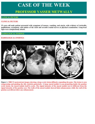
  • 1. CASE OF THE WEEK PROFESSOR YASSER METWALLY CLINICAL PICTURE CLINICAL PICTURE 15 years old male patient presented with symptoms of nausea, vomiting, and ataxia, with evidence of torticollis, papilledema, nystagmus, and palsies of the sixth and seventh cranial nerves at physical examination. Long-tract signs were conspicuously absent. RADIOLOGICAL FINDINGS RADIOLOGICAL FINDINGS   Figure 1. MRI T1 postcontrast images showing a large cystic lesion diffusely expanding the pons. The lesion is seen compressing and pushing the 4th ventricle posteriorly. A large mural nodule is seen projecting inside the pontine cystic lesion, the mural nodule itself is cystic. The signal intensity of the mural nodule cyst is different from the signal intensity of the pontine cyst. The walls of the mural nodule showed faint enhancement, while the wall of the pontine cyst did not show any enhancement.
  • 2. Figure 2. MRI T1 postcontrast images showing a large cystic lesion diffusely expanding the pons. The lesion is seen compressing and pushing the 4th ventricle posteriorly. A large mural nodule is seen projecting inside the pontine cystic lesion, the mural nodule itself is cystic. The signal intensity of the mural nodule cyst is different from the signal intensity of the pontine cyst. The walls of the mural nodule showed faint enhancement, while the wall of the pontine cyst did not show any enhancement. Figure 3. The intrapontine cystic lesion and the cystic mural nodule. Notice that the lesion is seen extending (exophytosis) into the right cerebellopontine ojn the right side
  • 3. Figure 4. MRI T2 images showing the intrapontine cystic lesion and the large cystic mural nodule projecting inside the pontine cyst, notice the upward extension of the mass up to the level of midbrain (C). The basilar artery is encased by the pontine lesion. The wall of the cystic mural nodule is hypointense probably because of the existence of calcification Stereotactic biopsy showed that the lesion is a pilocytic astrocytoma  Radiological pathology of pilocytic astrocytoma The capillaries may be abnormal and can be Grossly, PA is a well-circumscribed yet unencapsulated mass. The coiled (angiomatous) and thick-walled. lesion grows primarily by expansion rather than the infiltration Apparently the blood-brain barrier is not well characteristic of most astrocytomas. In many cases, lesions are easily formed in these tumors. The proteinaceous fluid separated from the adjacent uninvolved cerebellar folia. Most PA that accumulates as both microcysts and have a significant grossly visible cystic component. In many cases, macrocysts probably leaks from the abnormal the tumor has the classically described cyst with nodule morphology- vessels. Mitosis and necrosis are distinctly in which neoplasm is confined to a nubbin of tissue embedded in the uncommon. Despite this, occasional PA show wall of a fluid-filled cavity. In these cases, the cyst fluid is microscopic hemorrhages or brownish staining. surrounded by nonneoplastic compressed or gliotic tissue. Calcification can be seen in 25% of cases. Microscopically, there is a biphasic pattern of dense areas with elongated bipolar hairlike (pilocytic) astrocytes alternating with looser regions that may have microcysts. One distinctive feature is the presence of eosinophilic curvilinear Rosenthal fibers within the dense regions. The capillaries may be abnormal and can be coiled (angiomatous) and thick-walled. Apparently the blood-brain barrier is not well formed in these tumors. The proteinaceous fluid that accumulates as both microcysts and macrocysts probably leaks from the abnormal vessels. Mitosis and necrosis are distinctly uncommon. Despite this, occasional PA show microscopic hemorrhages or brownish staining. Table 1. The mural nodule is composed of two main parts as follows Part Appearance Histopathology Part I Dense compact appearance Composed of elongated bipolar hairlike (pilocytic) astrocytes with high nuclear to cytoplasmic ratio and with minimal extracellular fluid. This part is relatively vascular. Part II Loose appearance Relatively acellular and composed mainly of microcysts and enlarged extracellular fluid filled spaces. This part is relatively avascular. * The spatial distribution of each part within the mural nodule will determine the neuroimaging appearance of the mural nodule.
  • 4. Figure 5. Cystic (pilocytic) brain stem glioma.   Many astrocytomas, particularly in the cerebellum, hypothalamus, and optic pathways of children, exhibit a typical histologic appearance previously termed polar spongioblastoma and now universally referred to as juvenile pilocytic astrocytoma. These tumors frequently contain both macrocysts (as in cystic cerebellar astrocytoma) or microcysts. Rosenthal fibers, strongly eosinophilic coalescences of neurofibrillary elements, are characteristic of juvenile pilocytic astrocytoma, but may be found in other forms of tumor, particularly as a glial reaction surrounding craniopharyngioma. Endothelial proliferation is common in these tumors and has none of the ominous connotations in this context that it implies in other forms of astrocytomas. Figure 6. Pilocytic astrocytoma with hair-like cells From the pathological point of view pilocytic (hair cells) astrocytomas are composed of two main parts, a fluid- filled large cyst and a projecting mural nodule. The neoplastic cells are confined to the mural nodule and the cyst walls are composed of non-neoplastic compressed or gliotic neural tissues. Pilocytic astrocytomas do not have true capsule, yet they can easily be separated from the surrounding tissues. Calcification can be seen in 25% of cases. These tumors frequently have microcysts and macrocysts. Microscopically, there is a biphasic pattern of dense areas with elongated bipolar hairlike (pilocytic) astrocytes alternating with looser regions that may have microcysts. One distinctive feature is the presence of eosinophilic curvilinear Rosenthal fibers and strongly eosinophilic coalescences of neurofibrillary elements within the dense regions. The capillaries may be abnormal and can be coiled (angiomatous) and thick-walled. Apparently the blood-brain barrier is not well formed in these tumors. The proteinaceous fluid that accumulates as both microcysts and macrocysts probably leaks from the abnormal vessels. Necrosis, mitotic activity, endothelial proliferation are selectively absent in pilocytic astrocytomas. Pilocytic astrocytomas are very slowly growing tumors, with long premonitory symptoms before clinical presentation, that selectively grow by expansion rather by infiltration of the surrounding neural tissues that is more characteristic of diffuse astrocytomas. A pilocytic astrocytoma usually starts as hypercellular solid tumor with elongated bipolar hairlike (pilocytic) astrocytes with high nuclear to cytoplasmic ratio and with minimal extracellular fluid (purely solid tumors). Solid tumors are vascular and their capillaries may be abnormal and can be coiled (angiomatous) and thick-walled.
  • 5. Apparently the blood-brain barrier is not well formed in these tumors and proteinaceous fluid probably leaks from the abnormal vessels and accumulates in the tumors as microcysts, first, and macrocysts later on. With progressive enlargement of the macrocysts (microcysts enlarge and coalesce forming a single large cyst), the viable tumor tissues are progressively compressed into a smaller, dense and hypercellular peripheral mural nodule. Progressive leakage of proteinaceous fluid within the core of the mural nodule will result in progressive enlargement of the mural nodule, the core of which will be cystic with a thin outer cover of viable tumor tissues (pattern I). Although the typical appearance of a pilocytic tumor is a large single cyst with a mural nodule, however the spatial distribution of the solid (hypercellular/vascular) and the cystic components within the tumors can vary, also the share taken by the solid the cystic parts in the histopathological make-up of the tumors might vary. These histopathological variations might result in tumors that have quite atypical appearance with irregular cystic and solid parts and with irregular or patchy contrast enhancement. Figure 7. A pilocytic astrocytoma commonly starts as a solid mas (1), however due to defective blood brain barrier in the newly formed blood vessels proteinaceous fluid probably leaks and accumulates inside the tumor as microcysts, first (2), and macrocysts later on (3). With progressive enlargement of the macrocysts (microcysts enlarge and coalesce forming a single large cyst), the viable tumor tissues are progressively compressed into a smaller, dense and hypercellular peripheral mural nodule (pattern II) against a large cyst (3). Progressive leakage of proteinaceous fluid within the core of the mural nodule will result in progressive enlargement of the mural nodule, the core of which will be cystic with a thin outer cover of viable tumor tissues (pattern I) (4,5,6). (Blue = cystic parts and brown = solid parts of the tumor) Figure 8. This figure shows a gross specimen of a pilocytic astrocytoma of the posterior fossa. Identify the cerebellar hemispheres, the pons and the fourth ventricle. The fourth ventricle is nearly obliterated due to the large cystic tumor in the midline. Note the white nodule to one side of the cyst. This is the actual tumor. Many pilocytic astrocytomas in the posterior fossa will have an associated cyst and a contrast enhancing quot;muralquot; nodule. Pilocytic astrocytomas are one of the most common pediatric brain tumors and most occur in the posterior fossa, but in children with Neurofibromatosis type I, they may occur in the optic tracts.
  • 6. Juvenile pilocytic astrocytomas tend to be well circumscribed and to grow slowly with long periods of premonitory symptoms before presentation. This pattern is especially true of tumors that arise in the cerebellum. Tumors of the anterior third ventricle tend to be well-defined superiorly but diffusely infiltrating the optic mechanisms and hypothalamus inferiorly. The course of these tumors is normally benign but may be unpredictable thereby making treatment decisions extremely difficult. The presence of a juvenile pilocytic astrocytoma that extends into the subarachnoid space is common. DIAGNOSIS: DIAGNOSIS: BRAIN STEM JUVENILE PILOCYTIC ASTROCYTOMA DISCUSSION DISCUSSION Pilocytic astrocytoma is the most common pediatric central nervous system glial neoplasm and the most common pediatric cerebellar tumor. This tumor has a noteworthy benign biologic behavior that translates into an extremely high survival rate—94% at 10 years—that is by far the best of any glial tumor. Most patients present in the first 2 decades, and clinical symptoms and signs are usually of several months duration and directly related to the specific location of the tumor. The cerebellum, optic nerve and chiasm, and hypothalamic region are the most common locations, but the tumor can also be found in the cerebral hemisphere, ventricles, and spinal cord. Surgical resection is the treatment of choice for all tumors, except for those involving the optic pathway and hypothalamic region, which may be treated with radiation therapy and chemotherapy. Cross-sectional imaging often demonstrates a classic appearance: a cystic mass with an enhancing mural nodule. Less common appearances are quite nonspecific. Surrounding vasogenic edema is rarely present, and this feature provides a valuable clue to the correct diagnosis. Accurate interpretation of imaging studies plays an essential role in directing treatment of these tumors, particularly when they arise in the optic pathway of patients with neurofibromatosis type 1. Disseminated disease and recurrence are extremely rare. Originally identified in a series of 76 cases of cerebellar astrocytomas by Harvey Cushing in 1931, the pilocytic astrocytoma occupies a unique place among cerebral neoplasms (1). With its notable indolent biologic behavior, pilocytic astrocytoma carries one of the highest survival rates of any brain tumor and certainly the highest rate for any astrocytoma. Yet, as reviewed herein, there are numerous oddities about this neoplasm. It appears well circumscribed, yet occasionally it infiltrates the surrounding brain tissue, as seen at histologic examination. It enhances intensely, sometimes with a ringlike pattern that is more commonly seen in highly malignant astrocytomas, yet it is not a high-grade neoplasm. In rare cases, it can even produce widespread dissemination, which seems incongruous for a brain tumor with slow growth and fairly bland histologic characteristics. Even more fascinating, such metastatic spread can occur without associated increased mortality, in contrast to the poor prognosis so common in patients with metastatic high-grade tumors. In view of these many contradictions, pilocytic astrocytoma qualifies as quot;the tumor that is the exception to the rule.quot;  Epidemiologic Characteristics Pilocytic astrocytoma is the most common pediatric cerebellar neoplasm and the most common pediatric glioma, constituting 85% of all cerebellar astrocytomas and 10% of all cerebral astrocytomas in this age group (2). Overall, it accounts for 0.6%–5.1% of all intracranial neoplasms and 1.7%–7% of all glial tumors (3). Pilocytic astrocytoma occurs most commonly in children and young adults, with most cases (75%) manifesting in the first 2 decades of life (2,4). No gender predilection is reported (2). Most of the lesions occur in or near the midline, usually arising from the cerebellum, the optic nerve and chiasm, or the region of the hypothalamus- thalamus. Less common locations include the cerebral hemispheres, the cerebral ventricles, velum interpositum, and spinal cord (5–9). In adults, the tumor more frequently occurs in the cerebral hemisphere (8,10). The association of pilocytic astrocytoma with neurofibromatosis type 1 (NF1) is well documented. Pilocytic astrocytoma is the most common tumor seen in this population, occurring in up to 15%–21% of all NF1 patients, and typically involves the optic nerve or chiasm (3,11–16). Because the vast majority of optic pathway gliomas are histologically regarded as pilocytic astrocytoma, it has been suggested that the most appropriate term for this entity
  • 7. should be quot;pilocytic astrocytoma of the optic pathwayquot; (16). Nearly all optic pathway gliomas in NF1 patients manifest before the age of 6 years and females are more commonly affected by a 2:1 ratio (16,17). Of all patients with an optic pathway glioma, about one-third have NF1, and, of all tumors in this region, 40%–70% occur in NF1 patients (14,16,18). Pilocytic astrocytomas account for 1.5%–3.5% of all orbital neoplasms and two-thirds of all neoplasms of the optic nerve (3). Most optic pathway pilocytic astrocytomas (75%) arise in children less than 12 years old; in addition, the tumor is more likely to arise in the optic nerve in children, whereas it is more commonly located in the optic chiasm in adolescents and young adults (19,20). Involvement of the chiasm and hypothalamus has been reported to occur in 25%–60% of patients (11,19,20). Optic pathway gliomas much less commonly manifest in adult patients without NF1; when they do, these tumors have a dramatically different biologic behavior and are regarded histologically as anaplastic astrocytoma or glioblastoma multiforme (21).  Clinical Features Clinical presentation of patients with a pilocytic astrocytoma varies with its site of origin. Headache, vomiting, gait disturbance, blurred vision, diplopia, and neck pain are common symptoms in patients with a cerebellar pilocytic astrocytoma (22–24). Clinical signs usually include hydrocephalus, papilledema, truncal ataxia, appendicular dysmetria, head tilt, sixth nerve palsy, and nystagmus (22,24). When a pilocytic astrocytoma arises in the brainstem, it typically extends exophytically from its dorsal margin and causes symptoms of nausea, vomiting, and ataxia, with evidence of torticollis, papilledema, nystagmus, and palsies of the sixth and seventh cranial nerves at physical examination (25–27). Long-tract signs are conspicuously absent (25,27). Pilocytic astrocytomas arising from the tectum characteristically manifest with headache, vomiting, paresis, abnormal gait, somnolence, Parinaud syndrome, and diplopia (28,29). Pilocytic astrocytoma of the optic pathway frequently produces visual loss or visual-field deficit, with optic disk pallor and optic nerve atrophy in the involved eye secondary to axonal damage and ischemia (3,21,30). Proptosis may be seen with larger masses. Papilledema is common for lesions arising from the optic nerve but unusual for those originating from the optic chiasm (21). Some patients may show spasmus nutans, a nystagmus characterized by high frequency and low amplitude and associated with head nodding movements (31). Precocious puberty is commonly seen (39% of cases) in NF1 patients with an optic pathway glioma and has not been reported in its absence (16). Less specific manifestations include amblyopia (30). Smaller lesions may not be associated with any symptoms at all (2). Pilocytic astrocytoma of the hypothalamus may produce symptoms related to obesity, diabetes insipidus, and other symptoms of hypothalamic-pituitary dysfunction (2). On occasion, these masses may produce the so-called diencephalic syndrome, which is characterized by emaciation despite a normal to slightly decreased caloric intake, alert appearance, hyperkinesis, irritability, and normal to accelerated growth (20,32,33). Although this syndrome may occur with any hypothalamic mass, the vast majority of reported cases are secondary to a pilocytic astrocytoma, with widespread dissemination noted in some cases (32). When the tumor involves the thalamus, hydrocephalus or hemiparesis secondary to compression of the corticospinal tract within the nearby internal capsule is typical (2,34). Headache, seizure activity, hemiparesis, ataxia, nausea, and vomiting are common clinical manifestations for pilocytic astrocytomas arising in the cerebral hemispheres (7). The occurrence of seizure activity generally indicates cortical gray matter involvement (2). Papilledema is noted in about one-third of these patients (7).  Pathologic Characteristics Pilocytic astrocytoma has been known by many names over the years. Because of its resemblance histologically to the spongioblastic cells of the fetus, German pathologists called it quot;spongioblastoma polare,quot; a term that has now been abandoned (35,36). Russell and Rubenstein (37) distinguished the tumor into adult and juvenile forms. In addition, many generic pseudonyms and euphemisms based on the geographic location of the tumor have persistently appeared in the prior pathology lexicon to describe these lesions. Hence, a variety of names—optic nerve glioma, hypothalamic glioma, cerebellar astrocytoma, microcystic astrocytoma, cystic astrocytoma, and others—have inadvertently blurred the true identity of the pilocytic astrocytoma and led to confusion (26).
  • 8. Although this tumor may be found in many central nervous system locations and may account for a significant proportion of the tumors in those locations, pilocytic astrocytoma is now regarded by the latest World Health Organization (WHO) classification as a distinct clinicopathologic entity, and use of these less specific terms should be avoided if the pathologic findings warrant the diagnosis (26). The macroscopic appearance of pilocytic astrocytoma varies with its location within the central nervous system. Tumors of the cerebellum and cerebral hemisphere are typically well-circumscribed, cystlike masses with a discrete mural nodule, whereas those arising in the hypothalamus and optic chiasm tend to be large, soft, cystlike masses. When the tumor arises within the optic nerve, it infiltrates and engulfs the nerve to produce fusiform enlargement of that structure, with peripheral extension into the surrounding leptomeninges. Brainstem pilocytic astrocytomas are usually peripheral and are attached to the brainstem surface without extensive infiltration (38). At histologic examination, pilocytic astrocytoma classically manifests in a noteworthy biphasic pattern composed of a combination of loose glial tissue punctuated by numerous vacuoles, microcysts, occasional macrocysts, and compacted piloid tissue (38). The piloid tissue component is composed of dense sheets of elongated bipolar cells that demonstrate fine fibrillary (hairlike) processes, a highly distinctive feature, and typically an abundance of Rosenthal fibers (38). In contrast, the multipolar cells of the loose glial tissue, frequently called protoplasmic astrocytes, are much less fibrillated and commonly are intermixed with degenerative products of astrocyte formation, known as eosinophilic granular bodies or quot;protein dropletsquot; (2). These quot;classicquot; but nonspecific granular bodies are believed to indicate slow growth and low histologic grade and are associated with a favorable prognosis (38). Calcification is an uncommon feature, usually occurring in those tumors arising from the optic nerve or hypothalamic-thalamic region, but it can be extensive (2,38). The tumor is suspected to arise from reactive astrocytes (2). There is considerable variability in the contribution of each pattern to the overall histologic appearance of pilocytic astrocytomas. Some tumors have a predominance of the glial component, whereas others show more of the piloid tissue. A minority of pilocytic astrocytomas demonstrates both components equally (38). More recently, a subset of pilocytic astrocytomas known as the pilomyxoid variant has been identified with monomorphous histologic features and lack of Rosenthal fibers. The few reported cases have occurred in the chiasmatic-hypothalamic region in children less than 2 years of age; these tumors were associated with a higher rate of recurrence and cerebrospinal fluid dissemination (2,39). These tumors have some ultrastructural features that have fueled speculation that this subtype may be of tanycytic origin, leading some authorities to describe it as a quot;tanycytoma.quot; However, more evidence is needed to prove this cellular lineage and justify use of this term (40). Slow growth is the rule for most pilocytic astrocytomas (41). However, some tumors, particularly those of the optic nerve and chiasm, may show a propensity for periods of accelerated growth (19). Growth of the tumor, especially those occurring in the cerebellum, may overrun normal ganglion cells (neurons), producing the quot;trapped neuronquot; appearance (2,38). The growth pattern of optic pathway glioma (including pilocytic astrocytoma) correlates with the presence or absence of NF1 in the patient. When the tumor arises in patients with NF1, it tends to grow within the nerve, whereas a circumferential pattern is observed when it occurs in patients without NF1 (42). Although many pilocytic astrocytomas appear well circumscribed macroscopically, they may infiltrate into the surrounding brain parenchyma for several millimeters (2). Infiltration occurs more commonly for those tumors that arise from the optic nerve and chiasm, where there is frequently no clear demarcation between the tumor and normal tissue (2). Infiltration of the adjacent leptomeninges is a signature feature of a cerebellar pilocytic astrocytoma, causing fixation of the cerebellar folia and filling of the sulci (2). This infiltration, along with perivascular space extension, may also be seen with tumors in other locations (2). Pilocytic astrocytomas are highly vascular (2). Some long-standing tumors, such as those of the cerebellum and cerebral hemisphere, frequently have markedly hyalinized and glomeruloid vessels (2). This new vessel formation is particularly evident within the cyst wall, and it is believed that the fluid within the cyst directly supports vascular proliferation (2) The presence of these glomeruloid vessels accompanied by extensive nuclear pleomorphism may mimic some of the features of high-grade astrocytomas (2,38). The increased vascularity of pilocytic astrocytoma may explain why it may even be a target for metastatic disease, as in one case of a breast carcinoma that spread to a pilocytic astrocytoma (43). The overwhelming majority of pilocytic astrocytomas are regarded as grade I tumors in the WHO classification (2,38). Most tumors demonstrate sufficient characteristic histologic features that a definitive diagnosis can be
  • 9. rendered with confidence. However, the variety of histologic patterns among pilocytic astrocytomas and the lack of specific immunohistochemical, cytogenetic, and molecular markers may occasionally make the diagnosis difficult (2). Occasional hyperchromasia and pleomorphism, especially when they are noted in a diffuse tumor, may cause pilocytic astrocytoma to be confused with a higher-grade astrocytoma (2). In rare cases, some tumors, especially those arising in patients who underwent radiation therapy, may have frankly malignant features (increased mitotic activity, hypercellularity, endothelial proliferation, and necrosis with pseudopalisading) and also demonstrate aggressive biologic behavior (38). Very rarely, a pilocytic astrocytoma may undergo malignant transformation with an aggressive histologic appearance; such a tumor is called an anaplastic (malignant) pilocytic astrocytoma. Although fatal cases have been reported, this event does not necessarily carry a less favorable outcome (2,8,44–46). Many cases of malignant transformation have occurred after administration of radiation therapy, leading to speculation that irradiation is at least a contributing factor in the development of this change, which may manifest up to 52 years after initial treatment (45). Cytogenetic studies of pilocytic astrocytomas have revealed loss of genetic material involving the long arm of chromosome 17 (17q) near the same locus for the NF1 tumor suppressor gene (47). Furthermore, lack of expression of an NF1 gene product, neurofibromin, has been documented in NF1-associated pilocytic astrocytomas (48). These findings have fueled speculation that the NF1 tumor suppressor gene is linked with the expression of pilocytic astrocytoma. Additional chromosomal mutations have been identified on chromosomes 7, 8, 11, 19, and 21 (2,49). Pilocytic astrocytomas differ from the more common diffuse astrocytomas from the pathological, nosological, genetic and prognostic point of view. From the pathological point of view diffuse astrocytomas are neoplasms of widely varying potential that are unencapsulated, poorly marginated and diffusely infiltrate into the surrounding brain. These diffuse astrocytomas appear to form a continuum of both biological and histological aggression. They vary from lesions with almost normal cytology (grade II astrocytomas) through intermediate stages (grade III, anaplastic astrocytomas) and up to the most aggressive of all human brain tumours (grade IV astrocytomas or glioblastoma multiforme). (99) From the nosological point of view, and according to the WHO classification of brain tumours, pilocytic astrocytomas are ranked as grade I benign gliomas while diffuse astrocytomas are ranked as grade II, grade III (anaplastic astrocytomas) and grade IV (glioblastoma multiforme). The following pathological differences are present between diffuse and pilocytic astrocytomas. (99)  Diffuse astrocytomas, unlike pilocytic astrocytomas, have a peculiar tendency to change its grade over time and the condition is age dependant. A change in the grade of diffuse astrocytoma is more likely to occur in the older age group. In older age group (over the age of 40 years) diffuse low grade astrocytomas (grade II astrocytoma according to WHO) have a bad prognosis because they have a great tendency for anaplastic transformation (to grade III or grade IV astrocytoma according to WHO), while at a younger age group anaplastic transformation of diffuse low grade astrocytomas (grade II astrocytoma according to WHO) is extremely uncommon, also the probability for diffuse low grade astrocytomas to have a highly malignant component (i.e, grade III or IV mixed with grade II) is higher in the older age group. On the other hand pilocytic astrocytomas (grade I astrocytoma according to WHO) never change its grade over time.(99)  Diffuse astrocytomas (grade II, III or grade IV astrocytomas) grow by infiltration of the nearby neural tissues (commonly in the form of remote neoplastic cells radiating from the mother tumour) and so they are poorly marginated and, practically, complete surgical resection is not possible and some neoplastic cells are almost invariably left behind after surgical resection. On the other hand pilocytic astrocytomas grow by expansion and so they are well circumscribed and subsequently complete surgical resection is possible. (99)  Diffuse astrocytomas, unlike pilocytic astrocytomas, are highly cellular neoplasms with cells that range from normal appearing astrocytes (grade II) to cells with marked pleomorphism and hyperchromatic nuclei ( grade III, and IV). On the other hand pilocytic astrocytomas are histopathologically composed of scanty elongated cells, Rosenthal fibers and microcysts and this combination constitutes the classic of pilocytic astrocytomas. Cells are only confined to the mural nodule in pilocytic astrocytomas and subsequently the mural nodule is the only neoplastic part of the tumour.(99)  Pilocytic astrocytomas are truly benign gliomas while diffuse astrocytomas are, at best, of low grade
  • 10. malignancy. (99) Radiologically pilocytic astrocytomas differ from diffuse astrocytomas in the following points 1. Pilocytic astrocytomas are typically quot;cystic tumours with a mural nodulequot; and with prominent mass effect, while diffuse astrocytomas with a lower grade (grade II) are typically solid tumours with minimal or no mass effect that appear homogeneously hypodense on CT scan, hyperintense on the MRI T2 images and hypointense on the T1 MRI images. The neuroimaging appearance of diffuse astrocytomas is due to increased cell count. The mural nodule of pilocytic astrocytomas might or might enhance while diffuse low grade astrocytomas usually do not enhance on postcontrast scan. Diffuse low grade astrocytomas (grade II) diffusely expand the affected part of the brain with poor margin, while pilocytic astrocytomas are well circumscribed rounded or oval tumours.(99) 2. Although central necrosis in highly malignant glioblastoma multiforme might, morphologically, creates the appearance of a cyst with a projecting mural nodule, however this can easily be differentiated from pilocytic astrocytomas by the fact that the walls of the cyst in pilocytic astrocytomas, being composed of non-neoplastic compressed neural tissues, never enhance while the walls of the cyst in glioblastomas with central necrosis invariably enhance because it is composed of viable tumour tissues. Wall enhancement is characteristic of glioblastomas with central necrosis and when it is observed radiologically should shift the tumour grade from the most benign pilocytic astrocytoma to the most malignant glioblastoma multiforme,The presence of significant edema and short history before clinical presentation favor the diagnosis of glioblastoma multiforme. In general glioblastoma multiforme occurs at an older age compared with pilocytic astrocytoma. (99) From the genetic point of view pilocytic astrocytoma is different from diffuse astrocytomas in the following points (99)  The analyses of the genetic lesions of pilocytic astrocytoma have targeted the TP53 gene on chromosome 17. Investigations have not confirmed a critical role for alterations of this gene in the development of these tumors, however. In one series, the cytogenetic analysis of 14 pilocytic tumor cultures did not identify a specific pattern of chromosomal aberration. Patients with tumors characterized by normal stem line karyotypes had the most favorable outcomes. The presence of clonal structural abnormalities and the presence of markers were associated with a high risk of early recurrence. (99)  On the other hand genetic lesions associated with the development and malignant transformation of diffuse astrocytomas have been well described in the cytogenetic literature. To date, three distinct clinical, histologic, and genetic patterns of glioblastoma multiforme have been characterized. In younger patients, most diffuse astrocytomas are believed to begin as low-grade astrocytoma, with progression to glioblastoma multiforme through a stepwise acquisition of genetic lesions. These secondary glioblastoma multiforme often contain areas of well-differentiated residual tumor. The most frequent chromosomal abnormality identified in diffuse astrocytomas is the abnormal gain of chromosome 7 with an associated loss of one of the sex chromosomes. Additionally, allelic loss or mutation of 17p, resulting in critical alterations of the TP53 gene, has been targeted as an essential step in the early development of glioma. Mutant TP53, identified in at least one third of all astrocytomas, may contribute to the formation of these tumors by inhibiting programmed cell death. glioblastoma multiforme in older patients are usually primary-that is, they occur as glioblastoma multiforme from their inception, without progression from a lower- grade tumor. In this group, the development of glioblastoma multiforme involves a parallel sequence of genetic alterations, including amplifications and deletions, that up-regulate growth factor receptors and drive cell proliferation. (99)  Imaging Characteristics At computed tomography (CT), most cerebellar and cerebral pilocytic astrocytomas have a well-demarcated appearance with a round or oval shape smaller than 4 cm in size, cystlike features, smooth margins, and occasional calcifications (8,50,51). Most tumors (82% in one series) are located near the ventricular system, and almost all (94%) enhance, typically intensely, on postcontrast images obtained after intravenous administration of contrast material (51). At MR imaging, pilocytic astrocytoma is typically isointense to hypointense relative to normal brain with T1-
  • 11. weighted pulse sequences and hyperintense compared with normal brain with T2-weighted pulse sequences (50). As expected for a tumor of low biologic activity, the degree of surrounding vasogenic edema is diminished in comparison with that seen in high-grade (WHO grade III and IV) glial neoplasms (8,9). When it does occur, the area of edema is smaller in size than the diameter of the tumor (51). Postcontrast imaging features are similar to those reported with CT. Because of its superiority to CT in assessing the posterior fossa and the usefulness of multiplanar imaging, MR imaging is regarded as the imaging study of choice for evaluation of pilocytic astrocytomas (24). The well-demarcated appearance of virtually all (96%) pilocytic astrocytomas is misleading, as most (64%) show infiltration into the surrounding brain parenchyma at histologic examination (51). Four predominant imaging patterns of pilocytic astrocytoma have been described: (a) mass with a nonenhancing cyst and an intensely enhancing mural nodule (21% of cases), (b) mass with an enhancing cyst wall and an intensely enhancing mural nodule (46%), (c) necrotic mass with a central nonenhancing zone (16%), and (d) predominantly solid mass with minimal to no cystlike component (17%) (24). Hence, two-thirds of all cases demonstrate the classic imaging manifestation of a cystlike mass with an enhancing mural nodule. Although most cyst walls do not enhance, some may enhance intensely, even as much as the mural nodule (52). Although most cyst walls do not enhance, some may enhance intensely, even as much as the mural nodule; however, cyst wall enhancement is not necessarily indicative of tumor involvement (52). Beliefs vary among neurosurgeons regarding whether to resect the cyst itself in cases of pilocytic astrocytoma. Some advocate complete resection, others biopsy, and still others no resection (52). Removal of the cyst wall has not been linked with improved survival (53). There is contradictory evidence in the literature regarding the most common location of pilocytic astrocytoma in the cerebellum. In one series of 132 patients, 16% of the tumors arose in the vermis, 53% in the cerebellar hemisphere, and 26% in both; 34% also involved the brainstem (54). However, in another review of 168 cases, 71% of the tumors were located in the vermis, whereas 29% occurred in the hemisphere (24). Those tumors arising in the cerebral hemisphere are more likely to occur in the temporal lobe (7,53). Location, enhancement, and the presence or absence of calcification seen at the time of initial presentation are not reliably predictive of the future clinical behavior of a pilocytic astrocytoma (55). In contrast to the more biologically aggressive fibrillary astrocytoma (which is most common in the pons), pilocytic astrocytoma may be found throughout the brainstem and frequently extends in an exophytic fashion from its dorsal surface (56,57). Pilocytic astrocytoma typically demonstrates sharp delineation with cystlike regions and fourth ventricle obliteration (25,27,57). The distinction between these two entities is more than an academic curiosity, since the fibrillary brainstem astrocytoma carries a dismal prognosis compared with the excellent outlook associated with the pilocytic variety (57). Because available tissue for histologic analysis from this exquisitely sensitive region of the central nervous system is generally limited, knowledge of the differences in imaging manifestations of the two diseases may help establish the correct diagnosis (26). When pilocytic astrocytoma arises within the optic nerve, it manifests on both CT and MR images with enlargement of the optic nerve as an intraconal mass with characteristic kinking or buckling of the nerve secondary to the neoplasm itself and vascular congestion (21). Isointensity on T1-weighted MR images and heterogeneous hyperintensity on T2-weighted MR images are typical, with variable enhancement following intravenous contrast material administration (21). Differences in the imaging appearance of optic pathway gliomas in patients with NF1 versus that of tumors in patients without NF1 have been noted in one study (58). NF1-associated tumors affected the optic nerve and chiasm with nearly equal prevalence, preserved the optic nerve shape, rarely extended beyond the optic pathway, and usually were not cystic. In contrast, the majority of tumors not associated with NF1 involved the optic chiasm rather than the optic nerve, extended extraoptically, and frequently contained cystic areas. The imaging findings did not statistically correlate with the biologic behavior of the tumors (58). Atypical imaging manifestations may be seen occasionally. Multiple cystlike masses and association with an area of gray matter heterotopia have been reported (59,60). Hemorrhage, either within the tumor or within the subarachnoid space, is a rare phenomenon (61–63). Pilocytic astrocytoma may masquerade as a cerebellopontine angle mass that mimics the imaging appearance of a vestibular schwannoma with widening of the internal auditory canal (64,65). When the tumor is located in or near the optic chiasm, a bilobed shape may be seen occasionally (50). The small number of pilomyxoid astrocytomas reported have occurred in the chiasmatic-hypothalamic region, with a tendency to have more intense homogeneous enhancement and homogeneous hyperintensity on T2-weighted images compared with that seen in pilocytic astrocytomas (39).
  • 12. Reports of MR spectroscopy performed on the soft-tissue portions of pilocytic astrocytomas have documented elevation in the choline (Cho) to N-acetylaspartate (NAA) ratio, ranging from 1.80 to 3.40 (compared with 0.53–0.75 for normal cerebellum) (61,66). Elevated Cho/creatine (Cr) and lactate/Cr ratios have also been noted, whereas the NAA/Cr ratio has not been significantly elevated (61,66). There is some controversy regarding whether the Cho resonance in these tumors is truly elevated compared with that in normal tissue (67). The lipid resonance is minimally elevated, in contrast to significant elevation noted in glioblastoma multiforme and metastatic disease (61). Elevated lactate doublet resonance and diminished Cr and NAA peaks were observed in all eight patients of one study that used point-resolved spectroscopy (PRESS) (61). It is clear that this lactate elevation is not related to necrosis, which is a rare histologic feature in pilocytic astrocytomas. Instead, the lactate elevation most likely reflects alterations in mitochondrial metabolism or represents variability in glucose utilization rates among low- grade astrocytomas (61). Follow-up surveillance imaging of cerebellar pilocytic astrocytoma is recommended at every 3 months for 2 years, followed by every 6 months for an additional 2 years, and then annually after 4 years from the time of treatment (68). A similar follow-up pattern is used to assess changes in patients with pilocytic astrocytoma of the optic pathway (16). Routine contrast-enhanced MR imaging to exclude spinal dissemination is not indicated in patients with a single primary mass and without corroborative symptoms (69). Screening imaging for this tumor in NF1 patients without symptoms is not currently recommended, because the disease rarely progresses and early detection does not currently affect the long-term outcome (17).  Therapy Just as its clinical manifestations vary according to its different locations, treatment of pilocytic astrocytoma can also vary, depending on where it arises. Surgical resection of cerebellar and cerebral pilocytic astrocytomas is considered the treatment of choice and is generally regarded as curative when a gross total resection is attained (4,6,22,24,54,68,70–72). For lesions in less favorable locations (eg, basal ganglia), stereotactic resection may be used (34). Radiation therapy is strictly avoided, given its risk of causing significant morbidity in children younger than 5 years of age and the absence of clinical proof that it prevents recurrence (71,72). Resection of the mural nodule, when present, is the key surgical objective, since the surrounding cyst occurs as a simple reactive change in most cases (4). However, neoplastic changes in the cyst wall, even in the absence of findings at gross inspection or at imaging studies, have been documented; these observations have led to considerable debate among neurosurgeons regarding the optimal surgical management of this structure (24). No statistical difference in survival has been noted in patients who have undergone resection of the cyst wall compared with those in which the cyst is left alone (53). Direct neurosurgical evaluation of the extent of resection is surprisingly unreliable, and postoperative contrast-enhanced cross-sectional imaging, preferably MR imaging, is essential for definitive assessment (4,68,73,74). Patients with optic pathway gliomas and stable symptoms are managed conservatively with clinical and imaging follow-up (15,16,20). In contrast, patients who have progressive symptoms or evidence of hydrocephalus may be treated with surgical resection or radiation therapy, with the former recommended for those whose tumor is located in the optic nerve and the latter for those with an optic chiasm tumor (75,76). Large cystic masses of the chiasm may be amenable to surgical resection, thus allowing radiation therapy to be delayed (15). To avoid the deleterious effects of radiation therapy on the developing brain, chemotherapy is recommended for children younger than 5 years of age (15). Intraarterial delivery of chemotherapy to treat recurrent disease has also been reported (77).  Prognosis, Recurrence, and Dissemination Overall, the prognosis for patients with a pilocytic astrocytoma is excellent, with a 10-year survival rate of up to 94% and a 20-year survival rate of 79%, which is in marked contrast to the more guarded prognosis seen with the diffuse (WHO grade II) astrocytoma (4,54,78,79). Other factors besides the native biologic behavior of the tumor influence this favorable outcome. The increase in 10-year survival rate (about 90% after 1970, compared with about 70% before 1970) has been attributed to improvements in neurosurgical techniques and equipment (80). The location of the pilocytic astrocytoma directly affects the prognosis for the patient. For patients with non–optic pathway pilocytic astrocytomas, the most important factors for improved survival appear to be good neurologic status at the time of diagnosis and gross total surgical resection (6,54). Pilocytic astrocytomas arising in the optic
  • 13. pathway or hypothalamus have the least favorable prognosis (75). For patients whose tumor was confined to the optic pathway, the 17-year survival rate was 85%, compared with only 44% who survived 19 years when the tumor extended into the hypothalamus (81). It is believed that many, if not all, of the tumors in this latter group may represent the pilomyxoid subtype (82). Pilocytic astrocytoma appears to have a more benign biologic behavior when it occurs in patients with NF1 (20). Only a small number of patients with NF1 and an isolated intraorbital glioma showed progression of the tumor 10 years after the time of diagnosis (16). For patients with an optic pathway tumor and NF1, the interval between primary diagnosis and first recurrence tends to be longer, compared with that for patients who do not have NF1 (11,19,76). In contrast to the generally poor outcome (a 5-year survival of usually only 30%) for patients with an infiltrating brainstem glioma (WHO grade II), those with a dorsally exophytic brainstem pilocytic astrocytoma enjoy a much brighter outlook, with stable neurologic status and long-term survival likely (25,27). Subtotal resection and cerebrospinal fluid diversion are the recommended treatment for these tumors. Recurrence may be treated with a second surgical intervention (25,27). Although recurrence rates are low if gross total resection has been attained, they are substantially increased when only partial resection is achieved (24). Most recurrences are noted within 4 years of the initial surgery, although recurrent disease has been documented as late as 36 years after initial removal (71,83). Survival rates among patients with partial resection, compared with those for patients with gross total resection, are not statistically different (22,24,68). Distant dissemination from pilocytic astrocytoma is rare, with a reported prevalence of 2%–12% (72). The occurrence of distant dissemination appears increased in three settings: (a) for those tumors located in the hypothalamus (presumably because of their proximity to the ventricular system or because of the tendency of the pilomyxoid subtype to occur in this region); (b) for tumors that have been partially resected; and (c) for tumors occurring in patients less than 4 years of age at initial diagnosis (35,36,72). Dissemination in reported cases tends to manifest within 3 years of initial diagnosis (72). Dissemination from a pilocytic astrocytoma, unlike virtually all other glial neoplasms, does not necessarily correlate with a poorer prognosis, and many patients are asymptomatic with long-term survival (36,72,84–86). Reoperation for recurrent disease is preferred, whereas radiation therapy or chemotherapy is recommended for the treatment of multicentric or surgically inaccessible dissemination (24,72). There are numerous reports of spontaneous regression of pilocytic astrocytomas arising in the optic pathway, diencephalon, and tectal region (75,87–92). Most of these cases have occurred in children with NF1, but spontaneous regression of the tumor may also occur in patients without that phakomatosis and in adults (18,75,91,93). Numerous theories have been proposed for this finding and include postsurgical apoptosis (programmed cell death), host immune reaction, thrombosis or infarction of tumor vessels, alteration of growth kinetics, removal of the offending carcinogen, genetic programming, and hormonal factors (89,94–98). SUMMARY The pilocytic astrocytoma has exceptionally slow growth and a usually indolent biologic behavior that directly effects an extraordinarily promising prognosis for patients with the disease. Characterized by its classic quot;cystic mass with enhancing mural nodulequot; imaging appearance, the tumor is easily recognizable in most circumstances. MR imaging is essential in facilitating appropriate therapeutic management.  Because pilocytic astrocytomas grow by expansion rather by infiltration of the nearby neural structure (infiltration results in tumour cells being found histologically radiating diffusely from the mother tumour to the surrounding
  • 14. normal neural structures), they remain circumscribed and can be separated from normal neural tissues , thus allowing complete surgical removal without leaving behind any residual tumour cells and it is because of this that postoperative radiotherapy or chemotherapy are not indicated and probably even contraindicated The prognosis in pilocytic astrocytoma is good with a five year survival rate reaching up to 95% to 100% in many studies after complete surgical removal. Recurrence was attributed in most studies to incomplete surgical removal in technically difficult anatomical sites as it was the case in our patient with hypothalamic neoplasm.(99) Because of the very good prognosis of this neoplasm it is important to be familiar with its clinical and neuroimaging pictures. The presence of a cystic component in these lesions is suggestive. The lesions may appear on both CT and MR as a classic cyst with a nodular mass. Purely solid masses are not common. Complex shapes (e.g., multiloculated) can occur and may create a misleading appearance. With contrast infusion, both on CT and on MR, the mural nodule might or might not enhance, however the cyst wall does not enhance. Histopathological confirmation is invariably needed for the ultimate diagnosis of this neoplasm. Although the word astrocytoma is typed in the name of this neoplasm, however it should not be equated with the more common diffuse astrocytoma. quot;Overgradingquot; occurs when the generic name quot;astrocytomaquot; is applied to pilocytic astrocytomas, so it is very important to distinguish between diffuse astrocytoma and pilocytic astrocytoma as there is a real chance that pilocytic astrocytomas are cured by surgery alone once the neoplasms are completely removed. (99) Finally it must be mentioned that brain stem pilocytic astrocytoma is indeed a rare occurrence and it is of interest to report a case with pontine pilocytic astrocytoma. (99)  Addendum  A new version of this PDF file (with a new case) is uploaded in my web site every week (every Saturday and remains available till Friday.)  To download the current version follow the link quot;http://pdf.yassermetwally.com/case.pdfquot;.  You can also download the current version from my web site at quot;http://yassermetwally.comquot;.  To download the software version of the publication (crow.exe) follow the link: http://neurology.yassermetwally.com/crow.zip  The case is also presented as a short case in PDF format, to download the short case follow the link: http://pdf.yassermetwally.com/short.pdf  At the end of each year, all the publications are compiled on a single CD-ROM, please contact the author to know more details.  Screen resolution is better set at 1024*768 pixel screen area for optimum display.  For an archive of the previously reported cases go to www.yassermetwally.net, then under pages in the right panel, scroll down and click on the text entry quot;downloadable case records in PDF formatquot; REFERENCES References 1. Cushing H. Experiences with the cerebellar astrocytomas: a critical review of 76 cases. Surg Gynecol Obstet 1931; 52:129-191. 2. Burger PC, Scheithauer BW, Paulus W, Szymas J, Giannini C, Kleihues P. Pilocytic astrocytoma. In: Kleihues P, Cavenee W, eds. Pathology and genetics of tumours of the nervous system. Lyon, France: IARC, 2000; 45-51. 3. Dutton JJ. Gliomas of the anterior visual pathway. Surv Ophthalmol 1994; 38:427-452. 4. Wallner KE, Gonzales MF, Edwards MSB, Wara WM, Sheline GE. Treatment of juvenile pilocytic astrocytoma. J Neurosurg 1988; 69:171-176.
  • 15. 5. Ideguchi M, Nishizaki T, Harada K, Kwak T, Murakami T, Ito H. Pilocytic astrocytoma of the velum interpositum. Neurol Med Chir (Tokyo) 1998; 38:283-286. 6. Forsyth PA, Shaw EG, Scheithauer BW, O’Fallon JR, Layton DD, Jr, Katzmann JA. Supratentorial pilocytic astrocytomas: a clinicopathologic, prognostic, and flow cytometric study of 51 patients. Cancer 1993; 72:1335-1342. 7. Clark GB, Henry JM, McKeever PE. Cerebral pilocytic astrocytoma. Cancer 1985; 56:1128-1133. 8. Katsetos CD, Krishna L. Lobar pilocytic astrocytomas of the cerebral hemispheres. I. Diagnosis and nosology. Clin Neuropathol 1994; 13:295-305. 9. Brat DJ, Burger PC. Cerebral pilocytic astrocytoma: distinction from infiltrating fibrillary astrocytomas. Pathol Case Rev 1998; 3:290-295. 10. Garcia DM, Fulling KH. Juvenile pilocytic astrocytoma of the cerebrum in adults: a distinctive neoplasm with favorable prognosis. J Neurosurg 1985; 63:382-386. 11. Deliganis AV, Geyer JR, Berger MS. Prognostic significance of type 1 neurofibromatosis (von Recklinghausen disease) in childhood optic glioma. Neurosurgery 1996; 38:1114-1119. 12. Mulvihill JJ, Parry DM, Sherman JL, Pikus A, Kaiser-Kupfer MI, Eldridge R. National Institutes of Health Conference. Neurofibromatosis 1 (Recklinghausen disease) and neurofibromatosis 2 (bilateral acoustic neurofibromatosis): an update. Ann Intern Med 1990; 113:39-52. 13. Garner A, Klinworth GK. Tumors of the orbit, optic nerve, and lacrimal sac. Pathobiology of ocular disease. New York, NY: Marcel Dekker, 1982; 741. 14. Lewis RA, Gerson LP, Axelson KA, Riccardi VM, Whitford RP. von Recklinghausen neurofibromatosis. II. Incidence of optic nerve gliomata. Ophthalmology 1984; 91:929-935. 15. Packer RJ, Bilaniuk LT, Cohen BH, et al. Intracranial visual pathway gliomas in children with neurofibromatosis. Neurofibromatosis 1988; 1:212-222. 16. Listernick R, Louis DN, Packer RJ, Gutmann DH. Optic pathway gliomas in children with neurofibromatosis 1: consensus statement from the NF1 Optic Pathway Glioma Task Force. Ann Neurol 1997; 41:143-149. 17. Listernick R, Charrow J, Greenwald M, Mets M. Natural history of optic pathway tumors in children with neurofibromatosis type 1: a longitudinal study. J Pediatr 1994; 125:63-66. 18. Balkhoyor KB, Bernstein M. Involution of diencephalic pilocytic astrocytoma after partial resection. J Neurosurg 2000; 93:484-486. 19. Alvord EC, Lofton S. Gliomas of the optic nerve or chiasm: outcome by patients’ age, tumor age, and treatment. J Neurosurg 1988; 68:85-98. 20. Rodriguez LA, Edwards MSB, Levin VA. Management of hypothalmic gliomas in children: an analysis of 33 cases. Neurosurgery 1990; 26:242-247. 21. Hollander MD, FitzPatrick M, O’Connor SG, Flanders AE, Tartaglino LM. Optic gliomas. Radiol Clin North Am 1999; 37:59-71. 22. Abdollahzadeh M, Hoffman HJ, Blazer SI, et al. Benign cerebellar astrocytoma in childhood: experience at the Hospital for Sick Children 1980–1992. Childs Nerv Syst 1994; 10:380-383. 23. Gol A, McKissock W. The cerebellar astrocytomas: a report on 98 verified cases. J Neurosurg 1959; 16:287- 296.
  • 16. 24. Pencalet P, Maixner W, Sainte-Rose C, et al. Benign cerebellar astrocytomas in children. J Neurosurg 1999; 90:265-273. 25. Pollack IF, Hoffman HJ, Humphreys RP, Becker L. The long-term outcome after surgical treatment of dorsally exophytic brain-stem gliomas. J Neurosurg 1993; 78:859-863. 26. Burger PC. Pathology of brain stem astrocytomas. Pediatr Neurosurg 1996; 24:35-40. 27. Khatib Z, Heidemann R, Kovnar E, et al. Pilocytic dorsally exophytic brainstem gliomas: a distinct clinicopathological entity. Ann Neurol 1992; 32:458-459. 28. Bowers DC, Georgiades C, Aronson LJ, et al. Tectal gliomas: natural history of an indolent lesion in pediatric patients. Pediatr Neurosurg 2000; 32:24-29. 29. Boydston WR, Sanford RA, Muhlbauer MS, et al. Gliomas of the tectum and periaqueductal region of the mesencephalon. Pediatr Neurosurg 1991; 17:234-238. 30. Roh S, Mawn L, Hedges TR, 3rd. Juvenile pilocytic astrocytoma masquerading as amblyopia. Am J Ophthalmol 1997; 123:692-694. 31. King RA, Nelson LB, Wagner RS. Spasmus nutans: a benign clinical entity? Arch Ophthalmol 1986; 104:1501-1504. 32. Perilongo G, Carollo C, Salviati L, et al. Diencephalic syndrome and disseminated juvenile pilocytic astrocytomas of the hypothalamic-optic chiasm region. Cancer 1997; 80:142-146. 33. Poussaint TY, Barnes PD, Nichols K, et al. Diencephalic syndrome: clinical features and imaging findings. AJNR Am J Neuroradiol 1997; 18:1499-1505. 34. McGirr SJ, Kelly PJ, Scheithauer BW. Stereotactic resection of juvenile pilocytic astrocytomas of the thalamus and basal ganglia. Neurosurgery 1987; 20:447-452. 35. Versari P, Talamonti G, D’Aliberti G, Fontana R, Columbo N, Casadei G. Leptomeningeal dissemination of juvenile pilocytic astrocytoma: case report. Surg Neurol 1994; 41:318-321. 36. Obana WG, Cogen PH, Davis RL, Edwards MSB. Metastatic juvenile pilocytic astrocytoma: case report. J Neurosurg 1991; 75:972-975. 37. Russell DS, Rubenstein LJ. Pathology of tumours of the nervous system 5th ed. Baltimore, Md: Williams & Wilkins, 1989. 38. Burger PC, Scheithauer BW. The brain: tumors. In: Burger P, Scheithauer B, eds. Surgical pathology of the nervous system and its coverings. 4th ed. Philadelphia, Pa: Churchill Livingstone, 2002; 203-215. 39. Arslanoglu A, Cirak B, Horska A, et al. MR imaging characteristics of pilomyxoid astrocytomas. AJNR Am J Neuroradiol 2003; 24:1906-1908. 40. Petito CK. Suprasellar monomorphous pilomyxoid gliomas. AJNR Am J Neuroradiol 2003; 24:1931-1932. 41. Behin A, Hoang-Xuan K, Carpentier AF, Delattre JY. Primary brain tumours in adults. Lancet 2003; 361:323-331. 42. Stern J, Jacobiek FA, Housepian EM. The architecture of optic nerve gliomas with and without neurofibromatosis. Arch Ophthalmol 1980; 98:505-511. 43. Muller W, Schroder R. Spreading of metastases into cranial tumors: metastasis of a breast carcinoma to a pilocytic astrocytoma. Clin Neuropathol 1999; 18:109-112.
  • 17. 44. Kuroiwa T, Ohta T, Tsutsumi A. Malignant pilocytic astrocytoma in the medulla oblongata: case report. Brain Tumor Pathol 1999; 16:81-85. 45. Dirks PB, Jay V, Becker LE, et al. Development of anaplastic changes in low-grade astrocytomas of childhood. Neurosurgery 1994; 34:68-78. 46. Alpers CE, Davis RL, Wilson CB. Persistence and late malignant transformation of childhood cerebellar astrocytoma. J Neurosurg 1982; 57:548-551. 47. von Deimling A, Louis DN, Menon AG, et al. Deletions on the long arm of chromosome 17 in pilocytic astrocytoma. Acta Neuropathol (Berl) 1993; 86:81-85. 48. Gutmann DH, Donahoe J, Brown T, James CD, Perry A. Loss of neurofibromatosis 1 (NF1) gene expression in NF1-associated pilocytic astrocytomas. Neuropathol Appl Neurobiol 2000; 26:361-367. 49. Zattara-Cannoni H, Gambarelli D, Lena G, et al. Are juvenile pilocytic astrocytomas benign tumors? A cytogenetic study in 24 cases. Cancer Genet Cytogenet 1998; 104:157-160. 50. Lee YY, Van Tassel P, Bruner JM, Moser RP, Share JC. Juvenile pilocytic astrocytomas: CT and MR characteristics. AJR Am J Roentgenol 1989; 152:1263-1270. 51. Coakley KJ, Huston J, 3rd, Scheithauer BW, Forbes G, Kelly PJ. Pilocytic astrocytomas: well-demarcated magnetic resonance appearance despite frequent infiltration histologically. Mayo Clin Proc 1995; 70:747-751. 52. Beni-Adani L, Gomori M, Spektor S, Constantini S. Cyst wall enhancement in pilocytic astrocytoma: neoplastic or reactive phenomenon. Pediatr Neurosurg 2000; 32:234-239. 53. Palma L, Guidetti B. Cystic pilocytic astrocytomas of the cerebral hemispheres: surgical experience with 51 cases and long-term results. J Neurosurg 1985; 62:811-815. 54. Hayostek CJ, Shaw EG, Scheithauer B, et al. Astrocytomas of the cerebellum: a comparative clinicopathologic study of pilocytic and diffuse astrocytomas. Cancer 1993; 72:856-869. 55. Strong JA, Hatten HP, Jr, Brown MT, et al. Pilocytic astrocytoma: correlation between the initial imaging features and clinical aggressiveness. AJR Am J Roentgenol 1993; 161:369-372. 56. Khatib ZA, Heiderman RL, Kovnar EH, et al. Predominance of pilocytic histology in dorsally exophytic brain stem tumors. Pediatr Neurosurg 1994; 20:2-10. 57. Burger PC, Breiter SN, Fisher PG. Pilocytic and fibrillary astrocytomas of the brain stem: a comparative clinical, radiological, and pathological study (abstr). J Neuropathol Exp Neurol 1996; 55:640. 58. Kornreich L, Blaser S, Schwarz M, et al. Optic pathway glioma: correlation of imaging findings with the presence of neurofibromatosis. AJNR Am J Neuroradiol 2001; 22:1963-1969. 59. Amagasa M, Kojima H, Yuda F, Ohtomo S, Numagami Y, Sato S. Pilocytic astrocytoma arising from an area of nodular heterotopia located in the white matter of the temporal lobe: case report. Brain Tumor Pathol 2000; 17:147-151. 60. Senaratna S, Hanieh A, Manson J, Toogood I. Multiple cystic brain lesions in a patient with pilocytic astrocytoma. J Clin Neurosci 2001; 8:363-366. 61. Hwang JH, Egnaczyk GF, Ballard E, Dunn RS, Holland SK, Ball WS, Jr. Proton MR spectroscopic characteristics of pediatric pilocytic astrocytomas. AJNR Am J Neuroradiol 1998; 19:535-540. 62. Golash A, Thorne J, West CG. Low grade pilocytic astrocytoma presenting as a spontaneous intracerebral haemorrhage in a child. Br J Neurosurg 1998; 12:59-62.
  • 18. 63. Matsumoto K, Akagi K, Abekura M, et al. Hypothalamic pilocytic astrocytoma presenting with intratumoral and subarachnoid hemorrhage. Neurol Med Chir (Tokyo) 1997; 37:849-851. 64. Beutler AS, Hsiang JK, Moorhouse DF, Hansen LA, Alksne JF. Pilocytic astrocytoma presenting as an extra- axial tumor in the cerebellopontine angle: case report. Neurosurgery 1995; 37:125-128. 65. Takada Y, Ohno K, Tamaki M, Hirakawa K. Cerebellopontine angle pilocytic astrocytoma mimicking acoustic schwannoma. Neuroradiology 1999; 41:949-950. 66. Sutton LN, Wang Z, Gusnard D, et al. Proton magnetic resonance spectroscopy of pediatric brain tumors. Neurosurgery 1992; 31:195-202. 67. Lazareff JA, Olmstead C, Bockhurst K, Alger JR. Proton magnetic resonance spectroscopic imaging of pediatric low-grade astrocytomas. Child Nerv Syst 1996; 12:130-135. 68. Schneider JH, Jr, Raffel C, McComb JG. Benign cerebellar astrocytomas of childhood. Neurosurgery 1992; 30:58-63. 69. Pollack IF, Hurtt M, Pang D, Albright AL. Dissemination of low grade intracranial astrocytomas in children. Cancer 1994; 73:2869-2878. 70. Geissinger JD, Bucy PC. Astrocytomas of the cerebellum in children. Arch Neurol 1971; 24:125-135. 71. Austin EJ, Alvord EC, Jr. Recurrences of cerebellar astrocytomas: a violation of Collins’ law. J Neurosurg 1988; 68:41-47. 72. Mamelak AN, Prados MD, Obana WG, Cogen PH, Edwards MS. Treatment options and prognosis for multicentric juvenile pilocytic astrocytoma. J Neurosurg 1994; 81:24-30. 73. Sioutos PJ, Hamilton AJ, Narotam PK, Weinand ME. Unusual early recurrence of a cerebellar pilocytic astrocytoma following complete surgical resection. J Neurooncol 1996; 30:47-54. 74. Kondziolka D, Lunsford LD, Martinez AJ. Unreliability of contemporary neurodiagnostic imaging in evaluating suspected adult supratentorial (low-grade) astrocytoma. J Neurosurg 1993; 79:533-536. 75. Colosimo C, Cerase A, Maira G. Regression after biopsy of a pilocytic opticochiasmatic astrocytoma in a young adult without neurofibromatosis. Neuroradiology 2000; 42:352-356. 76. Jenkin D, Angyalfi S, Becker L, et al. Optic glioma in children: surveillance, resection, or irradiation? Int J Radiat Oncol Biol Phys 1993; 25:215-225. 77. Osztie E, Varallyay P, Doolittle ND, et al. Combined intraarterial carboplatin, intraarterial etoposide phosphate, and IV cytoxan chemotherapy for progressive optic-hypothalamic gliomas in young children. AJNR Am J Neuroradiol 2001; 22:818-823. 78. Gjerris F, Klinken L. Long-term prognosis in children with benign cerebellar astrocytoma. J Neurosurg 1978; 49:179-184. 79. Winston K, Gilles FH, Leviton A, Fulchiero A. Cerebellar gliomas in children. J Natl Cancer Inst 1977; 58:833-838. 80. Kehler U, Arnold H, Muller H. Long-term follow-up of infratentorial pilocytic astrocytomas. Neurosurg Rev 1990; 13:315-320. 81. Rush JA, Youngee BR, Campbell RJ, MacCarty CS. Optic glioma: long-term follow-up of 85 histopathologically verified cases. Ophthalmology 1982; 89:1213-1219.
  • 19. 82. Tihan T, Burger PC. A variant of quot;pilocytic astrocytomaquot;: a possible distinct clinicopathological entity with a less favorable outcome (abstr). J Neuropathol Exp Neurol 1998; 57:500. 83. Pagni CA, Giordana MT, Canavero S. Benign recurrence of a pilocytic cerebellar astrocytoma 36 years after radical removal: case report. Neurosurgery 1991; 28:606-609. 84. Patt S, Haberland N, Graupner H, Schreiber D, Kalff R. May 1999–16 year old male with an unexpected MRI finding. Brain Pathol 1999; 9:743-744. 85. Mishima K, Nakamura M, Nakamura H, Nakamura O, Funata N, Shitara N. Leptomeningeal dissemination of cerebellar pilocytic astrocytoma: case report. J Neurosurg 1992; 77:788-791. 86. Boch AL, Cacciola F, Mokhtari K, Kujas M, Phillippon J. Benign recurrence of a cerebellar pilocytic astrocytoma 45 years after gross total resection. Acta Neurochir (Wien) 2000; 142:341-346. 87. Brzowski AE, Bazan C, 3rd, Mumma JV, Ryan SG. Spontaneous regression of optic glioma in a patient with neurofibromatosis. Neurology 1992; 42:679-681. 88. Hoffman HJ, Humphreys RP, Drake JM, et al. Optic pathway/hypothalamic gliomas: a dilemma in management. Pediatr Neurosurg 1993; 19:186-195. 89. Takeuchi H, Kabuto M, Sato K, Kubota T. Chiasmal gliomas with spontaneous regression: proliferation and apoptosis. Childs Nerv Syst 1997; 13:229-233. 90. Parazzini C, Tiriulzi F, Bianchini E, et al. Spontaneous involution of optic pathway lesions in neurofibromatosis type 1: serial contrast MR evaluation. AJNR Am J Neuroradiol 1995; 16:1711-1718. 91. Leisti EL, Pyhtinen J, Poyhonen M. Spontaneous decrease of a pilocytic astrocytoma in neurofibromatosis type I. AJNR Am J Neuroradiol 1996; 17:1691-1694. 92. Gottschalk S, Tavakolian R, Buske A, Tinschert T, Lehmann R. Spontaneous remission of chiasmatic/hypothalamic masses in neurofibromatosis type 1: report of 2 cases. Neuroradiology 1999; 41:199- 201. 93. Gallucci M, Catalucci A, Scheithauer BW, Forbes GS. Spontaneous involution of pilocytic astrocytoma in a patient without neurofibromatosis type I: case report. Radiology 2000; 214:223-226. 94. Firminger HI. A pathologist looks at spontaneous regression of cancer. Natl Cancer Inst Monogr 1976; 44:15- 18. 95. Cole WH. Spontaneous regression of cancer and the importance of finding its cause. Natl Cancer Inst Monogr 1976; 44:5-9. 96. Imaoka S, Sasaki Y, Masutani S, et al. Necrosis of hepatocellular carcinoma caused by spontaneously arising arterial thrombus. Hepatogastroenterology 1994; 41:359-362. 97. Nauts HC. Bacteria and cancer: antagonisms and benefits. Cancer Surv 1989; 8:713-723. 98. Cole WH. Efforts to explain spontaneous regression of cancer. J Surg Oncol 1981; 17:201-209. 99. Metwally, MYM: Textbook of neurimaging, A CD-ROM publication, (Metwally, MYM editor) WEB-CD agency for electronic publishing, version 9.1a January 2008