Case record...Cerebral sinus thrombosis with congestive encephalopathy
•
6 j'aime•2,128 vues

Case record...Cerebral sinus thrombosis with congestive encephalopathy

Recommandé
Contenu connexe
Tendances
Tendances (20)
En vedette
En vedette (20)
Similaire à Case record...Cerebral sinus thrombosis with congestive encephalopathy
Similaire à Case record...Cerebral sinus thrombosis with congestive encephalopathy (20)
Plus de Professor Yasser Metwally
Plus de Professor Yasser Metwally (20)
Dernier
Dernier (20)
Case record...Cerebral sinus thrombosis with congestive encephalopathy
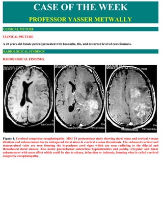
- 1. CASE OF THE WEEK PROFESSOR YASSER METWALLY CLINICAL PICTURE CLINICAL PICTURE A 40 years old female patient presented with headache, fits, and disturbed level of consciousness. RADIOLOGICAL FINDINGS RADIOLOGICAL FINDINGS Figure 1. Cerebral congestive encephalopathy. MRI T1 postcontrast study showing dural sinus and cortical venous dilations and enhancement due to widespread dural sinus & cerebral venous thrombosis. The enhanced cortical and transcerebral veins are seen forming the hyperdense cord signs which are seen radiating to the dilated and thrombosed dural sinuses. Also notice parenchymal subcortical hypointensities and patchy, irregular and linear enhancement with mass effect which could be due to edema, infarction or ischemia, forming what is called cerebral congestive encephalopathy.
- 2. Figure 2. Venous congestive encephalopathy due to venous hypertension secondary to venous thrombosis. MRI T2 images showing subcortical white matter hyperintensities with mass effect, mixed with linear and patchy hypointensity and signal void structures. Changes are due to edema, petechial hemorrhages and dilated veins. Figure 3. Cerebral congestive encephalopathy. MRI T1 postcontrast study showing dural sinus and cortical venous dilations and enhancement due to widespread dural sinus & cerebral venous thrombosis. The enhanced cortical and transcerebral veins are seen forming the hyperdense cord signs which are seen radiating to the dilated and thrombosed dural sinuses. Also notice parenchymal subcortical hypointensities and patchy, irregular and linear enhancement with mass effect which could be due to edema, infarction or ischemia, forming what is called cerebral congestive encephalopathy.
- 3. Figure 4. MRI T1 postcontrast study showing dural sinus and cortical venous dilations and enhancement due to widespread dural sinus & cerebral venous thrombosis. The enhanced cortical and transcerebral veins are seen forming the hyperdense cord signs which are seen radiating to the dilated and thrombosed dural sinuses. Also notice parenchymal subcortical hypointensities and patchy, irregular and linear enhancement which could be due to edema, infarction or ischemia, forming what is called cerebral congestive encephalopathy. Figure 5. MRI T1 postcontrast study showing enhancement and dilation of the thrombosed superior sagittal sinus with central hypointense filling defects which could be due to the intraluminal thrombi. Dilated enhanced cortical veins are seen pouring in the thrombosed sinus, subcortical parenchymal hypointensity could be due to edema or infarction
- 4. Pathophysiology of cerebral sinus thrombosis and its parenchymal changes The pathophysiology of brain parenchymal involvement in venous occlusion differs from that in arterial occlusion. Parenchymal changes may be secondary to cytotoxic edema, vasogenic edema, or intracranial hemorrhage. The primary underlying mechanism is likely to be increased venous pressure. If collateral pathways of venous drainage are insufficient, especially in the presence of cortical venous involvement, subsequent parenchymal changes may occur. If venous pressure continues to increase, with a consequent diminishment in arterial perfusion pressure, cell death may ensue. If adequate collateral pathways develop or recanalization occurs before cell death or intracranial hemorrhage, the parenchymal changes may resolve partly or completely. Vasogenic and cytotoxic edema patterns may coexist Table 1. Effect of increased intracranial venous pressure due to sinovenous thrombosis. [31] Comment Venous thrombosis produce effects on the vein that include increase in diameter and length of the thin walled vein, kinking, folding, stenosis, and sometimes occlusion. This has been described as a hemodynamic effect caused by pressure and flow of arterial blood in a vein. In the presence of venous thrombosis, a subpial or cortical vein dilates, lengthens, and may kink at the junction with the sinus. It may also balloon to the point of rupture or thromboses The deep venous collector in the galenic malformations (the embryonic precursor to the vein of Galen) typically shows a dilatation and focal stenosis at the outlet to the straight sinus or a falcine sinus. Occasionally, this structure spontaneously thromboses. Under normal conditions, there is negative venous pressure in the dural sinuses relative to the heart. There are no valves and pulsatile flow in the sinuses. Blood is effectively sucked through the shunt rather than pushed. An increased resistance to flow occurs when the venous pressure rises. This may occur transiently during a Valsalva maneuver or chronically in severe right heart failure or when there is a stenosis in the venous sinuses due to sinus thrombosis. When there is increased venous pressure, there is a corresponding decrease in water resorption by the arachnoid granulations, which is reflected in an increased amount of water in the ventricles and subarachnoid spaces. The third and lateral ventricles become prominent, and the cerebral sulci enlarge. If the fontanelles are open, the head enlarges (macrocrania). Normally, the posterior fossa drainage relies on the petrosal sinuses draining anteriorly to the cavernous sinus and caudally to the jugular bulb. In the absence of anterior drainage and restricted caudal drainage, there is an increase in cerebellar water, which results in a small fourth ventricle and tonsillar herniation. With persistent increase of the venous pressure due to venous thrombosis, there will be reduction of the venous return, stasis of blood, or even reversal of flow of blood (cerebral venous reflux) depending on the degree of venous pressure rise. Stasis of blood in the venous sinuses predisposes to further thrombosis. Cerebral venous reflux predisposes to dilatation of superficial, deep venous system, transcerebral vein, white matter congestive encephalopathy with edema, petechial haemorrhages and mass effect. Increase of intravenous pressure often results in venous wall remodelling with increase in diameter and length of the thin walled vein, kinking, folding, stenosis, and sometimes occlusion of thrombosis. Venous infarction or ischemic cerebral changes can occur due to increased venous pressure -secondary to venous thrombosis, the mechanism for venous infarction is obstruction of venous drainage with increasing venous pressure in the affected region of the brain. The venous congestion results in significant extravasation of fluid into the brain, producing focal cerebral edema and hemorrhage. The edema may be transient, if venous flow is re-established, or be associated with permanent tissue infarction if the increased venous blood pressure eventually exceeds the arterial blood pressure. In the latter situation, there is insufficient delivery of arterial blood and regional ischemic infarction. MR imaging studies utilizing diffusion-weighted imaging (DWI) have demonstrated cytotoxic edema early in acute venous thrombosis, preceding the onset of vasogenic edema. These findings support the presence of primary neuronal injury early in venous infarction. Under normal conditions, there is negative venous pressure in the dural sinuses relative to the heart. There are no valves and pulsatile flow in the sinuses. Blood is effectively sucked through the shunt rather than pushed. An
- 5. increased resistance to flow occurs when the venous pressure rises. This may occur transiently during a Valsalva maneuver or chronically in severe right heart failure or when there is a stenosis in the venous sinuses due to sinus thrombosis. Venous hypertension probably passes into three stages depending upon the degree of venous hypertension and the chronicity of the condition. Table 2. Stages of cerebral venous hypertension [31] When there is increased venous pressure, there is a corresponding decrease in water resorption by the arachnoid granulations, which is reflected in an increased amount of water Stage I in the ventricles and subarachnoid spaces. The third and lateral ventricles become prominent, and the cerebral sulci enlarge. If the fontanelles are open, the head enlarges Absence of any (macrocrania). Normally, the posterior fossa drainage relies on the petrosal sinuses draining parenchymal anteriorly to the cavernous sinus and caudally to the jugular bulb. In the absence of anterior changes drainage and restricted caudal drainage, there is an increase in cerebellar water, which results in a small fourth ventricle and tonsillar herniation. At this stage the thrombosed sinus will show the characteristic MRI signal changes but without any parenchymal changes. With persistent increase of the venous pressure due to venous thrombosis, there will be Stage II reduction of the venous return, stasis of blood, or even reversal of flow of blood (cerebral venous reflux) depending on the degree of venous pressure rise. Stasis of blood in the venous Early cerebral sinuses predisposes to further thrombosis. Cerebral venous reflux predisposes to dilatation congestive of superficial, deep venous system, transcerebral vein, white matter congestive encephalopathy with encephalopathy with edema, petechial haemorrhages and mass effect. Increase of reversible intravenous pressure often results in venous wall remodelling with increase in diameter and parenchymal length of the thin walled vein, kinking, folding, stenosis, and sometimes occlusion of changes thrombosis. Parenchymal changes in this stage are due to reversible edema edema and petechial hemorrhage once venous flow is restored. Venous infarction or ischemic cerebral changes can occur due to increased venous pressure - secondary to venous thrombosis, the mechanism for venous infarction is obstruction of Stage III venous drainage with increasing venous pressure in the affected region of the brain. The venous congestion results in significant extravasation of fluid into the brain, producing focal Late cerebral cerebral edema and hemorrhage. The edema may be transient, if venous flow is re- congestive established, or be associated with permanent tissue infarction if the increased venous blood encephalopathy with pressure eventually exceeds the arterial blood pressure. In the latter situation, there is irreversible insufficient delivery of arterial blood and regional ischemic infarction. MR imaging studies parenchymal utilizing diffusion-weighted imaging (DWI) have demonstrated cytotoxic edema early in changes acute venous thrombosis, preceding the onset of vasogenic edema. These findings support the presence of primary neuronal injury early in venous infarction. Table 3. Biochemical stages of sinus thromboses [31] STAGE MRI PICTURE In the acute stage of thrombus formation (0–5 days), the signal is predominantly isointense on T1-weighted images and hypointense on T2-weighted images because of The acute deoxyhemoglobin in red blood cells trapped in the thrombus. A venous thrombus in the deoxyhemoglobin acute stage may have a signal intensity that mimics a normal flow state, and such a finding stage of blood may lead to diagnostic error. The signal may be very hypointense on T2-weighted images products and may be mistakenly thought to indicate a flow void. According to some estimates, in 10%–30% of cases of sinus thrombosis, the thrombus at initial presentation or imaging (days I through 5) examination is in the acute stage of formation. Contrast-enhanced MR venography or CT venography is usually necessary to achieve a definitive diagnosis at this stage. In the subacute stage of thrombus development (6–15 days), the signal is predominantly The subacute hyperintense on both T1-weighted images and T2-weighted images because of extracellular methemoglobin in the thrombus. Subacute-stage thrombus has been found in 55% of
- 6. methemoglobin stage patients at clinical presentation with cerebral venous thrombosis. This stage of formation is of blood products the easiest stage at which to detect a thrombus on MR images, as the signal intensity of the (from day 5 through sinus is most different from that in normal flow states. The finding of increased signal day 15) intensity on both T1-weighted images and T2-weighted images is almost always abnormal. The thrombus becomes hypointense and heterogeneous because of partial resolution and recanalization and might enhance after gadolinium administration. Enhancement within the occluded dural sinus is due to organization of the thrombus. Chronic thrombosis with incomplete recanalization of the sinus may present a diagnostic Chronic dural sinus challenge at MR imaging. As many as 15% of patients in whom sinus thrombosis is thrombosis diagnosed at MR imaging may have a chronic (>15-day-old) thrombus. Compared with the MR signal in normal brain parenchyma, the signal in a chronic thrombus is typically isointense or hyperintense on T2-weighted images and isointense on T1-weighted images; however, significant variability in thrombus signal intensity exists. The signal intensity may be similar to that of very slowly moving oxygenated blood. Sinus enhancement in sinus thrombosis is presumably secondary to an organized thrombus with intrinsic vascularization as well as to slow flow in dural and intrathrombus collateral channels. Parenchymal changes secondary to congestive encephalopathy are shown by MRI as subcortical white matter precontrast T1 hypointensity, with patchy, irregular and linear enhancement and T2 hyperintensity mixed with linear and patchy hypointensity and signal void structures. Changes are due to edema, petechial hemorrhages and dilated veins. Parenchymal changes commonly show positive mass effect and are usually focal rather than diffuse. Bilateral parenchymal changes are not uncommon. Although parenchymal changes may occur in areas of the brain that are directly drained by the occluded venous sinus, in some patients the parenchymal changes may not closely correlate with the location of venous occlusion. [31] Parenchymal swelling without abnormalities in attenuation or signal intensity on images may occur in as many as 42% of patients with cerebral venous thrombosis. Sulcal effacement, diminished cistern visibility, and a reduction in ventricular size may occur. Patients with brain swelling and without parenchymal signal intensity changes tend to have intrasinus pressures in the intermediate range (20–25 mm Hg); however, intrasinus pressures also may be markedly elevated. Such patients typically have more prominent clinical symptoms than would be expected on the basis of imaging findings. In view of the variable nature of the parenchymal abnormalities that may occur in cerebral venous thrombosis, the use of the term venous infarct in reference to these lesions should be discouraged because that term implies irreversibility. In contrast with arterial ischemic states, many parenchymal abnormalities secondary to venous occlusion are reversible. It is much better to refer to these parenchymal changes secondary to cerebral sinus thrombosis as cerebral venous encephalopathy. Persistence of parenchymal MRI signal changes over a long time might warrant the usage of the terminology venous infarction. [31] Parenchymal hemorrhage in cerebral sinus thrombosis Parenchymal hemorrhage can be seen in one-third of cases of cerebral venous thrombosis. Flame-shaped irregular zones of lobar hemorrhage in the parasagittal frontal and parietal lobes are typical findings in patients with superior sagittal sinus thrombosis and should prompt additional imaging evaluations (eg, with MR venography or CT venography). Hemorrhage in the temporal or occipital lobes is more typical of transverse sinus occlusion. Hemorrhage in cerebral venous thrombosis is typically cortical with subcortical extension. Smaller zones of isolated subcortical hemorrhage also may be seen and may be accompanied by minimal edema. MR imaging with GRE sequences is sensitive in the depiction of these zones of parenchymal hemorrhage. [31] The mechanism of hemorrhage in cerebral venous thrombosis is multifactorial. Hemorrhage may be precipitated by continued arterial perfusion in areas of cell death, as can be seen at reperfusion in arterial ischemia. Elevation of venous pressure beyond the limit of the venous wall also is likely operative. Hemorrhage was noted in patients with
- 7. intrasinus pressures higher than 42 mm Hg but not in those with lower pressures. Contrast Enhancement Parenchymal enhancement in 1%–29% of cases of cerebral venous thrombosis has been reported. The enhancement is typically gyral in location and may extend into the white matter. Parenchymal enhancement, which indicates disruption of the blood-brain barrier, may be seen in areas of cytotoxic or vasogenic edema and in the presence of either irreversible or reversible brain abnormalities. Increased tentorial enhancement (likely related to dural venous collaterals), adjacent leptomeningeal enhancement, and prominent cortical venous enhancement (secondary to venous congestion) also may be visible after the administration of contrast material. [31] Focal neurological signs and symptoms and disturbed level of consciousness are much more likely to be present when parenchymal involvement is demonstrated by neuroimaging studies. DIAGNOSIS: DIAGNOSIS: CEREBRAL MULTI-SINUS THROMBOSIS WITH SECONDARY CONGESTIVE ENCEPHALOPATHY DISCUSSION DISCUSSION In the last 30 years, the introduction and widespread use of cerebral angiography, CT of the brain, and MRI have allowed early diagnosis of CVT, completely modifying our knowledge of this condition. More common than previously thought, CVT is remarkable by its large spectrum of clinical presentation, its highly variable mode of onset, its numerous causes, and its unpredictable but usually favorable outcome. CVT does remain a diagnostic and therapeutic challenge for the clinician, however, because of its often misleading presentation and sometimes difficult treatment. Dural sinus thrombosis accounts for approximately 1% to 2% of acute strokes in young adults. Dural sinus thrombosis is associated with local and systemic diseases. Local diseases include infectious processes, such as mastoiditis, sinusitis, osteomyelitis, and meningitis; trauma involving a dural sinus; neoplasms such as meningioma and calvarial and meningeal metastases; and subarachnoid hemorrhage. Systemic processes include pregnancy, puerperium, and oral contraceptives; collagen vascular diseases such as systemic lupus erythematosus; and hematologic disorders, such as polycythemia, leukemia/lymphoma, sickle cell anemia, and other coagulopathies. Systemic diseases that cause a hypercoagulable state are among the most common causes of dural sinus thrombosis. At least one third of cases are associated with pregnancy. Depending on the degree and rate of the involvement of the cerebral veins, degree of recanalization, and collateral venous formation, the presentation can vary from a slow process to an acute episode. Signs and symptoms are nonspecific. Headache is the most common presenting symptom and is seen in about 75% of patients. Other symptoms include nausea and vomiting, papilledema, and decreased level of consciousness. Involvement of the cerebral veins may cause hemorrhagic infarction, hemiplegia, and seizures. Rarely, patients may present with symptoms simulating transient ischemic attacks or subarachnoid hemorrhage. Dural sinuses are formed by dural duplications and are fixed to the osseous skull. Because of absence of valves, blood can flow in different directions. The superior sagittal sinus joins the straight and lateral sinuses posteriorly forming the confluence of the sinuses. Lateral sinuses drain blood from the cerebellum, brain stem, and posterior parts of the hemispheres. The basal vein of Rosenthal drains both cortical and deep territories. The cortical territory includes the posterior part of the frontal lobe, parahippocampal gyrus, anterior part of the cingulate gyrus, and part of the temporo-occipital cortex. The deep territory includes the thalamus, basal nuclei, and deep brain structures. The basal vein of Rosenthal and internal cerebral veins join and form the vein of Galen, which drains
- 8. into the straight sinus. RELEVANT VENOUS ANATOMY Blood from the brain is drained by cerebral veins which empty into dural sinuses, themselves drained mostly by internal jugular veins. Dural Sinuses Figure 6. The venous anatomy The most commonly affected by thrombosis are the superior sagittal sinus, lateral sinuses, cavernous sinuses, and straight sinus. Superior Sagittal Sinus (SSS). The SSS, triangular in cross-section, lies in the attached border of the falx cerebri. It starts at the foramen cecum and runs backward toward the internal occipital protuberance, where it joins with the straight sinus (SS) and lateral sinuses (LS) to form the torcular Herophili. Its anterior part is narrow or sometimes absent, replaced by two superior cerebral veins that join behind the coronal suture. This is why the anterior part of the sinus is often poorly visualized at angiography and its isolated lack of filling is not sufficient to indicate thrombosis . The SSS receives superficial cerebral veins and drains the major part of the cortex. It also receives diploic veins, themselves connected to scalp veins by emissary veins, which explains some cases of SSS thrombosis after cutaneous infections or contusions. SSS and other sinuses play a major role in CSF circulation because they contain most of the arachnoid villi and granulations (Pacchionian bodies) in which CSF absorption takes place. The clear-cut consequence is a direct dependency of CSF pressure upon the intracranial venous pressure, accounting for the frequently raised intracranial pressure in SSS thrombosis. Lateral Sinuses (LS) These extend from the torcular Herophili to jugular bulbs and consist of two portions: the transverse portion, which
- 9. lies in the attached border of the tentorium, and the sigmoid portion, which runs on the inner aspect of the mastoid process and is thus susceptible to infectious thrombosis in patients with mastoiditis or otitis media. LS drains blood from the cerebellum, brain stem, and posterior part of the cerebral hemispheres. They also receive some of the diploic veins and some small veins from the middle ear, another possible source of septic thrombosis. There are numerous LS anatomic variations that may be misinterpreted as sinus occlusion at angiography. In particular, the right LS is frequently larger than the left, which receives most of its supply from the straight sinus. An isolated lack of filling of the transverse portion of left LS is thus more suggestive of hypoplasia than thrombosis. Cavernous Sinuses Cavernous sinuses consist of trabeculated cavities formed by the separation of the layers of the dura and located on each side of sella turcica, superolaterally to the sphenoid air sinuses. The oculomotor and trochlear cranial nerves, along with the ophthalmic and maxillary branches of the trigeminal nerve, course along the lateral wall of the cavernous sinuses, whereas the abducent nerve and the carotid artery with its surrounding sympathetic plexus are located within the center of the sinus itself. Cavernous sinuses drain the blood from the orbits through the ophthalmic veins and from the anterior part of the base of the brain by the sphenoparietal sinus and the middle cerebral veins. They empty into both the superior and inferior petrosal sinuses and ultimately into the internal jugular veins. Because of their situation, cavernous sinuses are often thrombosed in relation to infections of the face or sphenoid sinusitis and, by contrast to other varieties of sinus thrombosis, infection is still the leading cause. Rarely injected on carotid angiograms, cavernous sinuses are now well visualized on CT scans and MRI. Straight Sinus Formed by the union of the inferior sagittal sinus and the great vein of Galen, it has a triangular lumen and runs caudally in the junction between the falx cerebri and the tentorium cerebella to join the torcular at the internal occipital protuberance. Figure 7. Sagittal contrast-enhanced MR venogram MIP image of the deep cerebral veins and dural sinuses in a normal patient. SSS = superior sagittal sinus; To = torcular herophili or confluence of sinuses; S = straight sinus; G = great vein of Galen; I = inferior sagittal sinus; TH = thalamostriate veins; ICV = internal cerebral veins; R = basal vein of Rosenthal; L = vein of Labbe; TS transverse sinus; SG = sigmoid sinus; SP superior petrosal sinus; IP = inferior petrosal sinus; CS = cavernous sinus; sps = spheno-parietal sinus; PP = pterygoid plexus of veins; J = internal jugular vein; sov superior ophthalmic vein; C = internal carotid artery; and B = basilar artery,
- 10. Figure 8. Coronal contrast enhanced MR venogram MIP image shows many of the dural sinuses and a few of the deep cerebral veins. SSS = superior sagittal sinus; To = torcular herophili or confluence of sinuses; R = basal vein of Rosenthal; L = vein of Labbe; DMV = deep middle cerebral vein; TS = transverse sinus; SG = sigmoid sinus; SPS = superior petrosal sinus; J = internal jugular vein; C internal carotid artery; V = vertebral artery; JB jugular bulb; IJ = internal jugular vein; CV cortical veins. Cerebral Veins Three groups of veins drain the blood supply from the brain: Superficial Cerebral Veins (or cortical veins) Some of these - the frontal, parietal, and occipital superior cerebral veins - drain the cortex upward into the SSS, whereas others, mainly the middle cerebral veins, drain downward into the cavernous sinuses. These veins are linked by the great anastomotic vein of Trolard, which connects the SSS to the middle cerebral veins, which are themselves connected to the LS by the vein of Labbe. These cortical veins have thin walls, no muscle fibers, and no valves, thereby permitting both dilation and reversal of the direction of blood flow when the sinus in which they drain is occluded. They are linked by numerous anastomoses, allowing the development of a collateral circulation (angiographically visible as quot;cork- screwquot; vessels) and probably explaining the good prognosis of some CVT. Since the number and location of cortical veins are inconstant, the angiographic diagnosis of isolated cortical vein thrombosis is extremely difficult and sometimes impossible. Deep Cerebral Veins Blood from the deep white matter of the cerebral hemispheres and from the basal ganglia is drained by internal cerebral and basal veins, which join to form the great vein of Galen that drains into the straight sinus. By contrast to the superficial veins, the deep system is constant and always visualized at angiography, so its thrombosis is easily recognized. Posterior Fossa Veins The veins of the posterior fossa may be divided into three groups ,superior draining into the galenic system, anterior draining into petrosal sinus, and posterior draining into the torcular and neighboring straight and lateral sinuses. They are variable in course, and angiographic diagnosis of their occlusion is extremely difficult. PATHOLOGY
- 11. Pathologic findings have been extensively described in the past. They vary with the site of thrombosis and the interval between the onset of symptoms and death. The thrombus itself is like other venous thrombi elsewhere in the body. When it is fresh, it is a red thrombus rich in red blood cells and fibrin and poor in platelets; when it is old, it is replaced by fibrous tissue sometimes showing recanalization. Its formation is due to the usual pathogenetic factors: venous stasis, increased clotting tendency, changes in the vessel wall, and, less frequently, embolization. Its location and extension are variable. In autopsy series, extensive thrombosis of SSS and tributary veins is the most frequent finding, but this pattern of involvement no longer reflects the real distribution of CVT. Figure 9. Bilateral hemorrhagic venous infarction (A) due to superior sagittal thromboses (B) The consequences of CVT on the brain are again highly variable. The classic picture is that of SSS thrombosis with extensive bilateral hemorrhagic infarcts affecting the cortex and adjacent white matter. CT scan and MRI studies have now convincingly shown, however, that sinus thrombosis can induce varying degrees of edema without infarction and can even have no detectable effect on the brain. Figure 10. Superior sagittal sinus thromboses (A), with dilated thrombosed cortical veins radiating to the thrombosed sinus and forming what is termed radiologically the quot;cord signquot;.
- 12. INCIDENCE The true incidence of CVT is totally unknown in the absence of specific epidemiologic studies. In most autopsy series, the incidence was found to be extremely low. It has been suggested that the incidence of CVT is higher in females and in the aged, reflecting the overall greater incidence of thromboembolic diseases in these categories. The age distribution is uniform in men, whereas in women it frequently occurred between 20 and 35. This probably reflects the frequency of specific causes such as pregnancy and oral contraceptive use in young women. ETIOLOGY Numerous conditions can cause or predispose to CVT. They include all surgical, gyneco-obstetric, and medical causes of deep vein thrombosis as well as a number of local or regional causes, either infective or noninfective, such as head trauma, brain tumors, and arterial infarcts. Although infection still constituted the major identifiable cause ,the incidence of septic CVT has greatly diminished in developed countries since the introduction of antibiotics. Cavernous sinus thrombosis remains the most common form of septic thrombosis, usually following an infection of the middle third of the face due to Staphylococcus aureus. Other sites of infection include sphenoid or ethmoid sinusitis, dental abscess, and, less often, otitis media. In chronic forms, gram-negative rods and fungi such as Aspergillus species are more commonly isolated. Among general causes, parasitic infections such as trichinosis and more recently HIV and CMV infections have been added to the long list of infective conditions possibly leading to CVT. In young women, CVT occurs more frequently during puerperium than pregnancy and remains very common in developing countries, whereas in developed countries the role of oral contraceptives is more important. Among the numerous noninfective medical causes of CVT, malignancies,and inflammatory diseases such as Behcet's disease and connective tissue diseases are the most frequent. Although rare, hereditary antithrombin III, protein C,and protein deficiencies should be systematically looked for in the absence of obvious cause because they imply a family study and a long-term treatment. In neonates and children, the etiology of CVT is characterized by the frequency of regional infections (otitis, mastoiditis), neonatal asphyxia, severe dehydration, and congenital heart disease. Despite the continuous description of new causes, the proportion of cases of unknown etiology constitute about one third of cerebral venous thrombosis. TOPOGRAPHIC DIAGNOSIS Thrombosis most frequently affects (in order of decreasing frequency) SSS, LS, and cavernous sinus. In most cases, thrombosis affects several sinuses or sinuses and cerebral veins. Thrombosis of the galenic system is rare. The frequent association of sinus and cerebral vein thrombosis explains the lack of well-defined topographic clinical syndromes, similar to those described in arterial occlusions. Thus, SSS thrombosis can present with any of the above described patterns; this also applies to LS thrombosis, in which isolated intracranial hypertension is probably even more frequent and, among focal signs, dysphasia is not unusual. Thrombosis of the petrosal sinuses was described in the old literature and was characterized mainly by a fifth nerve palsy for the superior sinus and by a sixth nerve palsy for the inferior one. As already stressed, angiographic diagnosis of isolated cortical vein thrombosis is extremely difficult, but there are old reports of anatomic or surgical cases in patients presenting with an acute or rapid onset of focal deficits, seizures, or both. The classic picture of deep cerebral venous thrombosis is that of an acute coma with decerebration or extrapyramidal hypertonia leading to death in a few days or resolving, but with heavy sequelae such as akinetic mutism, dementia, bilateral athetoid movements, vertical gaze palsy, and dystonia. Recent reports have illustrated benign forms presenting mainly with confusion. The few reported cases of cerebellar vein thrombosis are mainly anatomic but we reported a patient presenting with a 3-month history of cranial nerve palsies, cerebellar incoordination, and papilledema simulating a posterior fossa tumor.
- 13. MR IMAGING OF SINUS THROMBOSIS Empty delta sign 21% On contrast-enhanced computed tomography (CT) and MR imaging dural sinus thrombosis Contrast enhancement of falx or tentorium 19% typically appears as a filling defect in the Small ventricles 52% dural sinus, also known as empty delta sign. Enlarged ventricles 3% The empty delta sign is due to enhancement of the surrounding falx with the hypodense Spontaneous hyperintensity 20% central clot. A similar findings can be Hypointensity 33% observed in MR imaging. Gyral enhancement 25% The empty delta sign has high specificity but low sensitivity. It is seen in only 30% of cases of sagittal sinus thrombosis. Hyperdense cortical veins (cord sign) may also be present. CT and MR imaging may also detect causes such as infection, trauma, or neoplasm. Imaging studies can also be helpful in the detection of complications such as diffuse edema or venous infarctions, which are often hemorrhagic. On MR imaging, dural sinus thrombosis is most commonly manifested as lack of the normal flow void within the dural sinuses. Affected dural sinuses demonstrate abnormal intraluminal signal, which varies depending on the stage of the thrombus. In the acute stage (days I through 5), the thrombus is isointense to the brain on Tl-weighted images and strongly hypointense on T2-weighted images because of the deoxyhemoglobin stage of blood products. Because of the low signal of acute thrombus on T2-weighted images, acquisition of only T2-weighted images may give a false impression of normal flow void. Figure 11. MRI T1 postcontrast study showing widespread enhancement of the dural sinuses and cortical veins.
- 14. Intra-sinuses hypointense filling defects are due to nonenhancement of the thrombus. Signal changes in the upper brain stem is probably due to ischemia Figure 12. Coronal contrast-enhanced Tl -weighted images show isointense thrombus (arrow) within the superior sagittal sinus with increased enhancement of the superior sagittal sinus leaves indicating increased vascularization without evidence of recanalization. There is also enhancement of the left transverse sinus reflecting partial thrombosis. There is peripheral enhancement of the right parietal infarct (open arrows). The dura, falx cerebri, and tentorium cerebella show irregular enhancement. In the subacute stage (from day 5 through day 15), the thrombus is hyperintense on both Tl-weighted and T2- weighted images because of the extracellular methemoglobin stage of blood products. Signal changes evolve from the periphery to the central portion of the thrombus. By the third week, signal changes of the thrombus are different from an intracranial bleed. The thrombus becomes hypointense and heterogeneous because of partial resolution and recanalization. Figure 13. A, Sagittal T1 -weighted images show increased signal intensity in the superior sagittal sinus (arrowheads), anterior portion of the straight sinus (small arrow), and vein of Galen (big arrow), consistent with subacute thrombosis, B MRI T1 precontrast and , C, MRI T2 image showing right parasagittal subcortical hemorrhagic infarct in the parietal lobe. The superior sagittal sinus shows isointense signal intensity consistent with thrombus in methemoglobin stage a case of dural sinus thrombosis with subcortical hemorrhagic infarct of the right parietal region Infarctions resulting from thrombosis of the internal cerebral vein or Dural sinus thrombosis may be associated straight sinus are usually deep within the brain, such as the thalami. with venous infarctions, which are frequently hemorrhagic. Venous infarctions characteristically have a subcortical location and do not follow a major arterial vascular territory. Infarctions resulting from thrombosis of the internal cerebral vein or straight sinus are usually deep within the brain, such as the thalami. Dilated collateral cortical and medullary veins may be visible as prominent signal voids. On contrast- enhanced MR imaging, the empty delta sign representing the intraluminal clot may be seen. With organization and recanalization of the thrombus, enhancement of the thrombus may be seen. The tentorium and falx may also show enhancement resulting from vascular congestion in the collateral venous channels. With obstruction of the venous system, cerebral edema and infarction may develop. It can be manifested as increased signal intensity on T2- weighted images. It may be associated with hemorrhage, which is most commonly seen in the parietal and parieto- occipital areas. The underlying venous stasis can lead to abnormal enhancement of the cortical or deep venous structures.
- 15. Figure 14. MRI T2 (A,B,C) and FLAIR studies (D,E,F,G,H) showing bilateral deep cerebral, paraventricular, basal ganglionic and thalamic signal changes representing subacute venous infarctions due to thrombosis of the deep venous systems Flow in the dural sinuses may be depicted with MR venography using different techniques, such as time-of-flight, phase-contrast, or gradient-echo imaging sensitive to flow. Intraluminal hyperintensity seen with subacute thrombus cannot be distinguished from flow hyperintensity on time-of-flight images; therefore this technique should be used cautiously when there is intraluminal increased signal intensity on Tl-weighted images. To avoid saturation of the venous structures, contrast-enhanced three-dimensional time-of-flight MR angiography may improve the visibility of the venous structures. Table 4. Biochemical stages of sinus thromboses STAGE MRI PICTURE The thrombus is isointense to the brain on Tl-weighted images and The acute deoxyhemoglobin stage of blood strongly hypointense on T2-weighted images because of the products deoxyhemoglobin stage of blood products. Because of the low signal of acute thrombus on T2-weighted images, acquisition of (days I through 5) only T2-weighted images may give a false impression of normal flow void. The thrombus is hyperintense on both Tl-weighted and T2- weighted images because of the extracellular methemoglobin stage The subacute extracellular methemoglobin of blood products. Signal changes evolve from the periphery to the
- 16. stage of blood products (from day 5 through central portion of the thrombus. By the third week, signal changes day 15) of the thrombus are different from an intracranial bleed. The thrombus becomes hypointense and heterogeneous because of partial resolution and recanalization. The thrombus becomes hypointense and heterogeneous because of partial resolution and recanalization and might enhance after Chronic dural sinus thrombosis gadolinium administration. Enhancement within the occluded dural sinus is due to organization of the thrombus. Figure 15. MRI FLAIR study (A) showing bilateral deep cerebral, paraventricular signal changes representing subacute venous infarctions due to thrombosis of the deep venous systems. B, MRI T1 postcontrast showing the empty delta sign. There are a number of pitfalls in the diagnosis of dural sinus thrombosis that should be considered. Flow-related enhancement occurs when unsaturated protons enter the imaging plane and produce increased signal intensity relative to the more saturated protons in the adjacent soft tissues. It is identified on Tl- weighted images within dural venous structures oriented perpendicular to the scanning plane. It is more commonly seen in the sigmoid sinus and jugular bulb. The same findings may be seen in the cortical veins near the superior sagittal sinus on sagittal images. Changing of slice orientation with constant sequence parameters resolves the flow artifact. With normal flow, the signal intensity within the dural sinus changes . Extremely slow flow can also produce an intraluminal signal. Increasing TR and TE diminishes this artifact. The anterior portion of the superior sagittal sinus may be hypoplastic or completely absent. The transverse sinuses are typically asymmetric, with the right usually larger than the left. One of the transverse sinuses may be completely absent. Hypoplasia or absence of a dural venous structure may result in a false positive result. In patients with chronic dural sinus thrombosis, the thrombus enhances after gadolinium administration. Enhancement within the occluded dural sinus is due to organization of the thrombus. The thrombus is vascularized as a result of invasion by fibroblasts and capillaries. This vascularization could lead to false negative results in patients with chronic dural sinus thrombosis using contrast-enhanced MR and time-of-flight MR Angiography techniques. Phase-contrast (with or without contrast) and time-of-flight (without contrast) MR angiography are preferred methods for evaluation of patients with dural sinus thrombosis.
- 17. Figure 16. MRI T2 images (A,B,C) and FLAIR (D) showing diffuse left hemispherical cortical/subcortical hyperintensities and mass effect due to widespread dural sinuses & cortical veins thromboses. Signal changes are due to edema, ischemia and infraction. Most of the parenchymal signal changes are due to edema in the acute stage of sinus thromboses. SUMMARY Radiological sign Comment Empty delta sign Thrombosis typically appears as a filling defect in the dural sinus, also known as empty delta sign. The empty delta sign is due to enhancement of the surrounding falx with the hypodense central clot left unenhanced. Hyperintense (precontrast Dilated collateral cortical and medullary veins may be visible as prominent signal MRI T1 ) cortical veins voids when not thrombosed. However when these veins are thrombosed they follow (cord sign) the same time-sensitive signal changes of the thrombosed dural sinuses. In the subacute stage of extracellular methemoglobin these veins are dilated and hyperintense on noncontrast MRI T1 studies. enhancement of the cortical veins may also form the cord sign. Edema With obstruction of the venous system, cerebral edema may develop. It can be manifested as increased signal intensity on T2-weighted images and can result in herniations. Venous infarctions Hemorrhagic venous infarctions characteristically have a subcortical location and do not follow a major arterial vascular territory. Infarctions resulting from thrombosis of the internal cerebral vein or straight sinus are usually deep within the brain, such as the thalami, the basal ganglia or the paraventricular regions. Affected dural sinuses 1. In the acute stage (days I through 5), the thrombus is isointense to the brain demonstrate abnormal on Tl-weighted images and strongly hypointense on T2-weighted images intraluminal signal, which because of the deoxyhemoglobin stage of blood products. varies depending on the stage of the thrombus. 2. In the subacute stage (from day 5 through day 15), the thrombus is hyperintense. 3. The thrombus becomes hypointense and heterogeneous because of partial resolution and recanalization and might enhance after gadolinium administration. With organization and recanalization of the thrombus, enhancement of the thrombus may be seen. Dural enhancement The tentorium and falx may may show enhancement resulting from vascular
- 18. congestion in the collateral venous channels. Venous stasis The underlying venous stasis can lead to abnormal enhancement of the cortical or deep venous structures. It can also result in edema, ischemia of cerebral infarctions. Thrombus enhancement Enhancement within the occluded dural sinus is due to organization of the thrombus. The thrombus is vascularized as a result of invasion by fibroblasts and capillaries. Parenchymal enhancement Could be due to cerebral ischemic changes, or frank cerebral venous infarction (it occurs due to vascular endothelial damage) Parenchymal T2 Could be due to cerebral edema, ischemia of cerebral venous infarctions. hyperintensities, precontrast Parenchymal signal changes in the acute stage of sinus thromboses (especially when T1 hypointensities associated with mass effect) are mainly due to cerebral edema and might completely disappear later on. MEDICATION Heparin should be considered seriously in the management of CVT. Conversion to warfarin as maintenance therapy is then suggested. Subcutaneous low-molecular-weight heparin (Lovenox) also has been used in patients with venous sinus thrombosis. Thrombolytic therapy may be useful, but all studies so far describe its use only with local instillation by microcatheter or direct instillation at the time of surgical thrombectomy. Drug Category: Anticoagulants - These medications are used to prevent propagation of the clot to more extensive areas of the cerebral venous system. Studies indicate a tendency toward better outcome in patients treated with anticoagulant therapy than in those who are not treated with anticoagulants. In Einhaupl's study, even patients with cerebral hemorrhage appeared to benefit from anticoagulation. Heparin (Hep-Lock)- Increases the action of antithrombin III, leading to inactivation of coagulation enzymes thrombin, factor Xa, and factor IXa. Thrombin is the most sensitive to inactivation by heparin. Because heparin is not absorbed from the GI tract, it must be given parenterally. When given IV, effect is immediate. Metabolism of heparin is complex; rapid Drug Name zero-order metabolism is followed by slower first-order renal clearance. Zero-order process is saturable, leading to an increase in half-life from 30-150 min as dose increased. Weight-based protocol now often used for dosing. When choosing this therapy, risks of its contraindications must be weighed against potential benefits. Loading dose: 80 U/kg IV bolus followed by infusion Initial infusion: 18 U/kg/h IV; aPTT checked in 6 h and q6h after any dosage change, as well as qam; adjust dose according to following parameters aPTT = <1.2 times control: 80 U/kg bolus with increase of 4 U/kg/h Adult Dose aPTT = 1.2-1.5 times control: 40 U/kg bolus with increase of 2 U/kg/h aPTT = 1.5-2.3 times control: No change in infusion rate needed aPTT = 2.3-3 times control: Decrease infusion rate by 2 U/kg/h aPTT >3 times control: Hold infusion for 1 h and decrease rate by 3 U/kg/h
- 19. Loading dose: 50 U/kg IV; increase by 15-25 U/kg/h to Pediatric Dose maintain aPTT at 1.5-2.5 times baseline Documented hypersensitivity, aneurysm, active or recent bleeding, coagulopathy, endocarditis, hemophilia, hepatic Contraindications disease, hypertension, inflammatory bowel disease, lumbar puncture/spinal anesthesia, sulfite hypersensitivity, surgery, thrombocytopenia Digoxin, nicotine, tetracycline, and antihistamines may Interactions decrease effects; NSAIDs, aspirin, dextran, dipyridamole, and hydroxychloroquine may increase toxicity Pregnancy C - Safety for use during pregnancy has not been established. Monitor platelet count for development of thrombocytopenia; severe hyperkalemia may occur with concomitant use of ACE Precautions inhibitors; increased bleeding risk occurs with many drugs, including platelet inhibitors, NSAIDs, valproic acid, Ginkgo biloba, and probenecid Warfarin (Coumadin)- Interferes with action of vitamin K, a cofactor essential for converting precursor proteins into factors II, VII, IX, and X. Does not affect activity of coagulation factors synthesized prior to exposure to warfarin. Depletion of these mature factors by normal metabolism must occur before therapeutic effects of newly synthesized factors can be seen, Drug Name thus may take several days to become effective. Dose influenced by differences in absorption, metabolism, and hemostatic responses to given concentrations; dose must be monitored closely by following PT and INR. Higher initial doses do not appear to improve time required to achieve therapeutic levels but do increase bleeding risk. Initial: 5 mg PO qd; adjust dose by monitoring INR (target, Adult Dose 2.5) Initial: 0.2 mg/kg PO up to 10 mg Pediatric Dose Maintenance: 0.1 mg/kg/d; INR must be monitored to determine maintenance dose Documented hypersensitivity, alcoholism, aneurysm, bleeding, breastfeeding, endocarditis, pregnancy, hemophilia, lumbar Contraindications puncture, thrombocytopenia, hypertension, leukemia, polycythemia vera, intracranial bleeding, vitamin C deficiency, vitamin K deficiency Monitor INR whenever a medication is added or discontinued; drugs that may decrease anticoagulant effects include griseofulvin, carbamazepine, glutethimide, estrogens, nafcillin, phenytoin, rifampin, barbiturates, cholestyramine, colestipol, vitamin K, spironolactone, oral contraceptives, and sucralfate; medications that may increase anticoagulant effects include Interactions oral antibiotics, phenylbutazone, salicylates, sulfonamides, chloral hydrate, clofibrate, diazoxide, anabolic steroids, ketoconazole, ethacrynic acid, miconazole, nalidixic acid, sulfonylureas, allopurinol, chloramphenicol, cimetidine, disulfiram, metronidazole, phenylbutazone, phenytoin, propoxyphene, sulfonamides, gemfibrozil, acetaminophen, and sulindac; supplements such as ginger and Ginkgo biloba should
- 20. be avoided; green leafy vegetables have high levels of vitamin K, which may decrease INR Pregnancy X - Contraindicated in pregnancy May cause uncontrolled bleeding and should not be used in conditions in which bleeding would be difficult to control, leading to a more catastrophic outcome; medications that inhibit platelet function should be avoided, including aspirin, NSAIDs, and valproic acid; patients with protein S or C Precautions deficiency may become transiently hypercoagulable (anticoagulate patient with heparin and then convert to warfarin); do not switch brands after achieving therapeutic response; caution in active tuberculosis or diabetes; patients with protein C or S deficiency are at risk of developing skin necrosis Drug Category: Thrombolytics - These agents cause lysis of the clot. All studies concerning the use of these agents in CVT involve either direct instillation into the sinus at the time of surgery or the use of microcatheters to reach the venous sinus. Alteplase (Activase)- Biosynthetic form of human tissue plasminogen activator. Tissue plasminogen activator exerts effect on fibrinolytic system to convert plasminogen to Drug Name plasmin. Plasmin degrades fibrin, fibrinogen, and procoagulant factors V and VIII. Not given as IV infusion to treat CVT. Refer patient to facility with expertise to perform venous sinus catheterization. 1 mg/cm infused via venous sinus catheter throughout clot, Adult Dose then 1-2 mg/h Pediatric Dose Not established Documented hypersensitivity, aneurysm, arteriovenous malformation, bleeding, coagulopathy, endocarditis, diabetic Contraindications retinopathy, mitral stenosis, recent surgery, pregnancy, breastfeeding Drugs that alter platelet function (eg, aspirin, dipyridamole, abciximab) may increase risk of bleeding prior to, during, or Interactions after alteplase therapy; may give heparin with and after alteplase infusions to reduce risk of rethrombosis; either heparin or alteplase may cause bleeding complications Pregnancy C - Safety for use during pregnancy has not been established. Monitor for bleeding, especially at arterial puncture sites, with coadministration of vitamin K antagonists; control and monitor BP frequently during and following alteplase administration Precautions (when managing acute ischemic stroke); do not use >0.9 mg/kg to manage acute ischemic stroke; doses >0.9 mg/kg may cause intracranial hemorrhage Urokinase (Abbokinase)- Produced by kidney, converts plasminogen to plasmin by cleaving arginine-valine bond in Drug Name plasminogen. Degradation products of fibrin and fibrinogen exert clinically significant anticoagulant effect. Erythrocyte aggregation and plasma viscosity also are reported to decrease. Given in CVT by catheterization of venous sinus or by direct
- 21. instillation at surgery during thrombectomy. Not currently available in US. 250,000 U/h instilled directly or via venous sinus catheter; Adult Dose additional doses of 50,000 U; total dose 1,000,000 U over 2 h Not currently available in the US Pediatric Dose Not established Documented hypersensitivity, aneurysm, arteriovenous malformation, bleeding, coagulopathy, endocarditis, diabetic Contraindications retinopathy, mitral stenosis, recent surgery, pregnancy, breastfeeding Effects increased with coadministration of aminocaproic acid, anticoagulants, antineoplastic agents, antithymocyte globulin, Interactions cefamandole, cefoperazone, Ginkgo biloba, NSAIDs, platelet inhibitors, porfimer, strontium-89 chloride, sulfinpyrazone, tranexamic acid, valproic acid Pregnancy B - Usually safe but benefits must outweigh the risks. Caution in patients receiving IM administration of medications or with severe hypertension or trauma or surgery in previous Precautions 10 d; do not measure BP in lower extremities, because may dislodge DVT; monitor therapy by performing PT, aPTT, TT, or fibrinogen approximately 4 h after initiation of therapy Streptokinase (Kabikinase, Streptase)- Facilitates thrombolysis through formation of an activator complex with plasminogen. Indirectly cleaves arginine-valine bond in plasminogen, Drug Name forming plasmin. Plasmin degrades fibrin, fibrinogen, and procoagulant factors V and VIII. Degradation products of fibrin and fibrinogen have significant anticoagulant effect. Adult Dose Instilled directly or via venous sinus catheter Only anecdotal reports describe use in children, and that in arterial occlusion; doses used were as follows Pediatric Dose Loading dose: 1000-3000 IU/kg; followed by infusion of 1000- 1500 IU/kg/h; in CVT, administered by direct infusion via catheter Documented hypersensitivity, aneurysm, arteriovenous malformation, bleeding, coagulopathy, endocarditis, diabetic Contraindications retinopathy, mitral stenosis, recent surgery, pregnancy, breastfeeding Effects are increased with coadministration of aminocaproic acid, anticoagulants, antineoplastic agents, antithymocyte Interactions globulin, cefamandole, cefoperazone, Ginkgo biloba, NSAIDs, platelet inhibitors, porfimer, strontium-89 chloride, sulfinpyrazone, tranexamic acid, valproic acid Pregnancy C - Safety for use during pregnancy has not been established. Caution in severe hypertension, IM administration of medications, trauma or surgery in previous 10 d; measure hematocrit, platelet count, aPTT, TT, PT, or fibrinogen levels before therapy is implemented; either TT or aPTT should be <2 times the normal control value following infusion of
- 22. streptokinase and before (re)instituting heparin; do not take BP in lower extremities, as possible DVT may be dislodged; PT, aPTT, TT, or fibrinogen should be monitored 4 h after Precautions initiation of therapy; in addition to bleeding complications inherent in thrombolytic agents, repeated administration of streptokinase can result in tolerance as well as hypersensitivity SUMMARY In venous stroke, even large parenchymal changes can resolve completely independent from recanalization of the thrombosed veins and sinuses. A plausible hypothesis is that venous infarcts largely consist of persistent edema and that the lesion volume is influenced by development of collateral veins. However, further investigations are necessary to understand the underlying abnormal mechanisms. The use of the term venous infarct in reference to these lesions should be discouraged because that term implies irreversibility. In contrast with arterial ischemic states, many parenchymal abnormalities secondary to venous occlusion are reversible. Addendum A new version of this PDF file (with a new case) is uploaded in my web site every week (every Saturday and remains available till Friday.) To download the current version follow the link quot;http://pdf.yassermetwally.com/case.pdfquot;. You can also download the current version from my web site at quot;http://yassermetwally.comquot;. To download the software version of the publication (crow.exe) follow the link: http://neurology.yassermetwally.com/crow.zip The case is also presented as a short case in PDF format, to download the short case follow the link: http://pdf.yassermetwally.com/short.pdf Screen resolution is better set at 1024*768 pixel screen area for optimum display REFERENCES References I .Robain 0, Floquet J, Heidt N, Rozemberg F. Hemimegalencephaly: a clinicopathological study of four cases. Neuropathol Appl Neurobiol 1988; 14:125-35. 2. Katifa GL, Chiron C, Sellier N, et al. Hemimegalencephaly MR imaging in five children. Radiology 1987;165:29- 33. 3. De Rosa MJ, Secor DL, Barsom M, Fisher RS, Vinters HV. Neuropathologic findings in surgically treated hemimegalencephaly: immunohistochemical, morphometric, and ultrastructural study. Acta Neuropathol (Berl) 1992;84:250-60. 4. Robain 0, Chiron C, Dulac 0. Electron, microscopic and Golgi study in a case of hemimegalencephaly. Acta
- 23. Neuropathol (Berl) 1989;77:664-6. 5. Squier M. White matter change in hemimegalencephaly. Rev Neurol 1993;149:370. 6. Dom R, Brucher JM. Hamartoblastome (gangliocytome diffus) unilateral de encore cerebrale, Rev Neurol 1969;120:317-8. 7. Townsend JJ, Nielsen SL, Malamud N. Unilateral megalencephaly hamartoma or neoplasm. Neurology 1975;25:448-53. 8. Davis RLM, Nelson E. Unilateral ganglioglioma in a tuberosclerotic brain. J Neuropathol Exp Neurol 1961;21:571-81. 9. Jervis GA. Spongioneuroblastoma and tuberous sclerosis. J Neuropathol Exp Neurol 1954;13:105- 16. 10. Fryer AE, Connor JM, Povey S, et al. Evidence that the gene for tuberous sclerosis is on chromosome 9. Lancet 1987;1:659-61. II. Szaro BG, Tompkins R. Effects of tetraploidy on dendritic branching in neurons and glial cells of the frog Xenopus laevis. J Comp Neurol 1987;258: 304--16. 12. Bignami A, Palladini G, Zappella M. Unilateral megalencephaly with cell hypertrophy. An anatomical and quantitative histochemical study. Brain Res 1968;9:103-14. 13. Manz H, Phillips T, Rowden G, McCullough DC. Unilateral megalencephaly, cerebral cortical dysplasia, neuronal hypertrophy and heterotopia: cytophotometric fluorometric cytochemical and biochemical analysis. Acta Neuropathol (Berl) 1979; 45:97-103. 14. David TJ. Hypomelanosis of Ito: a neurocutaneous syndrome. Arch Dis Child 1981;56:798-800. 15. Turleau C, Taillard F. Hypomelanosis of Ito (incontinentia pigmenti achromians) and mosaicism for a microdeletion of 15 ql. Hum Genet 1986;74: 185-7. 16. Choi BH, Kudo M. Abnormal neuronal migration and gliomatosis cerebri in epidermal naevus syndrome. Acta Neuropathol (Berl) 1981;53:319-25. 17. Vigevano F, Aicardi J, Lini M, Pasquinelli A. La sindrome del nevo sebaceo lineare presentazione di una casuistica multicentra. Boll Lega It Epil 1984;45-46:59-63. 18. Vles JSH, Degraeuwe P, De Cock P, Casaer P. Neuroradiological findings in Jadassohn's naevus phakomatosis: a report of two cases. Eur J Pediatr 1985;144:290-4. 19. Zaremba J, Wislawski J, Bidzinski J, Kansky J, Bogna S. ladassohn's naevus phakomatosis: a report of two cases. J Ment Deric Res 1978;22:91- 102. 20. Flores-Sarnat L. Hemimegalencephaly. I. Genetic, clinical, and imaging aspects. J Child Neurol 2002; 17:373- 384. 21. Barkovich AJ, Chuang SH. Unilateral megalencephaly: correlation of MR imaging and pathologic characteristics. AJNR Am J Neuroradiol 1990; 11:523-531. 22. Sims J. On hypertrophy and atrophy of the brain. Med Quir Trans 1835; 19:315-380. 22. Wolpert SM, Cohen A, Libenson MH. Hemimegalencephaly: a longitudinal MR study. AJNR Am J Neuroradiol
- 24. 1994; 15:1479-1482. 23. Vigevano F, Bertini E, Boldrini R, et al. Hemimegalencephaly and intractable epilepsy: benefits of hemispherectomy. Epilepsia 1989; 30:833-843. 24. Mathis JM, Barr JD, Albright AL, Horton JA. Hemimegalencephaly and intractable epilepsy treated with embolic hemispherectomy. AJNR Am J Neuroradiol 1995; 16:1076-1079. 25. Yagishita A, Arai N, Tamagawa K, Oda M. Hemimegalencephaly: signal changes suggesting abnormal myelination in MRI. Neuroradiology 1998; 40:734-738. 26. Rintahaka PJ, Chugani HT, Messa C, Phelps ME. Hemimegalencephaly: evaluation with positron emission tomography. Pediatr Neurol 1993; 9:21-28. 27. Adamsbaum C, Robain O, Cohen P, Delalande O, Fohlen M, Kalifa G. Focal cortical dysplasia and hemimegalencephaly: histological and neuroimaging correlations. Pediatr Radiol 1998; 28:583-590. 28. Woo C, Chuang S, Becker L, et al. Radiologic-pathologic correlation in focal cortical dysplasia and hemimegalencephaly in 18 children. Pediatr Neurol 2001; 25:295-303. 29. Carreno M, Wyllie E, Bingaman W, Kotagal P, Comair Y, Ruggieri P. Seizure outcome after functional hemispherectomy for malformations of cortical development. Neurology 2001; 57:331-333. 30. Di Rocco C, Iannelli A. Hemimegalencephaly and intractable epilepsy: complications of hemispherectomy and their correlation with the surgical technique—a report on 15 cases. Pediatr Neurosurg 2000; 33:198-207. 31. Metwally, MYM: Textbook of neurimaging, A CD-ROM publication, (Metwally, MYM editor) WEB-CD agency for electronic publishing, version 9.1a January 2008