Short case...Spinal metastasis
•
1 j'aime•392 vues

Short case...Spinal metastasis

Recommandé
Contenu connexe
Tendances
Tendances (20)
En vedette
En vedette (20)
Similaire à Short case...Spinal metastasis
Similaire à Short case...Spinal metastasis (20)
Plus de Professor Yasser Metwally
Plus de Professor Yasser Metwally (20)
Dernier
Dernier (20)
Short case...Spinal metastasis
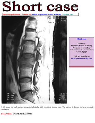
- 1. Short case publication... Version 3.1| Edited by professor Yasser Metwally | October 2009 Short case Edited by Professor Yasser Metwally Professor of neurology Ain Shams university school of medicine Cairo, Egypt Visit my web site at: http://yassermetwally.com A 60 years old male patient presented clinically with persistent lumbar pain. The patient is known to have prostatic carcinoma. DIAGNOSIS: SPINAL METASTASIS
- 2. Figure 1. MRI T2 image (A) and precontrast MRI T1 images (B) showing wedging of the vertebral body of L2 by a hypointense osteolytic lesion with preservation of the intervertebral discs above and below. The osteolytic lesion is seen extending posteriorly to the epidural spaces. Also noticed multiple osteolytic lesions in the body of L1 vertebra. A schorl's nodule is seen D11 vetrebra. Figure 2. MRI T2 images showing wedging of the vertebral body of L2 by a hyperinetse osteolytic lesion with preservation of the intervertebral discs above and below. The osteolytic lesion is seen extending posteriorly to the epidural spaces and encroaching upon the cauda roots.
- 3. Figure 3. MRI T2 images showing the L2 osteolytic lesion extending posteriorly to the epidural spaces. Facet degenerative changes are also seen Modern neurodiagnostic imaging studies are indispensable in the evaluation and treatment of patients with metastatic spinal tumors. These studies are used to diagnose, stage, and plan treatment in patients with metastatic lesions, as well as to follow them postoperatively. Spinal Radiography. Plain radiographs of the spine remain a valuable and readily available initial imaging study. Anteroposterior and lateral radiographs demonstrate abnormal findings in up to 90% of patients with symptomatic spinal metastasis. Lytic lesions and vertebral collapse are common however, both osteoblastic and -sclerotic alterations also occur, especially with breast and prostatic metastasis. Plain x-ray film findings include pedicle erosion (that is, the "winking owl" sign), paraspinal soft-tissue shadows, wedge compression, and pathological fracture-dislocation. Intervertebral disc margins are invariably spared in metastatic tumor invasion, contrasting with the disc erosion commonly observed with infectious entities. Local bone destruction of 50% is required before a lesion can be detected on plain x-ray films. In a high proportion of patients with symptomatic secondary spinal lesions abnormalities are demonstrated on plain radiographs, but in up to 26% of the cases early metastatic lesions can be missed. Consequently, clinicians investigating patients with a high suspicion of metastatic spinal neoplasms should arrange expeditious CT and/or MR imaging investigations, even if normal findings are observed on plain radiographs. Computerized Tomography Scanning. The principal use of CT scanning is in the assessment of the osseous architecture of the spinal axis because MR imaging does not provide optimum images of bone. Computerized tomography scanning provides important information regarding the degree of tumor association with cortical bone and the extent of neoplastic destruction. Thus, CT scanning is an important complement to MR imaging. In patients in whom MR imaging cannot be performed, CT scanning is particularly useful when conducted immediately
- 4. following myelography. Computerized tomography myelography provides better anatomical detail of the spinal axis than either CT or myelography alone. The information acquired from CT myelography is often comparable with that obtained using MR imaging. Magnetic Resonance Imaging Magnetic resonance imaging provides unparalleled visualization of the spinal column and spinal cord and is the neuroimaging method of choice in patients with suspected spinal tumors. It has been demonstrated to facilitate the earlier diagnosis of spinal metastases than other modalities. It is superior in depicting epidural and bone marrow tumor infiltration, and it delineates the extraosseous soft-tissue component of a neoplasm from the normal paraspinal soft tissue and neural structures. In addition to its diagnostic utility, MR imaging information is essential to the process of thorough surgical planning. Gadolinium enhancement further increases the sensitivity of MR imaging investigations, because metastases invariably enhance. Visualization of the entire vertebral axis in multiple orthogonal planes is easily accomplished, a goal that is important given the high incidence of multiple vertebral levels of tumor infiltration. Studies show that the results of MR imaging investigations alter therapeutic decisions in a significant number of patients, especially with regard to the addition or modification of radiotherapy. References 1. Metwally, MYM: Textbook of neurimaging, A CD-ROM publication, (Metwally, MYM editor) WEB-CD agency for electronic publishing, version 10.4a October 2009 Addendum A new version of short case is uploaded in my web site every week (every Saturday and remains available till Friday.) To download the current version follow the link "http://pdf.yassermetwally.com/short.pdf". You can download the long case version of this short case during the same week from: http://pdf.yassermetwally.com/case.pdf or visit web site: http://pdf.yassermetwally.com To download the software version of the publication (crow.exe) follow the link: http://neurology.yassermetwally.com/crow.zip At the end of each year, all the publications are compiled on a single CD-ROM, please contact the author to know more details. Screen resolution is better set at 1024*768 pixel screen area for optimum display For an archive of the previously reported cases go to www.yassermetwally.net, then under pages in the right panel, scroll down and click on the text entry "downloadable short cases in PDF format" Also to view a list of the previously published case records follow the following link (http://wordpress.com/tag/case- record/) or click on it if it appears as a link in your PDF reader