Short case..Subacute cerebral hemorrhage
•
1 j'aime•2,701 vues

Short case..Subacute cerebral hemorrhage

Recommandé
Recommandé
Contenu connexe
Similaire à Short case..Subacute cerebral hemorrhage
Similaire à Short case..Subacute cerebral hemorrhage (20)
Plus de Professor Yasser Metwally
Plus de Professor Yasser Metwally (20)
Dernier
Dernier (20)
Short case..Subacute cerebral hemorrhage
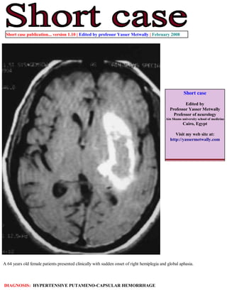
- 1. Short case publication... version 1.10 | Edited by professor Yasser Metwally | February 2008 Short case Edited by Professor Yasser Metwally Professor of neurology Ain Shams university school of medicine Cairo, Egypt Visit my web site at: http://yassermetwally.com A 64 years old female patients presented clinically with sudden onset of right hemiplegia and global aphasia. DIAGNOSIS: HYPERTENSIVE PUTAMENO-CAPSULAR HEMORRHAGE
- 2. Figure 1. Late subacute hematoma. A CT scan image showing the hematoma as an oval hyperdense space occupying lesion with positive mass effect. The lesion is surrounded by a hypodense edema rim. The increased protein content of the retracted clot is responsible for the high attenuation noted on noncontrast CT. B,C precontrast MRI T1 images. The subacute hematoma has a hyperintense peripheral rim (due to extracellular methemoglobin) and a central isointensity mostly due to Paramagnetic intracellular deoxyhemoglobin. Figure 2. Late subacute hematoma. MRI T2 images. The subacute hematoma has a hyperintense peripheral rim (due to extracellular methemoglobin) and a central hypointensity mostly due to Paramagnetic intracellular deoxyhemoglobin. Edema could also contribute to the peripheral T2 hyperintensity.
- 3. Figure 5. Figure 3. Late subacute hematoma. MRI FLAIR images. The subacute hematoma has a hyperintense peripheral rim (due to extracellular methemoglobin) and a central iso to hypointensity mostly due to deoxyhemoglobin. In this patient the appearance of the subacute hematoma on FLAIR images is similar to that seen on MRI T2 images. Edema could also contribute to the peripheral T2 hyperintensity. Figure 4. Diffusion weighted images. The appearance of hemorrhage on DW MR images is complex and involves many factors, including the relative amounts of different hemorrhagic products and the pulse sequence used. Oxyhemoglobin is hyperintense on DW images and has a lower ADC than does normal brain tissue; this may indicate the relative restriction of water movement inside the red blood cell (101). Extracellular methemoglobin has a higher ADC than does normal brain tissue, which indicates that the mobility of water in the extracellular space is increased. The prolongation of the T2 component of fluid with extracellular methemoglobin results in hyperintensity on DW images. Hemorrhage containing deoxyhemoglobin, intracellular methemoglobin, and hemosiderin are hypointense on DW images because of magnetic susceptibility effects. Because these products of hemorrhage have very low signal intensity on T2-weighted images, ADCs cannot be reliably calculated for them.
- 4. Figure 5. For comparison, A view showing the hematoma on CT scan, precontrast MRI T1 image, MRI T2, FLAIR and diffusion weighted images. The hematoma biochemical stages Addendum A new version of this software is uploaded in my web site every week (every Saturday and remains available till Friday.) To download the current version follow the link quot;http://pdf.yassermetwally.com/short.pdfquot;. You can download the long case version of this short case during the same week from: http://pdf.yassermetwally.com/case.pdf or visit web site: http://pdf.yassermetwally.com To download the software version of the publication (crow.exe) follow the link: http://neurology.yassermetwally.com/crow.zip At the end of each year, all the publications are compiled on a single CD-ROM, please contact the author to know more details. Screen resolution is better set at 1024*768 pixel screen area for optimum display For an archive of the previously reported cases go to www.yassermetwally.net, then under pages in the right panel, scroll down and click on the text entry quot;downloadable short cases in PDF formatquot;
- 5. References 1. Metwally, MYM: Textbook of neurimaging, A CD-ROM publication, (Metwally, MYM editor) WEB-CD agency for electronic publishing, version 9.1a January 2008