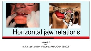
4. HORIZONTAL JAW RELATION
It is the relationship of the mandible to the maxilla in a
horizontal plane
The relationship of the mandible to the maxilla in the
anteroposterior direction
5. It can be of two type
Centric relation
Eccentric relation Protrusive relation
Lateral relation
6. CENTRIC RELATION
The most retruded relation of the mandible to the maxilla
when the condyle are in the most posterior unstrained
position in the glenoid fossa from which lateral movements
can be made, at any given degree of jaw separation
GPT 1
7. CENTRIC RELATION
A maxillomandibular relationship, independent of tooth contact,
in which the condyles articulate in the anterior-superior position
against the posterior slopes of the articular eminences in this
position, the mandible is restricted to a purely rotary movement;
from this unstrained, physiologic, maxillomandibular relationship,
the patient can make vertical, lateral or protrusive movements; it
is a clinically useful, repeatable reference position
GPT 9
8. MUSCLES INVOLVED IN
CENTRIC RELATION
The masseter, temporalis and medial
pterygoid muscle connects the mandible
to the lateral pterygoid plate in such a
way as to act as the steering mechanism
for the mandible and helps in elevating
mandible to centric relation position
9. Significance of Centric Relation
Centric relation position acts as
a proprioceptive center to guide
the mandibular movements
10. Purpose of recording centric relation
It is bone to bone relation and it is constant
It is repeatable and recordable and thus serves as reliable guide
for developing centric occlusion in complete dentures
It is related to terminal hinge axis
11. Purpose of recording centric relation
Functional movements like chewing and swallowing can be
carried out since it is the most unstrained position.
It is a definite entity, so used as reference point in establishing
centric occlusion
It is more definite than vertical relation since it is independent of
tooth contact
12. Theories of centric relation
The muscle theory
The ligament theory
The osteofiber theory
The meniscus theory
Saizar P. Centric relation and condylar movement: anatomic mechanism. J Prosthet Dent. 1971 Dec;26(6):581-91.
13. The Muscle Theory
Defence reflex
External pterygoid
muscles contract
Halts the joint
Saizar P. Centric relation and condylar movement: anatomic mechanism. J Prosthet Dent. 1971 Dec;26(6):581-91.
14. The Ligament Theory
Saizar P. Centric relation and condylar movement: anatomic mechanism. J Prosthet Dent. 1971 Dec;26(6):581-91.
Ligaments binds element of
articulation, limit the
possibilities of movement and
are also capable of determining
terminal border position
15. The Osteofiber Theory
Saizar P. Centric relation and condylar movement: anatomic mechanism. J Prosthet Dent. 1971 Dec;26(6):581-91.
Involves a retrusive terminal
stop formed by soft tissues of
posterior part of glenoid fossa
Fibrous stop act as buffer
16. The Meniscus Theory
Saizar P. Centric relation and condylar movement: anatomic mechanism. J Prosthet Dent. 1971 Dec;26(6):581-91.
The posterosuperior surface
unfolds along the roof of
the glenoid fossa
17. The Meniscus Theory
Saizar P. Centric relation and condylar movement: anatomic mechanism. J Prosthet Dent. 1971 Dec;26(6):581-91.
Disc with their retro meniscal fibrous
tissues- stop the retrusive condylar
movements
18. Factors influencing
centric relation records
Yurkstas AA, Kapur KK. Factors influencing centric relation records in edentulous mouths. 1964. J Prosthet Dent. 2005;93(4):305-310.
19. Yurkstas AA, Kapur KK. Factors influencing centric relation records in edentulous mouths. 1964. J Prosthet Dent. 2005;93(4):305-310.
The resiliency of the supporting tissues
Stability of the recording bases
Temporomandibular joint and associated neuromuscular mechanisms
Pressure applied in making the recording
The technique used in making the recording and the recording devices
used
Skill of the dentist
20. Yurkstas AA, Kapur KK. Factors influencing centric relation records in edentulous mouths. 1964. J Prosthet Dent. 2005;93(4):305-310.
The health and cooperation of the patient
The maxillomandibular relationship
Posture of the patient
Character and size of residual alveolar arch
The amount and character of saliva
Size and position of tongue
23. Difficulties in obtaining mandibular retrusion
Biological
Lack of muscle coordination
Lack of synchronization between the protruding and
Retruding muscles due to HABITUAL eccentric jaw
positions
24. Difficulties in obtaining mandibular retrusion
Psychological
Involves patient and dentist
Inability of the patient to follow
dentist’s instructions
25. Difficulties in obtaining mandibular retrusion
Mechanical
It is essential that the record bases on which the centric relation
are made, fit perfectly and not interfere with each other
Poorly fitting baseplates
26. Methods to retrude the mandible while recording centric relation
Relaxation of Jaw
Simplest, easiest and most efficient way of causing retrusion
by verbal instructions to the patient
Instruct the patient by saying “ Let your lower jaw relax, pull it
back, and close on your back teeth”
27. Methods to retrude the mandible while recording centric relation
Pushing Upper Jaw
Patient is instructed to “Get the feeling of pushing your upper
jaw out and close your back teeth together”
By getting the feeling of pushing the upper jaw forward, they
automatically pull the lower jaw backward.
28. Methods to retrude the mandible while recording centric relation
Stretch and Relax Movements
Patient is instructed to protrude and
retrude the mandible. Dentist can aid
by a slight pressure on the point of the
chin
29. Methods to retrude the mandible while recording centric relation
Retrusion of Tongue
Patient is instructed to keep the tip of tongue in contact with posterior border of
the maxillary record base and then patient is asked to close until the rims come
into contact
Bissasu M. Use of the tongue for recording centric relation for edentulous patients. J Prosthet Dent. 1999;82(3):369-370.
30. Methods to retrude the mandible while recording centric relation
Rapid Tapping of the Occlusal Rims
Gentle tapping of occlusal rims rapidly and repeatedly retrudes the mandible
31. Methods to retrude the mandible while recording centric relation
Head Position
Tilting the head backwards results in retrusion of the mandible as this
will place tension on the infra-mandibular muscles and tend to pull the
mandible to a retruded position
32. Methods to retrude the mandible while recording centric relation
Swallowing and Temporalis muscle check
Swallowing usually brings the mandible in a retruded position
The temporalis muscle show reduced function when the mandible is in
protruded position. So its contraction can be felt when the mandible is
in or near retruded position by placing finger tips on each side of the
head
34. Minimum closing pressure
The record should be made with minimal closing pressures so the
tissues supporting the bases will not be displaced while record is
being made.
Objective : For the opposing teeth to touch uniformly and
simultaneously at their first contact
35. Heavy closing pressure
The record should be made under heavy closing pressure so that
record bases will be displaced while the record is being made.
Objective : To produce the same displacement of the soft tissues as
would exist when heavy closing pressure were applied to denture
43. Commonly used materials
Waxes
Impression compound
Zinc oxide eugenol paste
Impression plaster
Additional silicone
Polyether
usually preferred because they offer
uniform resistance to pressure
44. Tentative jaw relation
Maxillary occlusal rim is inserted into patients
mouth
Vertical dimension at rest is established
Tentative centric relation is recorded by
Retruding the mandible
Occlusal rims articulated and artificial teeth
arranged
Trial dentures ready for making interocclusal
check record
45. Making inter-occlusal check record
The trial dentures are removed and the wax is allowed to cool
Aluwax is loaded onto the occlusal surface of teeth in the mandibular occlusal rim
The patient is asked to slowly retrude the mandible and close on the was till tooth contact occurs
The upper and lower trial dentures are inserted into patients mouth
Artificial teeth are prevented from contacting the opposing members by keeping a piece of cotton inter-occlusally
47. Factors affecting the success of interocclusal record method
Uniform consistency of the recording material.
Accurate vertical jaw relation records.
Stability and fit of the record base.
Presence of reference points embedded in the record like
metal pins or styli.
48. Static or Pressureless Method
The occlusal rims are customized as usual and the patient is trained to close at
centric relation position. Once the patient attains the centric relation position,
the denture bases with occlusal rims are indexed/sealed in this position
Nick and Notch
method
Stapler Pin
method
49. Nick and Notch method
• Most commonly used method of indexing the centric record
• Final centric jaw relation is carried out after establishing a
proper vertical jaw relation
• No occlusal check record is performed during try-in
50. The patient is seated in an upright position
Up to 3 mm of wax is removed on either side of the
mandibular occlusal rim from the premolar region
till the distal end. This depression created on the
occlusal rim due to removal of wax is called trough
51. One or two notches are cut on the
corresponding area on the maxillary occlusal
rim. The notch resembles a "V“ shaped valley
running totally across the width of the occlusal
rim
One nick is cut anterior to the notch. This is also
a "V“ shaped valley but it does not extend
throughout the width of the occlusal rim
52. the nick and notch on maxillary occlusal rim are
lubricated with petroleum jelly
The prepared occlusal rims are inserted into the
patient’s mouth and the patient is taught to close
his mandible in centric position
Mandibular occlusal rim removed from the
mouth
Bite registration material is placed in the trough
created in the mandibular occlusal rim
53. The mandibular occlusal rim is placed back in
patient’s mouth and patient is asked to close in
centric relation
The mouth should close such that the anterior parts
of the occlusal rim almost touch but not press
against it
Both the rims are removed, cooled and excess
material is trimmed
54. Stapler pin method
• In this method, after recording the
centric relation, the occlusal rims are
indexed using a bunch of stapler pin.
• The method is not preferred as centric
relation record cannot be verified
55. Pressure Method
• After establishing the vertical dimension, the maxillary occlusal rim is inserted
into patient’s mouth
• The mandibular rim is fabricated excess of height
• The entire mandibular rim is softened in water bath and inserted into
patient’s mouth
• Patient is guided to close in centric relation and asked to close on soft wax
• After patient closes mouth till the predetermined vertical dimension, both
rims are removed , cooled and articulated
56. Shanahan technique
The cones made up of soft wax were mounted on the mandibular
occlusal rim
Patient was advised to make swallowing movements several times,
while the tongue would force the mandible into its centric relation
position
The cones of soft wax were moulded during these movements
Shetty, Manoj & Shetty, Ganaraj. (2020). Comparative Evaluation of Various Techniques to Record Centric Relation- A Literature Review. Journal of Evolution
of Medical and Dental Sciences. 09. 53-59. 10.14260/jemds/2020/12.
58. • Utilizes the functional movements to record the
centric relation
• Patient is asked to perform border movements such
as protrusive and excursive movements , in order to
identify the most retruded position of mandible
59. Factors common to all functional methods
Tentative centric relation and vertical
dimension are measured for determining an
accurate centric relation
Occlusal rims are reduced in excess than that
required for tentative vertical dimension
Exact vertical dimension at occlusion is
determined only when the patient closes on
the occlusal rims and their attachments
Needles House
Patterson
Meyer
60. Disadvantages
Inaccuracy can result from:
• Displaceable basal seat tissues
• Resistance of recording mediums
• Lack of equalized pressures
Patients must have very good neuromuscular coordination and
be capable of following instructions
61. Needleshouse Method
Involves fabrication of occlusal rims
made from impression compound
4 metal beads or styli are embedded
into premolar and molar areas of
maxillary occlusal rim
65. As these movements are made, the height of the plaster carborundum mix
is also reduced.
The patient is asked to continue these movements till a predetermined
vertical dimension is obtained.
Finally the patient is asked to retrude his jaw and the occlusal rims are
fixed in this position with metal staples.
66. Meyer method ( modified functional technique )
Soft wax was utilized to make occlusal rims
The occlusal surface of the wax was coated with tinfoil
Mandible was guided to perform functional movement and
later plaster index was made on wax rims.
Plaster index was used as a guide to set the teeth
Shetty, Manoj & Shetty, Ganaraj. (2020). Comparative Evaluation of Various Techniques to Record Centric Relation- A Literature Review. Journal of Evolution
of Medical and Dental Sciences. 09. 53-59. 10.14260/jemds/2020/12.
68. Uses graphs or tracings to record the centric jaw relation
Graphic
methods
Arrow point
tracing
Measured along
a single plane
Pantograph
Measured three-
dimensionally
70. Why “ Gothic Arch “ ?
Arrow point tracing was first proposed by Hesse 1897,
and later popularized by Gysi, 1908
71. Historical review of graphical recordings
Balkwill (1866)
• Earliest graphic recordings based on mandibular movements
• The intersection of the arcs produced by the right and left
condyles formed the apex of what is known as the Gothic
arch tracing.
Myers ML. Centric relation records-historical review. J Prosthet Dent.
1982;47(2):141-145
72. Historical review of graphical recordings
Gysi (1910)
• Extraoral incisal tracer on maxillary rim traced onto the
tracing plate, coated with wax attached with mandibular rim
• The rims were made of modeling compound to maintain the
vertical dimension of occlusion
Myers ML. Centric relation records-historical review. J Prosthet Dent.
1982;47(2):141-145
73. Historical review of graphical recordings
Sears
• Used lubricated rims for easier movement.
• He placed the needle point tracer on the mandibular rim and the plate
on the maxillary rim.
• He believed this made the angle of the tracing more acute and more
easily discernible. He would then cement the rims together for
removal.
Myers ML. Centric relation records-historical review. J Prosthet Dent.
1982;47(2):141-145
74. Historical review of graphical recordings
Phillips
• Recognized that any lateral movements of the jaw would cause
interference of the rims which could result in distorted record.
• Developed a plate for the upper rim and a tripoded ball bearing
mounted on a jackscrew for the lower rim
“Central Bearing Point “
This innovation supposedly produced equalization of pressure on the
edentulous ridge
Myers ML. Centric relation records-historical review. J Prosthet Dent.
1982;47(2):141-145
75. Historical review of graphical recordings
Stansbery (1929)
• Used a curved plate corresponding to monson’s curve mounted on
upper rim. A central bearing screw was attached to lower plate with a
3inch radius curve. After extraoral tracing , plaster was used to form a
biconcave centric registration
Myers ML. Centric relation records-historical review. J Prosthet Dent.
1982;47(2):141-145
76. Historical review of graphical recordings
Silverman
• Used an intraoral gothic arch tracer to locate the “biting point” of the
patient.
• The patient was asked to bite on hard tracing plate. This developed a
functional resultant of the closing muscles which would retrude the
mandible.
• The indentation made by the patient would be used for centric record
whether it corresponds to gothic apex or not
Myers ML. Centric relation records-historical review. J Prosthet Dent.
1982;47(2):141-145
77. Arrow point tracing
The pattern obtained on the horizontal plate with
a central bearing tracing device
78. Concept of arrow point tracing
The concept consists of attaching a stylus (a writing
device with a pointed end) to one occlusal rim and a
plate to the other rim.
The stylus traces or marks the path in the plate as
the mandible performs excursive movements from
the centric position.
The tracing is typically in the shape of a ‘gothic arch’
or ‘arrow head’ if the patient is trained to move the
mandible from centric to protrusive, right and left
lateral positions
79. Central bearing tracing device
A device that provides a central point of bearing or
support between the maxillary and mandibular dental
arches. It consists of a contacting point attached to
one dental arch and a plate attached to the opposing
dental arch. The plate provides the surface on which
the bearing point rests or moves and on which the
tracing of the mandibular movement is recorded. It
may be used to distribute the occlusal forces evenly
during jaw relation and/or for the correction of
disharmonious occlusal contacts.
87. Gothic arch tracing
Advantages
• Most accurate method of
recording CR
• Allows equalisation of
pressure on supporting tissues
• Easy verifiable
• Can also be used to record
eccentric relations
Disadvantages
• May be difficult to locate the
centre of the arches
• More time consuming
• Training patient in making
mandibular movements is
strenuous
88. Important factors in graphical recordings
Displacement of the record bases may result from pressure if the central
bearing point is off center, when the mandible moves into eccentric
relations to the maxillae
If a central bearing device is not used, the occlusion rims offer more
resistance to horizontal movements
It is difficult to locate the center of the true arches to centralize the
forces with a central bearing device when the jaws are in favorable
relation and far more difficult if the jaws are in excessive protrusive or
retrusive relation.
Bansal S, Palaskar J. Critical evaluation of various methods of recording centric jaw
relation. J Indian Prosthodont Soc 2008;8:185-9
89. Important factors in graphical recordings
It is difficult to stabilize a record base against horizontal force on
residual ridge that have no vertical height.
It is difficult to stabilize a record base against horizontal forces on
tissues that are pendulous or otherwise easily displaceable.
It is difficult to stabilize a record base or bearing device with patients
who have large awkward tongues.
Bansal S, Palaskar J. Critical evaluation of various methods of recording centric jaw
relation. J Indian Prosthodont Soc 2008;8:185-9
90. Important factors in graphical recordings
Recording devices are not usually considered compatible with normal
physiologic simulation in mandibular movement.
The tracing is not acceptable unless a pointed apex is developed, a blunt
apex usually indicates an acquired functional relationship and a sharp
apex usually indicates the position of centric relation.
Double tracings usually indicate lack of coordinated movements or
recordings at a different vertical dimension of jaw separation. In either
event, additional tracings are necessary
Bansal S, Palaskar J. Critical evaluation of various methods of recording centric jaw
relation. J Indian Prosthodont Soc 2008;8:185-9
91. Important factors in graphical recordings
A graphic tracing to determine Centric Relation is made at the
predetermined vertical dimension of occlusion. This harmonizes Centric
Relation with centric occlusion and the antero-posterior bone to bone
relation with the tooth-to-tooth contact
Graphic methods can record eccentric relations of the mandible to the
maxillae.
Graphic methods are the most accurate visual means of making a
Centric Relation record with mechanical instruments; however, all
graphic tracings are not necessarily accurate
Bansal S, Palaskar J. Critical evaluation of various methods of recording centric jaw
relation. J Indian Prosthodont Soc 2008;8:185-9
92. Indications
Broad edentulous sides
Adequate interarch space
In patients with habitual centric (a more anterior position of
the jaws due to prolonged edentulous period without tooth
replacement), the use of the graphic method eliminates all
occlusal contacts on the rims, thus breaking the neuromuscular
reflex and allows the patient to record his true centric
93. Contraindications
Severely resorbed ridges and excessively flabby ridges as they
lead to instability of denture bases
Decreased interarch space – difficult to place central bearing
device without raising the vertical dimension
TMJ arthropathy
Abnormal jaw relations
95. In 1927, Hanau conceded that the Gysi tracing was satisfactory to check records,
but that universal usage was not good
Tech stated that the Gysi tracing technique was the only means that should be
used for centric records, all other methods were “mere deceptions and playthings”.
Kingery pointed out several drawbacks in the use of the central bearing point and
added that the “central bearing point allows for no control over the amount of
closing pressure applied by the patient.”
Bansal S, Palaskar J. Critical evaluation of various methods of recording centric jaw
relation. J Indian Prosthodont Soc 2008;8:185-9
96. Kapur and A. Albert Yurkstas told that intraoral tracing procedure and
extraoral tracing procedure were more consistent as compared to wax
registration method
Phillips pointed to various errors produced by GYSI’s technique and
stated that, “if one occlusal rim is allowed to touch the other during the
lateral extreme positions, undue pressure is bound to be exerted on the
contact side, and on account of resiliency of the underlying tissues the
side not in contact will be unseated just enough to cause a false
reading for the horizontal inclination of the condylar path”.
Bansal S, Palaskar J. Critical evaluation of various methods of recording centric jaw
relation. J Indian Prosthodont Soc 2008;8:185-9
97. A. Albert Yurkstas, and Krishan K. Kapur. Factors influencing centric relation records in
edentulous mouths., Journal of Prosthetic Dentistry 1957
Trapozzano stated “the use of the central bearing point is based on the
fallacious assumption that the central bearing point will produce
equalization of pressure.
Trapozzano maintained that the wax recording method was the most
accurate method because of the greater ability to equalize or centralize
pressure with this technique.
99. Coble balancer
The central bearing pin is a
small cylinder attached to
maxillary denture base
The central bearing plate is
narrow and is designed like a
bridge across lower occlusal
rim
100. Condylator
The central bearing pin is
attached to mandibular
occlusal rim
The central bearing plate is
trapezoidal in shape
101. Ballard intraoral tracer
The tracing pin is attached to
mandibular rim and plate to
maxillary rim
• Palatal bearing plate
• Rounded head of correlator
pin
• Tension spring
• Mounting plate
• Pointed end of correlator pin
102. Swiss dent ball bearing bite recorder
The central bearing
point is a ball with a
single point of
contact .
The ball add weight
and stability to the
tracer
103. Microtracer
It is a circular tracer with
a semi-circular central
bearing point.
It is more comfortable for
the patient
104. Procedure for intraoral tracing
The record bases attached to tracing point and plate are inserted
into patients mouth
The central bearing point is adjusted such that it contacts the plate
at predetermined vertical dimension
105. Procedure for intraoral tracing
The patient is asked to make
anteroposterior and lateral
movements. While making these
movements, the central bearing
point will draw the tracing pattern on
the plate
106. Procedure for intraoral tracing
When the patient closes the
mouth, the central bearing
point contacts the metal
plate
108. Hight tracer
The Hight tracer has an upper
tracing pin and a lower recording
plate. These tracers have ‘toothed’
extensions which are used to
attach the tracers to the side of the
occlusal rims
109. Sears tracer
It is a central bearing point tracer with
two tracing apparatus. It produces two
tracing simultaneously.
It is the only extra oral tracer which has
the tracing pin attached to the mandible
and the plate attached to the maxilla
110. Phillips extraoral tracer
Extraoral device with two tracing
apparatus
The tracing pin is attached to the
maxilla and the tracing plate is
attached to the mandible
111. Procedure for extraoral tracing
The maxillary cast is mounted on the articulator with
face bow transfer
The mandibular cast is oriented to the maxillary cast at
the established vertical dimension with a static CR record
The condylar elements of the articulator are secured
against the centric stops
112. Procedure for extraoral tracing
The central bearing and
tracing devices are mounted
on the respective rims
113. Procedure for extraoral tracing
The patient is seated with
head upright, and record
bases with attached devices
are inserted into patients
mouth.
Record bases are checked for
stability, contact during
movements and interferences
114. Procedure for extraoral tracing
The stylus is retracted and patient
is trained to make various excursive
movements passively and actively
(if needed). Patient is instructed to
move the jaw forwards, right and
left from centric position.
Ney Excursion Guide
115. Procedure for extraoral tracing
When the patient is well trained in making
the movements, the recording plate is
coated with a thin coating of lacquer,
precipitated chalk or dark coloured wax
The stylus is made to contact the recording
plate and the patient is instructed to make
the specific movements.
116. Procedure for extraoral tracing
When an acceptable tracing is made with a single sharp apex, a centric
record is obtained
The rims and tracing are prepared to receive the centric record
117. Procedure for extraoral tracing
Before making the records,
the undercuts are blocked
with wax and orientation
grooves made in the wax.
119. Procedure for extraoral tracing
The patient is instructed to retrude the
mandible such that the stylus contacts
the apex of the tracing
Quick setting plaster is injected between
the rims and allowed to harden; thus, the
centric record is obtained
120. Procedure for extraoral tracing
The occlusal rims are seated over
the casts and with the centric
record in place, and the mandibular
cast is remounted with the new
record.
121. Gerbers classification of arrow point tracing
Well defined apex with a symmetrical left and
right lateral component.
The mean gothic arch angle is about 120 degrees.
It reflects healthy TMJ without interferences in
condylar path and balanced muscle guidance
Typical
122. Gerbers classification of arrow point tracing
Similar to typical form except that it has more
obtuse left and right lateral tracings
The gothic arch angle is more than 120 degrees.
It signifies marked lateral movement of condyle in
the fossa.
Flat form
123. Gerbers classification of arrow point tracing
The tracing should be repeated till a defined
arrow point head is obtained.
Patient training is necessary
Round form / apex absent
124. Gerbers classification of arrow point tracing
Similar to typical form, however the extension
of tracing is very limited.
This can be due to restricted mandibular
movements, improper seating of denture
bases and painful fitting denture bases.
It is also an indication of long period of
edentulousness with an inhibition in condylar
movements
Miniature arrow point
125. Gerbers classification of arrow point tracing
It is a record of habitual and retruded centric
relation
Also seen when vertical dimension is altered
during registration
Double arrow point
126. Gerbers classification of arrow point tracing
The protrusive path of mandibular movement
extended beyond the apex of the Gothic arch.
This signifies a forced strained retrusive
movement of lower jaw either by patient or
operator.
It is also seen when the patient head is tilted
too far posteriorly
Dorsally extended arrow point
127. Gerbers classification of arrow point tracing
It is break or loss of continuity of lateral insical
path of gothic arch
This happens due to posterior interferences at
the heels of the denture base
Interrupted arrow point
128. Gerbers classification of arrow point tracing
The left and right lateral tracings meet in an
arrow point; however, the inclination to the
protrusive movement obtained is not
symmetrical.
This form of tracing indicates an error or
interference in forwarding movement of the
condyle.
Asymmetrical arrow point
130. Uses graphs or tracings to record the centric jaw relation
Graphic
methods
Arrow point
tracing
Measured along
a single plane
Pantograph
Measured three-
dimensionally
131. Pantographic tracing
A graphic record of mandibular movement
usually recorded in the horizontal, sagittal
and frontal planes as registered by styli on
the recording tables of a pantograph or by
means of electronic sensors
Pantogram
132. Pantograph
An instrument used to graphically record in, one or more planes,
paths of mandibular movement and to provide information for the
programming of an articulator
133. It resembles a complicated
facebow
The surface over which the
tracing is done is called a “
flag “
A stylus (tracing pointer) is
present for each flag and it
draws tracing patterns on
the flags
135. CEPHALOMETRIC RADIOGRAPHS
Pyott and Schaeffer used cephalometric
radiographs to record centric relation and
vertical dimension of occlusion
However this method never gained
widespread due to errors in recording
centric relation
136. Conclusion
Recording of centric relation is the most important factor in
dentistry. It is a very critical step.
Therefore, it is recommended to discuss regarding various
methods and material aspects used in recording centric relation
more precisely.
Skills of the operator and patient’s good neuromuscular
coordination are probably the most important factors in securing
an accurate Centric Relation record.
Centric relation is not a resting position or postural position of the mandible
contraction of muscles is necessary to move and fix the mandible in CR position
As we know that the proprioceptive impulses(impulses of 3 dimensional spatial orientation) guide the mandibular movements
in dentulous patients the proprioceptive impulses are obtained from pdl
Dentulous patient does not have any proprioceptive guidance from the teeth to guide mandibular movements. The source of impulses is transferred to tmj.
3. In CR condyles exhibit pure rotation without any translation
3. In CR condyles exhibit pure rotation without any translation
The anatomic mechanism responsible for CR is not unknown and therefore several theories have been developed
Or the myologic theory
This theory considers CR to be product of a defense reflex
Which halts the joint everytime the condyles approaches the posterosuperior depths of glenoid fossa
Ferrin was first to present the theory
According to this , when the ligamnets become tense they determine the limits of retrusive movement.
Proved by meyer
zenker believed that
he named this structure as retroarticular cushion
The mandible should be retruded to it posterior position before recording the centric jaw relation.
Some patients may show difficulties in Retruding due to certain systemic conditions
Disadvantage : likelihood of displacing the mandibular record base by the action of tongue.
Disadvantage : difficult to record and patient can easily tap in a slightly protrusive or lateral position
Disadvantage : insertion and removal of occlusal rim from mouth is difficult
Disadvantage of swallowing: unreliable since person can swallow when mandible is not completely retruded but 1-2 mm anterior to maxilla.
There are 2 basic concepts of closing pressure while recording centric relations each one having its own objective
Disadvantage- if soft tissue have uneven thickness, the teeth contact unevenly at first contact
Uneven contacts may cause clenching in nervous patients .
Advantage- occlusal pressure are evenly distributed over residual ridges under heavy load.
They are called physiological because they are based on
Waxes are technique sensistive and do not provide uniform resisitance to pressure they do not cool uniformly
Advantage of waxes – they harden quickly and record can be made immediately
Disadvantage of plaster and zoe- they take long time to set
The names nick and notch is from the shape of the indices made on the occlusal rims
1. As it is easier to retrude the mandible in this position
1. nick prevents lateral movement and the notch prevents anteroposterior movement
The arrow point resembles the high pointed arches of the Gothic architecture and was hence called as 'Gothic Arch Tracing’.
Also known as needle point tracing
When a good tracing was recorded, the patient held the rims in the apex of the tracing while notches were scored in the rims for orientation.
The pointer can be adjusted to the height
stylus (tracing device) and central bearing plate attached to the maxillary rim.
2. recording plate (tracing device) and central bearing point (central bearing device) is attached to the mandibular rim.
Intraoral tracer components: (a) Recording plate and central bearing plate are combined as one component and attached to maxillary rim.
(b) The stylus and central bearing point are combined as one component and attached to the mandibular rim.
The intraoral tracing procedure has also been criticized by many prosthodontists. Their main objections were based on the general disadvantages of a central bearing point device
Popular compact intraoral tracer
The tracer are easy to place because they are supplied with snap on spacer that provide even space for bite registration
After completing the movements, the tracing is removed and examined .
The tracing should resemble an arow point with sharp apex. If apex is blunt, the record is discarded and the procedure is freshly repeated.
These tracers were originally designed without a central bearing device. They were later modified to include a central bearing device to equalize biting pressure during jaw relation procedures
Occlusal rims with the extraoral tracer inserted in the patient’s mouth.
1. The Ney Excursion Guide has been used as an aid in training the patient but patient responds better to specific locations than numbers
2. The patient is trained to make the mandibular movements in the numerical order.
The coating material should not provide any resistance to movement and produce a clearly visible tracing
Before making the records, the tracing is protected by a transparent plastic film (b).
This film is secured over the recording plate using sticky wax (a).
The centric point of the arrow head is viewed and using a sharp heated needle, a hole is made (c) which will guide the needle in position while making records. 6 mm from the centric point, another slot is made, to stabilize the needle while making the protrusive record.
Gerber stated that there are six different types of tracing obtained from Gothic arch tracers based on the direction of movement of condylar head and also on neuromuscular coordination of muscle of mastication and jaw movements.