SlideShare une entreprise Scribd logo
1  sur  46
1
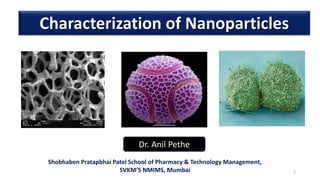
Characterization of Nanoparticles
Dr. Anil Pethe
Shobhaben Pratapbhai Patel School of Pharmacy & Technology Management,
SVKM’S NMIMS, Mumbai
 Introduction
 Nanoparticle characterization techniques
 Electron Microscope
 Scanning electron microscope
 Transmission electron Microscope
 X-ray powder diffraction
 Nuclear Magnetic Resonance
Contents
 Characterization refers to the study of material’s features such as its
composition, structure,& various properties like physical, electrical,
magnetic etc.
Nano = 10-9 (extremely small) Particle = Small piece of matter
 Nanoparticle is a microscopic particle whose size is measured in
nanometers (nm).
 These particles can be spherical, tubular, or irregularly shaped and can
exist in fused, aggregated or agglomerated forms.
Scale of Nano particles
Optical (Imaging) Probe Characterization Techniques
Electron Probe Characterization Techniques
Scanning Probe Characterization Techniques
Photon(Spectroscopic) Probe Characterization Techniques
Ion-particle probe Characterization Techniques
Thermodynamic Characterization Techniques
Nano particles Characterization Techniques
Optical(Imaging) Probe Characterization Techniques
Optical (Imaging) Probe Characterization Techniques
Acronym Technique Utility
CLSM Confocal laser-scanning
microscopy
Imaging/ultrafine
morphology
SNOM Scanning near-field optical
microscopy
Rastered images
2PFM Two-photon fluorescence
microscopy
Fluorophores/biological
systems
DLS Dynamic light scattering Particle sizing
BAM Brewster angle microscopy Gas-liquid interface
Imaging
Electron Probe Characterization Techniques
Acronym Technique Utility
SEM Scanning Electron Microscopy Imaging/ topology
morphology
EPMA Electron Probe Microanalysis Particle size/ local chemical
analysis
TEM Transmission Electron Microscopy Imaging/ Particle size shape
HRTEM High Resolution Transmission Electron
Microscopy
Imaging structure chemical
analysis
LEED Low Energy Electron Diffraction Surface/ adsorbate bonding
EELS Electron Energy Loss Spectroscopy Inelastic electron
interaction
AES Auger Electron Spectroscopy Chemical surface analysis
Scanning Probe Characterization Techniques
Acronym Technique Utility
AFM Atomic Force Microscopy Imaging/ topology/ surface
structure
CFM Chemical Force Microscopy Chemical/surface analysis
MFM Magnetic Force Microscopy Magnetic material analysis
STM Scanning Tunnelling Microscopy Topology/Imaging /surface
APM Atomic Probe Microscopy Three dimensional Imaging
FIM Field Ion Microscopy Chemical profiles/ atomic spacing
APT Atomic probe tomography Position sensitive lateral location
of atoms
Photon(Spectroscopic) Probe Characterization Techniques
Acronym Technique Utility
UPS Ultraviolet photoemission
spectroscopy
Surface analysis
UVVS UV Visible spectroscopy Chemical analysis
AAS Atomic absorption spectroscopy Chemical analysis
ICP Inductively coupled plasma
spectroscopy
Elemental analysis
FS Fluorescence spectroscopy Elemental analysis
LSPR Localized surface plasmon
resonance
Nanosized particle analysis
Ion-particle probe Characterization Techniques
Acronym Technique Utility
RBS Rutherford back scattering Quantitative- Qualitative elemental
analysis
SANS Small angle neutron scattering Surface characterization
NRA Nuclear reaction analysis Depth profiling of solid thin film
RS Raman Spectroscopy Vibration analysis
XRD X-ray diffraction Crystal structure
EDX Energy dispersive X-ray spectroscopy Elemental analysis
SAXS Small angle X-ray scattering Surface analysis/ particle sizing (1-100 nm)
CLS Cathodoluminescence Characteristics emission
NMR Nuclear magnetic resonance
spectroscopy
Analysis of odd no. of nuclear species
Thermodynamic Characterization Techniques
Acronym Technique Utility
TGA Thermal gravimetric analysis Mass loss Vs. Temperature
DTA Differential thermal analysis Reaction heat capacity
DSC Differential scanning calorimetry Reaction heat phase changes
NC Nanocalorimetry Latent heats of fusion
BET Brunauer-Emmett-Teller method Surface area analysis
Sears Sears method Colloid size, specific surface area
 Light microscopes cannot resolve structures closer than 200 nm
 Electron microscopes have greater resolving power and magnification
 Magnifies objects 10,000X to 100,000X
 Detailed views of bacteria, viruses, internal cellular structures,
molecules, and large atoms
 Two types
• Transmission electron microscopes
• Scanning electron microscopes
The Electron Microscope
• Energy source is a beam of electrons
• Image is created and viewed on a monitor
• TEM (transmission electron microscope) utilizes staining procedures
prior to use
• SEM (scanning electron microscope) adds three-dimensional viewing
The Electron Microscope
• Beams of electrons are used to
produce images
• Wavelength of electron beam is
much shorter than light,
resulting in much higher
resolution
The Electron Microscope
• In transmission electron microscopy electrons pass through thin
specimens (50-1000 nm). BULK BEAM
• In scanning electron microscopy signals emitted from the surface of
thick specimens. NARROW BEAM
Physical Limitations
• Uses electrons reflected from the surface of a specimen to create
image
• Produces a 3-dimensional image of specimen’s surface features
Scanning Electron Microscope (SEM)
Light Microscopy Vs. Electron Microscopy
 The SEM is an instrument that produces a largely magnified
image by using electrons instead of light to form an image.
 A beam of electrons is produced at the top of the microscope
by an electron gun.
 The electron beam follows a vertical path through the
microscope, which is held within a vacuum.
 The beam travels through electromagnetic fields and lenses,
which focus the beam down toward the sample.
 Once the beam hits the sample, electrons and X-rays are
ejected from the sample.
 Detectors collect these X-rays, backscattered electrons, and
secondary electrons and convert them into a signal that is
sent to a screen similar to a television screen. This produces
the final image.
Working of SEMWorking of Scanning Electron Microscope (SEM)
(Emiliania huxleyi, a haptophyte alga)
http://starcentral.mbl.edu/
SEM IMAGE
SEM IMAGES
Transmission Electron Microscope (TEM)
• Electrons scatter when they pass through thin sections of a
specimen
• Transmitted electrons (those that do not scatter) are used to
produce image
• Denser regions in specimen, scatter more electrons and appear
darker
Transmission Electron Microscope (TEM)
Working of Transmission Electron Microscope (TEM)
 Unlike SEM that bounces electrons off the surface of a sample
to produce an image, Transmission Electron Microscopes
(TEMs) shoot the electrons completely through the sample.
 TEMs work by using a tungsten filament to produce an
electron beam in a vacuum chamber.
 The emitted electrons are accelerated through an
electromagnetic field that also narrowly focuses the beam.
The beam is then passed through the sample material. The
specially prepared sample is a very thin (less than 100nm) slice
of material. The electrons that pass through the sample hit a
phosphor screen, CCD or film and produce an image. Where
the sample has less density, more electrons get through and
the image is brighter. A darker image is produced in areas
where the sample is more dense and therefore less electrons
pass through.
 TEMs can produce images with resolution down to 0.2nm. This
resolution is smaller than the size of most atoms and therefore
images can be produced using TEM that show the true
structural arrangement of atoms in the sample material.
• X-ray diffraction is used to obtain
structural information about crystalline
solids.
• Useful in biochemistry to solve the 3D
structures of complex biomolecules.
• Bridge between physics, chemistry, and
biology.
• X-ray diffraction is important for
 Solid-state physics
 Biophysics
 Medical physics
 Chemistry and Biochemistry
Introduction of X-ray Diffraction
• Most useful in the characterisation of
• Crystalline materials;
• Ceramics,
• Metals,
• Intermetallic,
• Minerals,
• Inorganic compounds
• Rapid and non destructive techniques
• Provide information on unit cell dimension
Structural Analysis
• X-ray diffraction provides most definitive
structural information
• Interatomic distances and bond angles
What is X-ray Diffraction
• Beams of electromagnetic radiation
• *smaller wavelength than visible light,
• *higher energy
• *more penetrative
• (1895) X-rays discovered by Roentgen
• (1914) First diffraction pattern of a crystal made by
Knipping and von Laue
• (1915) Theory to determine crystal structure from
diffraction pattern developed by Bragg.
• (1953) DNA structure solved by Watson and Crick
• Now Diffraction improved by computer technology;
methods used to determine atomic structures and in
medical applications
History of X-ray Diffraction
• The prime component in X-Ray tube are
filament (cathode), vacuum room, anode,
and high voltage
• When high energy electrons strike an anode
in a sealed vacuum, x-rays are generated.
Anodes are often made of copper, iron or
molybdenum.
• High energy electron come from heated
filament in X-Ray tube.
• X-rays are electromagnetic radiation.
• They have enough energy to cause
ionization.
X-ray Production
• English physicists Sir W.H. Bragg and his son Sir W.L.
Bragg developed a relationship in 1913 to explain
why the cleavage faces of crystals appear to reflect
X-ray beams at certain angles of incidence (theta, θ).
• d is the distance between atomic layers in a
crystal,
• lambda λ is the wavelength of the incident X-ray
beam;
• n is an integer.
• This observation is an example of X-ray wave
interference (Roentgen strahl interferenzen),
commonly known as X-ray diffraction (XRD), and
was direct evidence for the periodic atomic
structure of crystals postulated for several centuries.
Bragg’s Law
nλ = 2d sin θ
• Wave Interacting with a Single Particle
• Incident beams scattered uniformly in all directions
• Wave Interacting with a Solid
• Scattered beams interfere constructively in some directions, producing
diffracted beams
• Random arrangements cause beams to randomly interfere and no distinctive
pattern is produced
• Crystalline Material
• Regular pattern of crystalline atoms produces regular diffraction pattern.
• Diffraction pattern gives information on crystal structure
How Diffraction Works
• X-ray source
• Device for restricting wavelength
range “goniometer”
• Sample holder
• Radiation detector
• Signal processor and readout
Components of X-ray Diffraction
X-ray Diffraction Instrumentation
 X-ray diffractometers consist of three basic elements:
 an X-ray tube,
 a sample holder,
 an X-ray detector.
 X rays are generated in a cathode ray tube by heating a filament to produce electrons,
accelerating the electrons toward a target by applying a voltage, and bombarding the
target material with electrons.
 When electrons have sufficient energy to dislodge inner shell electrons of the target
material, characteristic X-ray spectra are produced. These spectra consist of several
components, the most common being Kα and Kβ.
 Kαconsists, in part, of Kα1 and Kα2.
 Kα1 has a slightly shorter wavelength and twice the intensity as Kα2.
 The specific wavelengths are characteristic of the target material (Cu, Fe, Mo, Cr).
Filtering, by foils or crystal monochrometers, is required to produce monochromatic X-
rays needed for diffraction. Kα1and Kα2 are sufficiently close in wavelength such that a
weighted average of the two is used.
X-ray Diffraction Instrumentation
 Copper is the most common target material for single-crystal diffraction, with
CuKα radiation = 1.5418Å.
 These X-rays are collimated and directed onto the sample.
 As the sample and detector are rotated, the intensity of the reflected X-rays is recorded.
 When the geometry of the incident X-rays impinging the sample satisfies the Bragg
Equation, constructive interference occurs and a peak in intensity occurs.
 A detector records and processes this X-ray signal and converts the signal to a count rate
which is then output to a device such as a printer or computer monitor.
 The geometry of an X-ray diffractometer is such that the sample rotates in the path of
the collimated X-ray beam at an angle θ while the X-ray detector is mounted on an arm
to collect the diffracted X-rays and rotates at an angle of 2θ.
 The instrument used to maintain the angle and rotate the sample is termed
a goniometer.
 For typical powder patterns, data is collected at 2θ from ~5° to 70°, angles that are
present in the X-ray scan.
• A continuous beam of X-rays is
incident on the crystal
• The diffracted radiation is very
intense in certain directions
• These directions correspond to
constructive interference from
waves reflected from the layers of
the crystal
• The diffraction pattern is detected
by photographic film
How X-ray Diffraction works
NaCl
How Diffraction Works: Schematic
NaCl
How Diffraction Works: Schematic
Used to determine
• Crystal structure
• Orientation
• Degree of crystalline perfection/imperfections
Sample is illuminated with monochromatic radiation
• Easier to index and solve the crystal structure because it
diffraction peak is uniquely resolved
Single Crystal X-ray Diffraction
 A single crystal at random orientations and its corresponding diffraction pattern.
 Just as the crystal is rotated by a random angle, the diffraction pattern
calculated for this crystal is rotated by the same angle
Single Crystal X-ray Diffraction
 More appropriately called polycrystalline X-ray diffraction, because it
can also be used for sintered samples, metal foils, coatings and films,
finished parts, etc.
 Used to determine
 phase composition (commonly called phase ID)-what phases are
present?
 quantitative phase analysis-how much of each phase is present?
 unit cell lattice parameters, crystal structure
 average crystallite size of nanocrystalline samples
 crystallite microstrain and texture
 residual stress (really residual strain)
X-ray Powder Diffraction
Fundamental Principle of X-ray Powder Diffraction
This law relates the wavelength of electromagnetic radiation to the diffraction angle and
the lattice spacing in a crystalline sample.
These diffracted X-rays are then detected, processed and counted.
By scanning the sample through a range of 2θangles, all possible diffraction directions of
the lattice should be attained due to the random orientation of the powdered material.
 Conversion of the diffraction peaks to d-spacings allows identification of the mineral
because each mineral has a set of unique d-spacings.
Typically, this is achieved by comparison of d-spacings with standard reference patterns.
All diffraction methods are based on generation of X-rays in an X-ray tube.
These X-rays are directed at the sample, and the diffracted rays are collected.
A key component of all diffraction is the angle between the incident and diffracted rays.
Single crystal Vs. Powder crystal XRD
 Although the single crystal and powder crystal XRD patterns essentially contain
the same Information, but in the former case the information is distributed in
three dimensional space whereas In the latter case the three dimensional data
are “compressed” into one dimension
X-ray Powder Diffraction Applications
 Characterization of crystalline materials
 Identification of fine-grained minerals such as clays and mixed layer clays that are
difficult to determine optically
 Determination of unit cell dimensions
 Measurement of sample purity
 Determine of modal amounts of minerals (quantitative analysis)
 Characterize thin films samples by:
 determining lattice mismatch between film and substrate and to inferring stress and
strain
 determining dislocation density and quality of the film by rocking curve
measurements
 measuring superlattices in multilayered epitaxial structures
 determining the thickness, roughness and density of the film
 make textural measurements, such as the orientation of grains, in a polycrystalline
sample
Strength of X-ray Powder Diffraction
1. Powerful and rapid (< 20 min) technique for
identification of an unknown mineral
2. In most cases, it provides an unambiguous
mineral determination
3. Minimal sample preparation is required
4. XRD units are widely available
5. Data interpretation is relatively straight forward

Contenu connexe

Tendances

Magnetic Nanomaterials
Magnetic NanomaterialsMagnetic Nanomaterials
Magnetic NanomaterialsHemanth kumar
 
Properties of Nano-materials
Properties of Nano-materialsProperties of Nano-materials
Properties of Nano-materialsPartha P. Mishra
 
Quantum Dots and its applications
Quantum Dots and its applicationsQuantum Dots and its applications
Quantum Dots and its applicationsArun Kumar
 
Synthesis and Characterization of nanoparticle
Synthesis and Characterization of nanoparticleSynthesis and Characterization of nanoparticle
Synthesis and Characterization of nanoparticleMohammad Azam
 
Synthesis of nanoparticles- physical,chemical and biological
Synthesis of nanoparticles- physical,chemical and biologicalSynthesis of nanoparticles- physical,chemical and biological
Synthesis of nanoparticles- physical,chemical and biologicalPriya Nanda
 
Characterization of nanoparticles
Characterization of nanoparticlesCharacterization of nanoparticles
Characterization of nanoparticlesRAM PRAKASH
 
scanning electron microscope (SEM)
scanning electron microscope (SEM)scanning electron microscope (SEM)
scanning electron microscope (SEM)Himanshu Dixit
 
Nanotechnology: Origin, History and Principles
Nanotechnology: Origin, History and PrinciplesNanotechnology: Origin, History and Principles
Nanotechnology: Origin, History and PrinciplesHANAMANT SAVALAGI
 
Application of Nanoparticles
Application of NanoparticlesApplication of Nanoparticles
Application of NanoparticlesLAKSHMI NARAYAN
 
Biological methods for nanoparticle synthesis
Biological methods for nanoparticle synthesisBiological methods for nanoparticle synthesis
Biological methods for nanoparticle synthesisVaibhav Maurya
 
nanomaterial and dimensional effect
nanomaterial and dimensional effect nanomaterial and dimensional effect
nanomaterial and dimensional effect sameerr98
 
Synthesis Of Nanomaterials: Physical Methods
Synthesis Of Nanomaterials: Physical MethodsSynthesis Of Nanomaterials: Physical Methods
Synthesis Of Nanomaterials: Physical MethodsMayur D. Chauhan
 
Biomedical applications of nanoparticles
Biomedical applications of nanoparticlesBiomedical applications of nanoparticles
Biomedical applications of nanoparticlesSwathi Babu
 
Nanoparticles and their medical applications
Nanoparticles and their medical applicationsNanoparticles and their medical applications
Nanoparticles and their medical applicationsMuhammad Mudassir
 
Synthesis of Nanomaterials: Chemical Methods
Synthesis of Nanomaterials: Chemical MethodsSynthesis of Nanomaterials: Chemical Methods
Synthesis of Nanomaterials: Chemical MethodsMayur D. Chauhan
 
Green synthesis of nanoparticles
Green synthesis of nanoparticlesGreen synthesis of nanoparticles
Green synthesis of nanoparticlesAmina Khan
 
Size effect of nanomaterials part1
Size effect of nanomaterials part1Size effect of nanomaterials part1
Size effect of nanomaterials part1Mugilan Narayanasamy
 
Properties of nano materials
Properties of nano materialsProperties of nano materials
Properties of nano materialsMohd. Bilal
 

Tendances (20)

Magnetic Nanomaterials
Magnetic NanomaterialsMagnetic Nanomaterials
Magnetic Nanomaterials
 
Properties of Nano-materials
Properties of Nano-materialsProperties of Nano-materials
Properties of Nano-materials
 
Quantum Dots and its applications
Quantum Dots and its applicationsQuantum Dots and its applications
Quantum Dots and its applications
 
Synthesis and Characterization of nanoparticle
Synthesis and Characterization of nanoparticleSynthesis and Characterization of nanoparticle
Synthesis and Characterization of nanoparticle
 
Synthesis of nanoparticles- physical,chemical and biological
Synthesis of nanoparticles- physical,chemical and biologicalSynthesis of nanoparticles- physical,chemical and biological
Synthesis of nanoparticles- physical,chemical and biological
 
Characterization of nanoparticles
Characterization of nanoparticlesCharacterization of nanoparticles
Characterization of nanoparticles
 
scanning electron microscope (SEM)
scanning electron microscope (SEM)scanning electron microscope (SEM)
scanning electron microscope (SEM)
 
Quantum dots ppt
Quantum dots pptQuantum dots ppt
Quantum dots ppt
 
Nanotechnology: Origin, History and Principles
Nanotechnology: Origin, History and PrinciplesNanotechnology: Origin, History and Principles
Nanotechnology: Origin, History and Principles
 
Application of Nanoparticles
Application of NanoparticlesApplication of Nanoparticles
Application of Nanoparticles
 
Biological methods for nanoparticle synthesis
Biological methods for nanoparticle synthesisBiological methods for nanoparticle synthesis
Biological methods for nanoparticle synthesis
 
nanomaterial and dimensional effect
nanomaterial and dimensional effect nanomaterial and dimensional effect
nanomaterial and dimensional effect
 
Synthesis Of Nanomaterials: Physical Methods
Synthesis Of Nanomaterials: Physical MethodsSynthesis Of Nanomaterials: Physical Methods
Synthesis Of Nanomaterials: Physical Methods
 
Biomedical applications of nanoparticles
Biomedical applications of nanoparticlesBiomedical applications of nanoparticles
Biomedical applications of nanoparticles
 
Nanoparticles and their medical applications
Nanoparticles and their medical applicationsNanoparticles and their medical applications
Nanoparticles and their medical applications
 
Synthesis of Nanomaterials: Chemical Methods
Synthesis of Nanomaterials: Chemical MethodsSynthesis of Nanomaterials: Chemical Methods
Synthesis of Nanomaterials: Chemical Methods
 
Green synthesis of nanoparticles
Green synthesis of nanoparticlesGreen synthesis of nanoparticles
Green synthesis of nanoparticles
 
Nano particles
Nano particlesNano particles
Nano particles
 
Size effect of nanomaterials part1
Size effect of nanomaterials part1Size effect of nanomaterials part1
Size effect of nanomaterials part1
 
Properties of nano materials
Properties of nano materialsProperties of nano materials
Properties of nano materials
 

Similaire à Characterization of nanoparticles

2018 HM-scanning electron microscope
2018 HM-scanning electron microscope2018 HM-scanning electron microscope
2018 HM-scanning electron microscopeHarsh Mohan
 
Electron microscopy ameena
Electron microscopy ameenaElectron microscopy ameena
Electron microscopy ameenaAnnakurian9
 
Chapter-3. Electron Microscopy.pptx
Chapter-3. Electron Microscopy.pptxChapter-3. Electron Microscopy.pptx
Chapter-3. Electron Microscopy.pptxShivayogiPatil2
 
Characterization of bionanomaterials
Characterization of bionanomaterialsCharacterization of bionanomaterials
Characterization of bionanomaterialstabirsir
 
Scanning electron microscope
Scanning electron microscopeScanning electron microscope
Scanning electron microscopeSenthil Arasan
 
Scanning Electron Microscopy (SEM)
Scanning Electron Microscopy (SEM)Scanning Electron Microscopy (SEM)
Scanning Electron Microscopy (SEM)Junaid Ahmad
 
Transmission Electron Microscope (TEM) for research (Full version)
Transmission Electron Microscope (TEM) for research (Full version)Transmission Electron Microscope (TEM) for research (Full version)
Transmission Electron Microscope (TEM) for research (Full version)Mohit Rajput
 
Scanning Electron Microscopy
Scanning Electron MicroscopyScanning Electron Microscopy
Scanning Electron MicroscopyDr. Siju N Antony
 
Powerpoint presentation on electron microscopy
Powerpoint presentation on electron microscopyPowerpoint presentation on electron microscopy
Powerpoint presentation on electron microscopykumar virbhadra
 
Scanning electron microscope
Scanning electron microscopeScanning electron microscope
Scanning electron microscopeSuganyaPaulraj
 
M3 Nano_RSET.pptx
M3 Nano_RSET.pptxM3 Nano_RSET.pptx
M3 Nano_RSET.pptxGodwinraj D
 
Scanning electron microscopy
Scanning electron microscopyScanning electron microscopy
Scanning electron microscopyGanesh Shinde
 
Scanning electron microscope
Scanning electron microscopeScanning electron microscope
Scanning electron microscopetaameb
 

Similaire à Characterization of nanoparticles (20)

Dr nh mat.char notes
Dr nh   mat.char  notesDr nh   mat.char  notes
Dr nh mat.char notes
 
SEM,TEM & AFM
SEM,TEM & AFMSEM,TEM & AFM
SEM,TEM & AFM
 
Microscoy description by praveen
Microscoy description by praveenMicroscoy description by praveen
Microscoy description by praveen
 
2018 HM-scanning electron microscope
2018 HM-scanning electron microscope2018 HM-scanning electron microscope
2018 HM-scanning electron microscope
 
Electron microscopy ameena
Electron microscopy ameenaElectron microscopy ameena
Electron microscopy ameena
 
Chapter-3. Electron Microscopy.pptx
Chapter-3. Electron Microscopy.pptxChapter-3. Electron Microscopy.pptx
Chapter-3. Electron Microscopy.pptx
 
Characterization of bionanomaterials
Characterization of bionanomaterialsCharacterization of bionanomaterials
Characterization of bionanomaterials
 
SEM- scanning electron microscope
SEM- scanning electron microscope SEM- scanning electron microscope
SEM- scanning electron microscope
 
Scanning electron microscope
Scanning electron microscopeScanning electron microscope
Scanning electron microscope
 
Scanning and Transmission Electron Microscope
Scanning and Transmission Electron MicroscopeScanning and Transmission Electron Microscope
Scanning and Transmission Electron Microscope
 
Scanning Electron Microscopy (SEM)
Scanning Electron Microscopy (SEM)Scanning Electron Microscopy (SEM)
Scanning Electron Microscopy (SEM)
 
Electron Microscope
Electron MicroscopeElectron Microscope
Electron Microscope
 
Transmission Electron Microscope (TEM) for research (Full version)
Transmission Electron Microscope (TEM) for research (Full version)Transmission Electron Microscope (TEM) for research (Full version)
Transmission Electron Microscope (TEM) for research (Full version)
 
Scanning Electron Microscopy
Scanning Electron MicroscopyScanning Electron Microscopy
Scanning Electron Microscopy
 
SEM & FESEM.pptx
SEM & FESEM.pptxSEM & FESEM.pptx
SEM & FESEM.pptx
 
Powerpoint presentation on electron microscopy
Powerpoint presentation on electron microscopyPowerpoint presentation on electron microscopy
Powerpoint presentation on electron microscopy
 
Scanning electron microscope
Scanning electron microscopeScanning electron microscope
Scanning electron microscope
 
M3 Nano_RSET.pptx
M3 Nano_RSET.pptxM3 Nano_RSET.pptx
M3 Nano_RSET.pptx
 
Scanning electron microscopy
Scanning electron microscopyScanning electron microscopy
Scanning electron microscopy
 
Scanning electron microscope
Scanning electron microscopeScanning electron microscope
Scanning electron microscope
 

Plus de Anil Pethe

Recent trends in healthcare technology
Recent trends in healthcare technologyRecent trends in healthcare technology
Recent trends in healthcare technologyAnil Pethe
 
Maintaining lab note book
Maintaining lab note bookMaintaining lab note book
Maintaining lab note bookAnil Pethe
 
Nanosuspension
NanosuspensionNanosuspension
NanosuspensionAnil Pethe
 
Glass as Pharmaceutical packaging material
Glass as Pharmaceutical packaging materialGlass as Pharmaceutical packaging material
Glass as Pharmaceutical packaging materialAnil Pethe
 
Dendrimers in drug delivery
Dendrimers in drug deliveryDendrimers in drug delivery
Dendrimers in drug deliveryAnil Pethe
 
Probe sonicator
Probe sonicatorProbe sonicator
Probe sonicatorAnil Pethe
 
High pressure homogenization
High pressure homogenizationHigh pressure homogenization
High pressure homogenizationAnil Pethe
 
Nano robotics future medicine
Nano robotics future medicineNano robotics future medicine
Nano robotics future medicineAnil Pethe
 
Corrugated fiberboard
Corrugated fiberboardCorrugated fiberboard
Corrugated fiberboardAnil Pethe
 
Drug and cosmetic act 1940
Drug and cosmetic act 1940Drug and cosmetic act 1940
Drug and cosmetic act 1940Anil Pethe
 
Ancillary material for packaging
Ancillary material for packagingAncillary material for packaging
Ancillary material for packagingAnil Pethe
 
Blister & strip packaging
Blister & strip packaging Blister & strip packaging
Blister & strip packaging Anil Pethe
 
Pilferproof packaging
Pilferproof packagingPilferproof packaging
Pilferproof packagingAnil Pethe
 
Plastic packaging material
Plastic packaging materialPlastic packaging material
Plastic packaging materialAnil Pethe
 

Plus de Anil Pethe (20)

Recent trends in healthcare technology
Recent trends in healthcare technologyRecent trends in healthcare technology
Recent trends in healthcare technology
 
Maintaining lab note book
Maintaining lab note bookMaintaining lab note book
Maintaining lab note book
 
Nanosuspension
NanosuspensionNanosuspension
Nanosuspension
 
Glass as Pharmaceutical packaging material
Glass as Pharmaceutical packaging materialGlass as Pharmaceutical packaging material
Glass as Pharmaceutical packaging material
 
Ethosomes
EthosomesEthosomes
Ethosomes
 
Nanosponges
NanospongesNanosponges
Nanosponges
 
Dendrimers in drug delivery
Dendrimers in drug deliveryDendrimers in drug delivery
Dendrimers in drug delivery
 
Liposomes
LiposomesLiposomes
Liposomes
 
Niosomes
NiosomesNiosomes
Niosomes
 
Phytosomes
PhytosomesPhytosomes
Phytosomes
 
Probe sonicator
Probe sonicatorProbe sonicator
Probe sonicator
 
Nanofibers
NanofibersNanofibers
Nanofibers
 
High pressure homogenization
High pressure homogenizationHigh pressure homogenization
High pressure homogenization
 
Nano robotics future medicine
Nano robotics future medicineNano robotics future medicine
Nano robotics future medicine
 
Corrugated fiberboard
Corrugated fiberboardCorrugated fiberboard
Corrugated fiberboard
 
Drug and cosmetic act 1940
Drug and cosmetic act 1940Drug and cosmetic act 1940
Drug and cosmetic act 1940
 
Ancillary material for packaging
Ancillary material for packagingAncillary material for packaging
Ancillary material for packaging
 
Blister & strip packaging
Blister & strip packaging Blister & strip packaging
Blister & strip packaging
 
Pilferproof packaging
Pilferproof packagingPilferproof packaging
Pilferproof packaging
 
Plastic packaging material
Plastic packaging materialPlastic packaging material
Plastic packaging material
 

Dernier

Call Girl Service Bidadi - For 7001305949 Cheap & Best with original Photos
Call Girl Service Bidadi - For 7001305949 Cheap & Best with original PhotosCall Girl Service Bidadi - For 7001305949 Cheap & Best with original Photos
Call Girl Service Bidadi - For 7001305949 Cheap & Best with original Photosnarwatsonia7
 
VIP Call Girls Lucknow Nandini 7001305949 Independent Escort Service Lucknow
VIP Call Girls Lucknow Nandini 7001305949 Independent Escort Service LucknowVIP Call Girls Lucknow Nandini 7001305949 Independent Escort Service Lucknow
VIP Call Girls Lucknow Nandini 7001305949 Independent Escort Service Lucknownarwatsonia7
 
Call Girls Service in Bommanahalli - 7001305949 with real photos and phone nu...
Call Girls Service in Bommanahalli - 7001305949 with real photos and phone nu...Call Girls Service in Bommanahalli - 7001305949 with real photos and phone nu...
Call Girls Service in Bommanahalli - 7001305949 with real photos and phone nu...narwatsonia7
 
Russian Call Girl Brookfield - 7001305949 Escorts Service 50% Off with Cash O...
Russian Call Girl Brookfield - 7001305949 Escorts Service 50% Off with Cash O...Russian Call Girl Brookfield - 7001305949 Escorts Service 50% Off with Cash O...
Russian Call Girl Brookfield - 7001305949 Escorts Service 50% Off with Cash O...narwatsonia7
 
Call Girls Hsr Layout Just Call 7001305949 Top Class Call Girl Service Available
Call Girls Hsr Layout Just Call 7001305949 Top Class Call Girl Service AvailableCall Girls Hsr Layout Just Call 7001305949 Top Class Call Girl Service Available
Call Girls Hsr Layout Just Call 7001305949 Top Class Call Girl Service Availablenarwatsonia7
 
Call Girls Service Nandiambakkam | 7001305949 At Low Cost Cash Payment Booking
Call Girls Service Nandiambakkam | 7001305949 At Low Cost Cash Payment BookingCall Girls Service Nandiambakkam | 7001305949 At Low Cost Cash Payment Booking
Call Girls Service Nandiambakkam | 7001305949 At Low Cost Cash Payment BookingNehru place Escorts
 
Glomerular Filtration and determinants of glomerular filtration .pptx
Glomerular Filtration and  determinants of glomerular filtration .pptxGlomerular Filtration and  determinants of glomerular filtration .pptx
Glomerular Filtration and determinants of glomerular filtration .pptxDr.Nusrat Tariq
 
Call Girls Viman Nagar 7001305949 All Area Service COD available Any Time
Call Girls Viman Nagar 7001305949 All Area Service COD available Any TimeCall Girls Viman Nagar 7001305949 All Area Service COD available Any Time
Call Girls Viman Nagar 7001305949 All Area Service COD available Any Timevijaych2041
 
High Profile Call Girls Kodigehalli - 7001305949 Escorts Service with Real Ph...
High Profile Call Girls Kodigehalli - 7001305949 Escorts Service with Real Ph...High Profile Call Girls Kodigehalli - 7001305949 Escorts Service with Real Ph...
High Profile Call Girls Kodigehalli - 7001305949 Escorts Service with Real Ph...narwatsonia7
 
Book Call Girls in Yelahanka - For 7001305949 Cheap & Best with original Photos
Book Call Girls in Yelahanka - For 7001305949 Cheap & Best with original PhotosBook Call Girls in Yelahanka - For 7001305949 Cheap & Best with original Photos
Book Call Girls in Yelahanka - For 7001305949 Cheap & Best with original Photosnarwatsonia7
 
See the 2,456 pharmacies on the National E-Pharmacy Platform
See the 2,456 pharmacies on the National E-Pharmacy PlatformSee the 2,456 pharmacies on the National E-Pharmacy Platform
See the 2,456 pharmacies on the National E-Pharmacy PlatformKweku Zurek
 
call girls in munirka DELHI 🔝 >༒9540349809 🔝 genuine Escort Service 🔝✔️✔️
call girls in munirka  DELHI 🔝 >༒9540349809 🔝 genuine Escort Service 🔝✔️✔️call girls in munirka  DELHI 🔝 >༒9540349809 🔝 genuine Escort Service 🔝✔️✔️
call girls in munirka DELHI 🔝 >༒9540349809 🔝 genuine Escort Service 🔝✔️✔️saminamagar
 
Call Girl Koramangala | 7001305949 At Low Cost Cash Payment Booking
Call Girl Koramangala | 7001305949 At Low Cost Cash Payment BookingCall Girl Koramangala | 7001305949 At Low Cost Cash Payment Booking
Call Girl Koramangala | 7001305949 At Low Cost Cash Payment Bookingnarwatsonia7
 
Case Report Peripartum Cardiomyopathy.pptx
Case Report Peripartum Cardiomyopathy.pptxCase Report Peripartum Cardiomyopathy.pptx
Case Report Peripartum Cardiomyopathy.pptxNiranjan Chavan
 
Call Girls Service Noida Maya 9711199012 Independent Escort Service Noida
Call Girls Service Noida Maya 9711199012 Independent Escort Service NoidaCall Girls Service Noida Maya 9711199012 Independent Escort Service Noida
Call Girls Service Noida Maya 9711199012 Independent Escort Service NoidaPooja Gupta
 
Call Girls ITPL Just Call 7001305949 Top Class Call Girl Service Available
Call Girls ITPL Just Call 7001305949 Top Class Call Girl Service AvailableCall Girls ITPL Just Call 7001305949 Top Class Call Girl Service Available
Call Girls ITPL Just Call 7001305949 Top Class Call Girl Service Availablenarwatsonia7
 
Noida Sector 135 Call Girls ( 9873940964 ) Book Hot And Sexy Girls In A Few C...
Noida Sector 135 Call Girls ( 9873940964 ) Book Hot And Sexy Girls In A Few C...Noida Sector 135 Call Girls ( 9873940964 ) Book Hot And Sexy Girls In A Few C...
Noida Sector 135 Call Girls ( 9873940964 ) Book Hot And Sexy Girls In A Few C...rajnisinghkjn
 
Russian Call Girls Chickpet - 7001305949 Booking and charges genuine rate for...
Russian Call Girls Chickpet - 7001305949 Booking and charges genuine rate for...Russian Call Girls Chickpet - 7001305949 Booking and charges genuine rate for...
Russian Call Girls Chickpet - 7001305949 Booking and charges genuine rate for...narwatsonia7
 
College Call Girls Vyasarpadi Whatsapp 7001305949 Independent Escort Service
College Call Girls Vyasarpadi Whatsapp 7001305949 Independent Escort ServiceCollege Call Girls Vyasarpadi Whatsapp 7001305949 Independent Escort Service
College Call Girls Vyasarpadi Whatsapp 7001305949 Independent Escort ServiceNehru place Escorts
 
Pharmaceutical Marketting: Unit-5, Pricing
Pharmaceutical Marketting: Unit-5, PricingPharmaceutical Marketting: Unit-5, Pricing
Pharmaceutical Marketting: Unit-5, PricingArunagarwal328757
 

Dernier (20)

Call Girl Service Bidadi - For 7001305949 Cheap & Best with original Photos
Call Girl Service Bidadi - For 7001305949 Cheap & Best with original PhotosCall Girl Service Bidadi - For 7001305949 Cheap & Best with original Photos
Call Girl Service Bidadi - For 7001305949 Cheap & Best with original Photos
 
VIP Call Girls Lucknow Nandini 7001305949 Independent Escort Service Lucknow
VIP Call Girls Lucknow Nandini 7001305949 Independent Escort Service LucknowVIP Call Girls Lucknow Nandini 7001305949 Independent Escort Service Lucknow
VIP Call Girls Lucknow Nandini 7001305949 Independent Escort Service Lucknow
 
Call Girls Service in Bommanahalli - 7001305949 with real photos and phone nu...
Call Girls Service in Bommanahalli - 7001305949 with real photos and phone nu...Call Girls Service in Bommanahalli - 7001305949 with real photos and phone nu...
Call Girls Service in Bommanahalli - 7001305949 with real photos and phone nu...
 
Russian Call Girl Brookfield - 7001305949 Escorts Service 50% Off with Cash O...
Russian Call Girl Brookfield - 7001305949 Escorts Service 50% Off with Cash O...Russian Call Girl Brookfield - 7001305949 Escorts Service 50% Off with Cash O...
Russian Call Girl Brookfield - 7001305949 Escorts Service 50% Off with Cash O...
 
Call Girls Hsr Layout Just Call 7001305949 Top Class Call Girl Service Available
Call Girls Hsr Layout Just Call 7001305949 Top Class Call Girl Service AvailableCall Girls Hsr Layout Just Call 7001305949 Top Class Call Girl Service Available
Call Girls Hsr Layout Just Call 7001305949 Top Class Call Girl Service Available
 
Call Girls Service Nandiambakkam | 7001305949 At Low Cost Cash Payment Booking
Call Girls Service Nandiambakkam | 7001305949 At Low Cost Cash Payment BookingCall Girls Service Nandiambakkam | 7001305949 At Low Cost Cash Payment Booking
Call Girls Service Nandiambakkam | 7001305949 At Low Cost Cash Payment Booking
 
Glomerular Filtration and determinants of glomerular filtration .pptx
Glomerular Filtration and  determinants of glomerular filtration .pptxGlomerular Filtration and  determinants of glomerular filtration .pptx
Glomerular Filtration and determinants of glomerular filtration .pptx
 
Call Girls Viman Nagar 7001305949 All Area Service COD available Any Time
Call Girls Viman Nagar 7001305949 All Area Service COD available Any TimeCall Girls Viman Nagar 7001305949 All Area Service COD available Any Time
Call Girls Viman Nagar 7001305949 All Area Service COD available Any Time
 
High Profile Call Girls Kodigehalli - 7001305949 Escorts Service with Real Ph...
High Profile Call Girls Kodigehalli - 7001305949 Escorts Service with Real Ph...High Profile Call Girls Kodigehalli - 7001305949 Escorts Service with Real Ph...
High Profile Call Girls Kodigehalli - 7001305949 Escorts Service with Real Ph...
 
Book Call Girls in Yelahanka - For 7001305949 Cheap & Best with original Photos
Book Call Girls in Yelahanka - For 7001305949 Cheap & Best with original PhotosBook Call Girls in Yelahanka - For 7001305949 Cheap & Best with original Photos
Book Call Girls in Yelahanka - For 7001305949 Cheap & Best with original Photos
 
See the 2,456 pharmacies on the National E-Pharmacy Platform
See the 2,456 pharmacies on the National E-Pharmacy PlatformSee the 2,456 pharmacies on the National E-Pharmacy Platform
See the 2,456 pharmacies on the National E-Pharmacy Platform
 
call girls in munirka DELHI 🔝 >༒9540349809 🔝 genuine Escort Service 🔝✔️✔️
call girls in munirka  DELHI 🔝 >༒9540349809 🔝 genuine Escort Service 🔝✔️✔️call girls in munirka  DELHI 🔝 >༒9540349809 🔝 genuine Escort Service 🔝✔️✔️
call girls in munirka DELHI 🔝 >༒9540349809 🔝 genuine Escort Service 🔝✔️✔️
 
Call Girl Koramangala | 7001305949 At Low Cost Cash Payment Booking
Call Girl Koramangala | 7001305949 At Low Cost Cash Payment BookingCall Girl Koramangala | 7001305949 At Low Cost Cash Payment Booking
Call Girl Koramangala | 7001305949 At Low Cost Cash Payment Booking
 
Case Report Peripartum Cardiomyopathy.pptx
Case Report Peripartum Cardiomyopathy.pptxCase Report Peripartum Cardiomyopathy.pptx
Case Report Peripartum Cardiomyopathy.pptx
 
Call Girls Service Noida Maya 9711199012 Independent Escort Service Noida
Call Girls Service Noida Maya 9711199012 Independent Escort Service NoidaCall Girls Service Noida Maya 9711199012 Independent Escort Service Noida
Call Girls Service Noida Maya 9711199012 Independent Escort Service Noida
 
Call Girls ITPL Just Call 7001305949 Top Class Call Girl Service Available
Call Girls ITPL Just Call 7001305949 Top Class Call Girl Service AvailableCall Girls ITPL Just Call 7001305949 Top Class Call Girl Service Available
Call Girls ITPL Just Call 7001305949 Top Class Call Girl Service Available
 
Noida Sector 135 Call Girls ( 9873940964 ) Book Hot And Sexy Girls In A Few C...
Noida Sector 135 Call Girls ( 9873940964 ) Book Hot And Sexy Girls In A Few C...Noida Sector 135 Call Girls ( 9873940964 ) Book Hot And Sexy Girls In A Few C...
Noida Sector 135 Call Girls ( 9873940964 ) Book Hot And Sexy Girls In A Few C...
 
Russian Call Girls Chickpet - 7001305949 Booking and charges genuine rate for...
Russian Call Girls Chickpet - 7001305949 Booking and charges genuine rate for...Russian Call Girls Chickpet - 7001305949 Booking and charges genuine rate for...
Russian Call Girls Chickpet - 7001305949 Booking and charges genuine rate for...
 
College Call Girls Vyasarpadi Whatsapp 7001305949 Independent Escort Service
College Call Girls Vyasarpadi Whatsapp 7001305949 Independent Escort ServiceCollege Call Girls Vyasarpadi Whatsapp 7001305949 Independent Escort Service
College Call Girls Vyasarpadi Whatsapp 7001305949 Independent Escort Service
 
Pharmaceutical Marketting: Unit-5, Pricing
Pharmaceutical Marketting: Unit-5, PricingPharmaceutical Marketting: Unit-5, Pricing
Pharmaceutical Marketting: Unit-5, Pricing
 

Characterization of nanoparticles

  • 1. 1 Characterization of Nanoparticles Dr. Anil Pethe Shobhaben Pratapbhai Patel School of Pharmacy & Technology Management, SVKM’S NMIMS, Mumbai
  • 2.  Introduction  Nanoparticle characterization techniques  Electron Microscope  Scanning electron microscope  Transmission electron Microscope  X-ray powder diffraction  Nuclear Magnetic Resonance Contents
  • 3.  Characterization refers to the study of material’s features such as its composition, structure,& various properties like physical, electrical, magnetic etc. Nano = 10-9 (extremely small) Particle = Small piece of matter  Nanoparticle is a microscopic particle whose size is measured in nanometers (nm).  These particles can be spherical, tubular, or irregularly shaped and can exist in fused, aggregated or agglomerated forms.
  • 4. Scale of Nano particles
  • 5. Optical (Imaging) Probe Characterization Techniques Electron Probe Characterization Techniques Scanning Probe Characterization Techniques Photon(Spectroscopic) Probe Characterization Techniques Ion-particle probe Characterization Techniques Thermodynamic Characterization Techniques Nano particles Characterization Techniques
  • 6. Optical(Imaging) Probe Characterization Techniques Optical (Imaging) Probe Characterization Techniques Acronym Technique Utility CLSM Confocal laser-scanning microscopy Imaging/ultrafine morphology SNOM Scanning near-field optical microscopy Rastered images 2PFM Two-photon fluorescence microscopy Fluorophores/biological systems DLS Dynamic light scattering Particle sizing BAM Brewster angle microscopy Gas-liquid interface Imaging
  • 7. Electron Probe Characterization Techniques Acronym Technique Utility SEM Scanning Electron Microscopy Imaging/ topology morphology EPMA Electron Probe Microanalysis Particle size/ local chemical analysis TEM Transmission Electron Microscopy Imaging/ Particle size shape HRTEM High Resolution Transmission Electron Microscopy Imaging structure chemical analysis LEED Low Energy Electron Diffraction Surface/ adsorbate bonding EELS Electron Energy Loss Spectroscopy Inelastic electron interaction AES Auger Electron Spectroscopy Chemical surface analysis
  • 8. Scanning Probe Characterization Techniques Acronym Technique Utility AFM Atomic Force Microscopy Imaging/ topology/ surface structure CFM Chemical Force Microscopy Chemical/surface analysis MFM Magnetic Force Microscopy Magnetic material analysis STM Scanning Tunnelling Microscopy Topology/Imaging /surface APM Atomic Probe Microscopy Three dimensional Imaging FIM Field Ion Microscopy Chemical profiles/ atomic spacing APT Atomic probe tomography Position sensitive lateral location of atoms
  • 9. Photon(Spectroscopic) Probe Characterization Techniques Acronym Technique Utility UPS Ultraviolet photoemission spectroscopy Surface analysis UVVS UV Visible spectroscopy Chemical analysis AAS Atomic absorption spectroscopy Chemical analysis ICP Inductively coupled plasma spectroscopy Elemental analysis FS Fluorescence spectroscopy Elemental analysis LSPR Localized surface plasmon resonance Nanosized particle analysis
  • 10. Ion-particle probe Characterization Techniques Acronym Technique Utility RBS Rutherford back scattering Quantitative- Qualitative elemental analysis SANS Small angle neutron scattering Surface characterization NRA Nuclear reaction analysis Depth profiling of solid thin film RS Raman Spectroscopy Vibration analysis XRD X-ray diffraction Crystal structure EDX Energy dispersive X-ray spectroscopy Elemental analysis SAXS Small angle X-ray scattering Surface analysis/ particle sizing (1-100 nm) CLS Cathodoluminescence Characteristics emission NMR Nuclear magnetic resonance spectroscopy Analysis of odd no. of nuclear species
  • 11. Thermodynamic Characterization Techniques Acronym Technique Utility TGA Thermal gravimetric analysis Mass loss Vs. Temperature DTA Differential thermal analysis Reaction heat capacity DSC Differential scanning calorimetry Reaction heat phase changes NC Nanocalorimetry Latent heats of fusion BET Brunauer-Emmett-Teller method Surface area analysis Sears Sears method Colloid size, specific surface area
  • 12.  Light microscopes cannot resolve structures closer than 200 nm  Electron microscopes have greater resolving power and magnification  Magnifies objects 10,000X to 100,000X  Detailed views of bacteria, viruses, internal cellular structures, molecules, and large atoms  Two types • Transmission electron microscopes • Scanning electron microscopes The Electron Microscope
  • 13. • Energy source is a beam of electrons • Image is created and viewed on a monitor • TEM (transmission electron microscope) utilizes staining procedures prior to use • SEM (scanning electron microscope) adds three-dimensional viewing The Electron Microscope
  • 14. • Beams of electrons are used to produce images • Wavelength of electron beam is much shorter than light, resulting in much higher resolution The Electron Microscope
  • 15. • In transmission electron microscopy electrons pass through thin specimens (50-1000 nm). BULK BEAM • In scanning electron microscopy signals emitted from the surface of thick specimens. NARROW BEAM Physical Limitations
  • 16. • Uses electrons reflected from the surface of a specimen to create image • Produces a 3-dimensional image of specimen’s surface features Scanning Electron Microscope (SEM)
  • 18. Light Microscopy Vs. Electron Microscopy
  • 19.
  • 20.  The SEM is an instrument that produces a largely magnified image by using electrons instead of light to form an image.  A beam of electrons is produced at the top of the microscope by an electron gun.  The electron beam follows a vertical path through the microscope, which is held within a vacuum.  The beam travels through electromagnetic fields and lenses, which focus the beam down toward the sample.  Once the beam hits the sample, electrons and X-rays are ejected from the sample.  Detectors collect these X-rays, backscattered electrons, and secondary electrons and convert them into a signal that is sent to a screen similar to a television screen. This produces the final image. Working of SEMWorking of Scanning Electron Microscope (SEM)
  • 21. (Emiliania huxleyi, a haptophyte alga) http://starcentral.mbl.edu/ SEM IMAGE
  • 24. • Electrons scatter when they pass through thin sections of a specimen • Transmitted electrons (those that do not scatter) are used to produce image • Denser regions in specimen, scatter more electrons and appear darker Transmission Electron Microscope (TEM)
  • 25. Working of Transmission Electron Microscope (TEM)  Unlike SEM that bounces electrons off the surface of a sample to produce an image, Transmission Electron Microscopes (TEMs) shoot the electrons completely through the sample.  TEMs work by using a tungsten filament to produce an electron beam in a vacuum chamber.  The emitted electrons are accelerated through an electromagnetic field that also narrowly focuses the beam. The beam is then passed through the sample material. The specially prepared sample is a very thin (less than 100nm) slice of material. The electrons that pass through the sample hit a phosphor screen, CCD or film and produce an image. Where the sample has less density, more electrons get through and the image is brighter. A darker image is produced in areas where the sample is more dense and therefore less electrons pass through.  TEMs can produce images with resolution down to 0.2nm. This resolution is smaller than the size of most atoms and therefore images can be produced using TEM that show the true structural arrangement of atoms in the sample material.
  • 26. • X-ray diffraction is used to obtain structural information about crystalline solids. • Useful in biochemistry to solve the 3D structures of complex biomolecules. • Bridge between physics, chemistry, and biology. • X-ray diffraction is important for  Solid-state physics  Biophysics  Medical physics  Chemistry and Biochemistry Introduction of X-ray Diffraction
  • 27. • Most useful in the characterisation of • Crystalline materials; • Ceramics, • Metals, • Intermetallic, • Minerals, • Inorganic compounds • Rapid and non destructive techniques • Provide information on unit cell dimension Structural Analysis • X-ray diffraction provides most definitive structural information • Interatomic distances and bond angles What is X-ray Diffraction
  • 28. • Beams of electromagnetic radiation • *smaller wavelength than visible light, • *higher energy • *more penetrative
  • 29. • (1895) X-rays discovered by Roentgen • (1914) First diffraction pattern of a crystal made by Knipping and von Laue • (1915) Theory to determine crystal structure from diffraction pattern developed by Bragg. • (1953) DNA structure solved by Watson and Crick • Now Diffraction improved by computer technology; methods used to determine atomic structures and in medical applications History of X-ray Diffraction
  • 30. • The prime component in X-Ray tube are filament (cathode), vacuum room, anode, and high voltage • When high energy electrons strike an anode in a sealed vacuum, x-rays are generated. Anodes are often made of copper, iron or molybdenum. • High energy electron come from heated filament in X-Ray tube. • X-rays are electromagnetic radiation. • They have enough energy to cause ionization. X-ray Production
  • 31. • English physicists Sir W.H. Bragg and his son Sir W.L. Bragg developed a relationship in 1913 to explain why the cleavage faces of crystals appear to reflect X-ray beams at certain angles of incidence (theta, θ). • d is the distance between atomic layers in a crystal, • lambda λ is the wavelength of the incident X-ray beam; • n is an integer. • This observation is an example of X-ray wave interference (Roentgen strahl interferenzen), commonly known as X-ray diffraction (XRD), and was direct evidence for the periodic atomic structure of crystals postulated for several centuries. Bragg’s Law nλ = 2d sin θ
  • 32. • Wave Interacting with a Single Particle • Incident beams scattered uniformly in all directions • Wave Interacting with a Solid • Scattered beams interfere constructively in some directions, producing diffracted beams • Random arrangements cause beams to randomly interfere and no distinctive pattern is produced • Crystalline Material • Regular pattern of crystalline atoms produces regular diffraction pattern. • Diffraction pattern gives information on crystal structure How Diffraction Works
  • 33. • X-ray source • Device for restricting wavelength range “goniometer” • Sample holder • Radiation detector • Signal processor and readout Components of X-ray Diffraction
  • 34. X-ray Diffraction Instrumentation  X-ray diffractometers consist of three basic elements:  an X-ray tube,  a sample holder,  an X-ray detector.  X rays are generated in a cathode ray tube by heating a filament to produce electrons, accelerating the electrons toward a target by applying a voltage, and bombarding the target material with electrons.  When electrons have sufficient energy to dislodge inner shell electrons of the target material, characteristic X-ray spectra are produced. These spectra consist of several components, the most common being Kα and Kβ.  Kαconsists, in part, of Kα1 and Kα2.  Kα1 has a slightly shorter wavelength and twice the intensity as Kα2.  The specific wavelengths are characteristic of the target material (Cu, Fe, Mo, Cr). Filtering, by foils or crystal monochrometers, is required to produce monochromatic X- rays needed for diffraction. Kα1and Kα2 are sufficiently close in wavelength such that a weighted average of the two is used.
  • 35. X-ray Diffraction Instrumentation  Copper is the most common target material for single-crystal diffraction, with CuKα radiation = 1.5418Å.  These X-rays are collimated and directed onto the sample.  As the sample and detector are rotated, the intensity of the reflected X-rays is recorded.  When the geometry of the incident X-rays impinging the sample satisfies the Bragg Equation, constructive interference occurs and a peak in intensity occurs.  A detector records and processes this X-ray signal and converts the signal to a count rate which is then output to a device such as a printer or computer monitor.  The geometry of an X-ray diffractometer is such that the sample rotates in the path of the collimated X-ray beam at an angle θ while the X-ray detector is mounted on an arm to collect the diffracted X-rays and rotates at an angle of 2θ.  The instrument used to maintain the angle and rotate the sample is termed a goniometer.  For typical powder patterns, data is collected at 2θ from ~5° to 70°, angles that are present in the X-ray scan.
  • 36. • A continuous beam of X-rays is incident on the crystal • The diffracted radiation is very intense in certain directions • These directions correspond to constructive interference from waves reflected from the layers of the crystal • The diffraction pattern is detected by photographic film How X-ray Diffraction works
  • 39.
  • 40. Used to determine • Crystal structure • Orientation • Degree of crystalline perfection/imperfections Sample is illuminated with monochromatic radiation • Easier to index and solve the crystal structure because it diffraction peak is uniquely resolved Single Crystal X-ray Diffraction
  • 41.  A single crystal at random orientations and its corresponding diffraction pattern.  Just as the crystal is rotated by a random angle, the diffraction pattern calculated for this crystal is rotated by the same angle Single Crystal X-ray Diffraction
  • 42.  More appropriately called polycrystalline X-ray diffraction, because it can also be used for sintered samples, metal foils, coatings and films, finished parts, etc.  Used to determine  phase composition (commonly called phase ID)-what phases are present?  quantitative phase analysis-how much of each phase is present?  unit cell lattice parameters, crystal structure  average crystallite size of nanocrystalline samples  crystallite microstrain and texture  residual stress (really residual strain) X-ray Powder Diffraction
  • 43. Fundamental Principle of X-ray Powder Diffraction This law relates the wavelength of electromagnetic radiation to the diffraction angle and the lattice spacing in a crystalline sample. These diffracted X-rays are then detected, processed and counted. By scanning the sample through a range of 2θangles, all possible diffraction directions of the lattice should be attained due to the random orientation of the powdered material.  Conversion of the diffraction peaks to d-spacings allows identification of the mineral because each mineral has a set of unique d-spacings. Typically, this is achieved by comparison of d-spacings with standard reference patterns. All diffraction methods are based on generation of X-rays in an X-ray tube. These X-rays are directed at the sample, and the diffracted rays are collected. A key component of all diffraction is the angle between the incident and diffracted rays.
  • 44. Single crystal Vs. Powder crystal XRD  Although the single crystal and powder crystal XRD patterns essentially contain the same Information, but in the former case the information is distributed in three dimensional space whereas In the latter case the three dimensional data are “compressed” into one dimension
  • 45. X-ray Powder Diffraction Applications  Characterization of crystalline materials  Identification of fine-grained minerals such as clays and mixed layer clays that are difficult to determine optically  Determination of unit cell dimensions  Measurement of sample purity  Determine of modal amounts of minerals (quantitative analysis)  Characterize thin films samples by:  determining lattice mismatch between film and substrate and to inferring stress and strain  determining dislocation density and quality of the film by rocking curve measurements  measuring superlattices in multilayered epitaxial structures  determining the thickness, roughness and density of the film  make textural measurements, such as the orientation of grains, in a polycrystalline sample
  • 46. Strength of X-ray Powder Diffraction 1. Powerful and rapid (< 20 min) technique for identification of an unknown mineral 2. In most cases, it provides an unambiguous mineral determination 3. Minimal sample preparation is required 4. XRD units are widely available 5. Data interpretation is relatively straight forward