Digestive system in human body
•Download as PPTX, PDF•
7 likes•250 views

An overview of the GIT with detailed study of the organs, along with their anatomy and physiology. It will find u easier to go through this complex function within our body.

Recommended
More Related Content
What's hot
What's hot (20)
Similar to Digestive system in human body
Similar to Digestive system in human body (20)
Recently uploaded
Recently uploaded (20)
Digestive system in human body
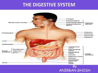
- 1. THE DIGESTIVE SYSTEM By ANIRBAN GHOSH
- 2. NUTRITION TYPES : * Ingestion * Digestion Types of Nutrients : * Micronutrients * Macronutrients FUNCTION OF DIGESTIVE SYSTEM * Acquires nutrients from environment * Anabolism (synthesize) * Catabolism (Decomposes & energy)
- 3. Actions of Digestive (GI) Tract & parts • Ingestion • Secretion • Mechanical process – crushing / shearing • Adsorption • Digestion – Chemical breakdown • Excretion
- 4. MOUTH – Mechanical digestion • teeth – breaking up food – Chemical digestion • saliva – amylase » enzyme digests starch – mucin » slippery protein (mucus) » protects soft lining of digestive system » lubricates food for easier swallowing – buffers » neutralizes acid to prevent tooth decay – anti-bacterial chemicals » kill bacteria that enter mouth with food Muscles of Mastication 1) Close the jaws 2) Slide or rock lower jaw from side to side 3) Chewing involves mandibular * Elevation and depression * Protraction and retraction * Medial and lateral movement • Function of Mouth 1. Break up food and digest starch . 2. Kills germs and moisten food .
- 5. Teeth & Tongue 1. Bitter 2. Sour 3. Salty 4. Sweet Function : Taste & Mastication * Upper surface - Epithelium * Dorsal surface - Keratinised
- 6. ESOPHAGUS Functions : 1.Secrete mucus 2.Moves food from the throat to the stomach using muscle movement called peristalsis • If acid from the stomach gets in here that’s heartburn. Peristalsis is series of involuntary wave-like muscle contractions which move food along the digestive tract . It is also called deglutition , it can be initiated voluntarily . This proceeds automatically and is divided into three phases i) Buccal phase , ii) Pharyngeal phase , iii) Esophageal phase . • A hollow muscular tube . • About 25 cm (10 in.) long and 2 cm (0.80 in.) wide . • Conveys solid food and liquids to the stomach . • Begins posterior to cricoids cartilage . • Is innervated by fibers from the esophageal plexus .
- 7. STOMACH * Stomach is a J-shaped muscular bag that stores the food we eat , breaks it down into tiny pieces. * The stomach takes around 4 hours to do it’s job on the food, depending on what kinds of food are digested. * Regions of the Stomach - Cardia - Fundus - Body - Pylorus Constantly being replaced, covered thick mucus, same simple columnar epithelium * Pyloric Sphincter – regulates gastric emptying * Gastric Glands - In fundus and body of stomach Extend deep into underlying lamina propria Each gastric pit communicates with several gastric glands Parietal cells Chief cells * Parietal Cells – Secrete intrinsic factor and hydrochloric acid * Chief Cells - Secrete hydrochloric acid (HCl) Are most abundant near base of gastric gland Secrete pepsinogen (inactive proenzyme) * Pepsinogen - Is converted by HCl in the gastric lumen To pepsin (active proteolytic enzyme)
- 8. •Functions of STOMACH – food storage - can stretch to fit ~2L food – disinfect food – (HCl = pH 2) , kills bacteria – chemical digestion • Pepsin • enzyme breaks down proteins • NOTES : Still, the epithelium is continually eroded, and the epithelium is completely replaced by mitosis every three days. • Gastric ulcers, lesions in the stomach lining, are caused by the acid-tolerant bacterium Heliobacter pylori. – Ulcers are often treated with antibiotics. • Pepsin is secreted in an inactive form, called pepsinogen by specialized chief cells in gastric pits. – Parietal cells, secrete hydrochloric acid converts pepsinogen to the active pepsin only when both reach the lumen of the stomach, minimizing self-digestion. – Also, in a positive-feedback system, activated pepsin can activate more pepsinogen molecules.
- 9. Small Intestines are roughly 7 meters long . Lining of intestine walls has finger-like projections called villi, to increase surface area. The villi are covered in microvilli which further increases surface area for absorption. Nutrients from the food pass into the bloodstream through the small intestine walls. Simple sugars and proteins are absorbed into the inner lining. Fatty acids and glycerol go to lymphatic system. Absorbs: 80% ingested water ,Vitamins , Minerals , Carbohydrates , Proteins Lipids * Secretes digestive enzymes Function * chemical digestion - major organ of digestion & absorption . * absorption through lining over 6 meters! small intestine has huge surface area = 300m2 (~size of tennis court) . Structure : duodenum = most digestion jejunum = absorption of nutrients & water ileum = absorption of nutrients & water
- 10. • The Duodenum – The segment of small intestine closest to stomach , is 25 cm (10 in.) long . – “Mixing bowl” that receives chyme from stomach and digestive secretions from pancreas and liver . Functions of the duodenum : 1)To receive chyme from stomach . 2) To neutralize acids before they can damage the absorptive surfaces of the small intestine . * About every 20 seconds, the stomach contents are mixed by the churning action of smooth muscles . A squirt at a time, it takes about 2 to 6 hours after a meal for the stomach to empty. The Jejunum - Is the middle segment of small intestine about 2.5 meters (8.2 ft) long . - Is the location of most Chemical digestion , Nutrient absorption . - Has few plicae circulares . The Ileum - The final segment of small intestine 3.5 meters (11.48 ft) long - Ends at the ileocecal valve, a sphincter that controls flow of material from the ileum into the large intestine Brush Border Enzymes Integral membrane proteins On surfaces of intestinal microvilli Break down materials in contact with brush border. Brush border enzymes break nucleotides into Sugars, Phosphates , Nitrogenous bases . Intestinal Glands Enteropeptidase A brush border enzyme Activates pancreatic proenzyme trypsinogen Enteroendocrine cells Produce intestinal hormones such as gastrin, cholecystokinin, and secretin Duodenal Glands Also called submucosal glands or Brunner glands Produce copious quantities of mucus with chyme
- 11. LARGE INTESTINE• Is horseshoe shaped • Extends from end of ileum to anus • Lies inferior to stomach and liver • Frames the small intestine • Also called large bowel • Is about 1.5 meters (4.9 ft) long and 7.5 cm (3 in.) wide Living in the large intestine is a community of helpful bacteria that ferments carbohydrates and breaks up proteins . Escherichia coli (E. coli) produce vitamins vitamin K ; B vitamins generate gases that are by-product of bacterial metabolism methane, hydrogen sulfide, Indole & skatole. Bacteria convert bilirubin to urobilinogens and stercobilinogens FUNCTION : • Re-absorption of water use ~ 9 liters of water every day in digestive juices > 90% of water reabsorbed If not enough water absorbed = diarrhea ; If too much water absorbed = constipation • Absorption of important vitamins produced by bacteria; Vitamins K ( fat soluble ) Required by liver for synthesizing four clotting factors, including prothrombin and B (Pantothenic acid B5) manufacture of steroid hormones and some neurotransmitters , Biotins ( Water soluble ) Important in glucose metabolismare reabsorbed with the water.
- 12. PARTS OF LARGE INTESTINEThe Cecum •Is an expanded pouch •Receives material arriving from the ileum •Stores materials and begins compaction Appendix •Also called vermiform appendix •Is a slender, hollow appendage about 9 cm (3.6 in.) long •Is dominated by lymphoid nodules (a lymphoid organ) The Colon •Has a larger diameter and thinner wall than small intestine •The wall of the colon forms a series of pouches (haustra) •Haustra permit expansion and elongation of colon
- 13. PARTS OF COLON • Ascending Colon – Begins at superior border of cecum – Ascends along right lateral and posterior wall of peritoneal cavity to inferior surface of the liver and bends at right colic flexure (hepatic flexure) • Transverse Colon – Crosses abdomen from right to left ; turns at left colic flexure (splenic flexure) – Is supported by transverse mesocolon – Is separated from anterior abdominal wall by greater omentum • The Descending Colon – Proceeds inferiorly along left side to the iliac fossa (inner surface of left ilium) – Is retroperitoneal, firmly attached to abdominal wall • The Sigmoid Colon – Is an S-shaped segment, about 15 cm (6 in.) long – Starts at sigmoid flexure – Lies posterior to urinary bladder – Is suspended from sigmoid mesocolon – Empties into rectum
- 14. PARTS OF LARGE INTESTINE • The Rectum – Forms last 15 cm (6 in.) of digestive tract – Is an expandable organ for temporary storage of feces – Movement of fecal material into rectum triggers urge to defecate • The anal canal is the last portion of the rectum – Contains small longitudinal folds called anal columns • Anus – Also called anal orifice – Is exit of the anal canal – Has keratinized epidermis like skin
- 15. Accessory Organs • Not part of the path of food, but play a critical role. • Include: Liver, gall bladder, and pancreas
- 16. Gall Bladder • Pouch structure located near the liver which concentrates and stores bile • Bile duct – a long tube that carries BILE. The top half of the common bile duct is associated with the liver, while the bottom half of the common bile duct is associated with the pancreas, through which it passes on its way to the intestine. Bile Bile emulsifies lipids (physically breaks apart FATS) Bile is a bitter, greenish-yellow alkaline fluid, stored in the gallbladder between meals and upon eating is discharged into the duodenum where it aids the process of digestion. Pancreas An organ which secretes both digestive enzymes (exocrine) and hormones (endocrine). Lies posterior to stomach from duodenum toward spleen is bound to posterior wall of abdominal cavity . It is wrapped in thin, connective tissue capsule Functions of the Pancreas 1)Endocrine cells of the pancreatic islets : Secrete insulin and glucagon into bloodstream . 2) Exocrine cells : Acinar cells and epithelial cells of duct system secrete pancreatic juice . Pancreatic Acini : It is a Blind pockets , they are lined with simple cuboidal epithelium , Contain scattered pancreatic islets (Endocrine tissues of pancreas , contains Scattered 1% of pancreatic cells) .
- 17. • Pancreatic Enzymes – Pancreatic alpha-amylase • A carbohydrase . • Breaks down starches . • Similar to salivary amylase ( i.e ; helps in chemical digestion ) . – Pancreatic lipase • Breaks down complex lipids . • Releases products (e.g., fatty acids) that are easily absorbed . – Nucleases • Break down nucleic acids . – Proteolytic enzymes • Break certain proteins apart . • Proteases break large protein complexes . • Peptidases break small peptides into amino acids . • 70% of all pancreatic enzyme production . • Secreted as inactive proenzymes . • Activated after reaching small intestine .
- 18. LIVER •Is the largest visceral organ (1.5 kg; 3.3 lb) •Lies in right hypochondriac and epigastric regions •Extends to left hypochondriac and umbilical regions •Performs essential metabolic and synthetic functions * Anatomy of the Liver Is wrapped in tough fibrous capsule Is covered by visceral peritoneum Is divided into lobes * Physiology of the Liver 1. Metabolic regulation 2. Hematological regulation 3. Bile production Metabolic Regulation The liver regulates: 1. Composition of circulating blood 2. Nutrient metabolism (carbohydrate, lipid & amino acid) 3. Waste product removal 4. Vitamin Storage (A, D, E & K) 5. Nutrient storage (iron) 6. Drug inactivation
- 19. HEMATOLOGICAL REGULATION • Composition of Circulating Blood – All blood leaving absorptive surfaces of digestive tract • Enters hepatic portal system • Flows into the liver – Liver cells extract nutrients or toxins from blood • Before they reach systemic circulation through hepatic veins – Liver removes and stores excess nutrients • Corrects nutrient deficiencies by mobilizing stored reserves or performing synthetic activities • Hematological Regulation : Largest blood reservoir in the body Removes damaged / old red blood cells and it receives 25% of cardiac output • Functions of Hematological Regulation 1. Phagocytosis and antigen presentation 2. Synthesis of plasma proteins : Plasma proteins: albumin – contrbutes to osmotic concentration 3. Removal of circulating hormones : Reabsorped Epinephrine, norepinephrine, insulin, thyroid & steroid hormones 4. Removal of antibodies : Removes antibodies and converts to amino acids 5. Removal or storage of toxins : Traps some lipid-soluble toxins (DDT) or breaks down and removes from blood 6. Synthesis and secretion of bile
- 20. BILE PRODUCTION 1. The Bile Duct System • Liver secretes bile fluid • Into a network of narrow channels (bile canaliculi) • Between opposing membranes of adjacent liver cells 2. Right and Left Hepatic Ducts • Collect bile from all bile ducts of liver lobes • Unite to form common hepatic duct that leaves the liver 3. Bile Flow • From common hepatic duct to either • The common bile duct, which empties into duodenal ampulla • The cystic duct, which leads to the gall-bladder 4. The Common Bile Duct •Is formed by union of - Cystic duct - Common hepatic duct •Passes within the lesser omentum toward stomach •Penetrates wall of duodenum •Meets pancreatic duct at duodenal ampulla
- 21. Coordination of Secretion & Absorption• Secretin Is released when chyme arrives in duodenum Increases secretion of bile and buffers by liver and pancreas • Cholecystokinin (CCK) Is secreted in duodenum When chyme contains lipids and partially digested proteins Accelerates pancreatic production and secretion of digestive enzymes Relaxes hepatopancreatic sphincter and gallbladder Ejecting bile and pancreatic juice into duodenum • Gastric Inhibitory Peptide (GIP) Is secreted when fats and carbohydrates enter small intestine • Vasoactive Intestinal Peptide (VIP) Stimulates secretion of intestinal glands Dilates regional capillaries Inhibits acid production in stomach • Gastrin Is secreted by G cells in duodenum When exposed to incompletely digested proteins Promotes increased stomach motility Stimulates acids and enzyme production • Enterocrinin Is released when chyme enters small intestine Stimulates mucin production by submucosal glands of duodenum
- 22. Digestive Homeostasis Disorders • ULCERS – erosion of the surface of the alimentary canal generally associated with some kind of irritant ; it is generally caused by HCl secretion for long period of time in the stomach .
- 23. Digestive Homeostasis Disorders CONSTIPATION – a condition in which the large intestine is emptied with difficulty. Too much water is reabsorbed and the solid waste hardens .
- 24. Digestive Homeostasis Disorders •DIARRHEA – a gastrointestinal disturbance characterized by decreased water absorption and increased peristaltic activity of the large intestine. •This results in increased, multiple, watery feces. •This condition may result in severe dehydration, especially in infants
- 25. Digestive Homeostasis Disorders APPENDICITIS – an inflammation of the appendix due to infection Common treatment is removal of the appendix via surgery
- 26. Digestive Homeostasis Disorders • GALLSTONES – an accumulation of hardened cholesterol and/or calcium deposits in the gallbladder • Can either be “passed” (OUCH!!) or surgically removed
- 27. Digestive Homeostasis Disorders • ANOREXIA NERVOSA - a psychological condition where an individual thinks they appear overweight and refuses to eat. • Weighs 85% or less than what is developmentally expected for age and height • Young girls do not begin to menstruate at the appropriate age.
- 29. Let’s go to the Video!