Anaesthesia equipment
•
364 j'aime•95,292 vues

Different types of anesthetic equipment and their use. Anaesthetic machine, Cylinders, Breathing systems, Ventilator, Air ways, Laryngoscope, Endotracheal tubes, Tracheostomy tube, I.V.Cannula, Syringe pumps, Magill’s forceps, Central Venous Pressure, Nasogastric tube, ect.

Recommandé
Contenu connexe
Tendances
Tendances (20)
Similaire à Anaesthesia equipment
Similaire à Anaesthesia equipment (20)
Plus de Arjuna Samaranayaka
Plus de Arjuna Samaranayaka (20)
Dernier
Dernier (20)
Anaesthesia equipment
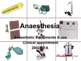
- 1. Anaesthesia Anaesthetic Equipments & use Clinical appointment 2002/03 A Devdas 2002 / 03 A
- 2. Anaesthetic machine • • • • • • • Cylinders / pipe line gas supply Reducing valves Vaporisers Flow meters Sodalime canister Breathing circuits Accessories – face masks, laryngoscopes, ET tubes, metal stylets, air ways
- 3. Anaesthesia machine Pipe line supply Vaporizers Reducing valves Flow meters Sodalime canister Cylinders Breathing circuit
- 4. Cylinders • Constructed from Molybdenum • Colour coded according to gas supplied • Safety – Pin index system – Oxygen – Nitrous oxide – Pipe line gas supply
- 6. Oxygen • In the form of gas – 130 bars • Pressure gauge is there • Outlet valve can only connect the O 2 cylinder to the machine • Alarm – when O2 is depleting
- 7. Nitrous oxide • Liquid form – pressure 51 bars until the liquid vaporizes • Heat vaporization • Pressure gauge – doesn’t indicate the measure of contents • Blue color cylinder
- 8. Pipe line gas supply • All gases in liquid form – economical • Pressure 4 bars • Identification – gas name, color code, shape • Convenient
- 10. Flow meters • Tapered glass tubes contains Spinning bobbins • Calibrated for a specific gas Vaporizers • Vaporizes inhalational anesthetics in liquid form at room T • Calibrated for a specific inhalational agent
- 11. Breathing systems • Delivers anaesthetic gases & oxygen to the patient & removes CO2 – Magill circuit – T-piece (Paediatric circuit) – Bain circuit – Circle circuit – commonly used
- 12. Mapleson classification • Mapleson system A • • – Magill system – most satisfactory with spontaneous respiration Mapleson system B – not common Mapleson system C – not common – Water’s system • Mapleson system D – Bain co-axial system • Mapleson system E – Ayre’s T piece – paediatric use • Efficiency of systems with spontaneous respiration A > D, E > C > B • Efficiency of systems with IPPV D, E > B > C > A • Most important closed circle or low flow system – Rebreathing system Refer more on Circle system
- 13. Ventilator circuit – circle circuit Re breathing system Sodalime is needed to absorb CO2 from exhaled gas Low fresh gas flows Economical Less theater pollution Humidification is better Valves – not ideal for pediatric use Heavy
- 14. Bain circuit Coaxial system – inspiratory limb inside & outer expiratory limb Used both in spontaneous & controlled ventilation Scavenging gases is easy Reservoir bag – 2 L
- 15. Pediatric – T-Piece No valves no resistance to respiration Fresh gas flow should be twice the minute volume Reservoir bag fixed on the open end – for controlled ventilation See different breathing circuits identify according to MAPLESON classification
- 16. Sodalime • Used with circle circuit – re-breathing systems • Calcium hydroxide(94%)Na(5%) & K(1%) hydroxides • Absorbs CO2 – color change when it becomes inactive Facemasks • Rubber or plastic • Transparent ones show vomitus & secretions
- 17. Ventilator
- 18. Setting up a simple ventilator • Connect the gas supply – usually N2O(66%) O2(33%) air… • Connect the outlet to the breathing circuit • Select the, – Tidal volume – 7-10ml/kg – Respiratory rate -12-16/min – Inspiration : expiration ratio (usually 1:2) • Keep upper airway pressure 10-20 cmH2O • Negative upper airway pressure means patient is voluntarily breathing
- 19. Oxygen mask Look how this is connected to gas supply & to ambu bag What happens if connection to ambu bag is lost?
- 20. Ambu bag Ideal valve should have No foreward leak No backward leak Low resistance Minimal dead space Self inflating bag Light weight Reservoir bag – 100% O2 Minimal opening pressure without sticking Valves – Transparency 1.Prevent forward leak Reliability & durability 2.Prevent backward leak 3.Expiratory 4.Inspiratory 5. Pressure releasing >40mmHg
- 21. Face mask Triple manure 1. Head tilt 2. Extended to straiten the airway & prevent tongue falling back 3. Jaw thrust Why DENITRATION is important? To increase the % of O2 in alveoli before intubation This enables enough time to intubate
- 22. Face Masks NOTE : How face mask is held preventing air leak Practice Triple Maneuver
- 23. Air ways • Air way maintenance by displacing the tongue from posterior pharynx • Oro-pharygeal airway • Nasal airway – when unable to open the mouth & in oral surgeries • Paediatric & adult types Dotted Color coding
- 24. Adult Guedel airways Look for the dotted coding in pediatric airway
- 25. Nasal airway What are the contraindications for nasal intubation? What are the complications of nasal intubation? Note : Why gauze pack is put into the oral cavity in nasal intubation?
- 26. Laryngeal mask airways • Short tube with a elliptical cuff • Placed in the laryngopharynx behind the inlet • Can avoid muscle relaxants no haemodynamic response like in ET tube • Relieves hypoxia in failed intubation • Blind intubation through mask is possible • Risk of regurgitation is there • Alternative to face mask • Needs minimum skills Learn the procedure of putting laryngeal mask
- 27. Laryngoscope Curved blade – Macintosh Straight blade – Magill (for children) Battery in the handle – check whether the bulb is working before use Use with the left hand Put it in from the right side Groove in the left side of the blade is for to keep the tongue apart & lifted What are the uses of laryngoscope? Macintosh laryngoscope – curved blade
- 28. Endotracheal tube - cuffed How to describe a endotracheal tube? Cuffed or not Internal diameter Main parts – from above downwards 1.Universal airway connector 2.Tube 3. Radio opaque line 7.Murfy’s eye 8. Black lines 2 Use of each part & there importance 4. Pilot tube 5.Pilot inflator bag 6.Cuff type – high P/ low V or low P / high V What are the uses of flexible & reinforced tubes? What is RAE tube?
- 29. Importance • Universal airway connector – external diameter is same in every tube (15mm) • Tube- internal diameter – female – 7.0 to 8.0 mm – male 8.0 to 10.0 mm • Radio opaque line – identify it radiologically • Pilot tube – to inflate the cuff • Inflating bag – 5ml • Cuff – High pressure / low volume – high risk of aspiration & pressure necrosis – Low pressure / high volume – vise versa • Two black lines – vocal cords should be in between – risk of damage is reduced • Murfy’s eye – to keep patancy when secretions block the tube – To keep the patancy of the upper right bronchus
- 30. Endotracheal tube – plain/non cuffed Indications for ET tubes 1.When muscle relaxants given 2.In patients with risk of aspiration 3.To provide controlled ventilation 4.For prolong operations Why non-cuffed is preferred in children? Their narrowest place is at cricoid. tube fits nicely.
- 31. Endotracheal tubes • What is I.T. on tube? – Implantation test for allergies has been done. • What is the importance of reinforced tube? – It is not liable to kink – in oral surgeries • What are the indications of Proper Intubation? 1. Feels expired air touching the dorsal surface of hand when it is neared to the airway connector 2. See vapor in the tube 3. Hold a piece of thread near the opening – it will move 4. Positive capnogram on monitor
- 32. Tracheostomy tube Sized according to the internal diameter Cuffed or uncuffed Introducer is available Winged flange to secured to skin Indications 1.In ICU patients who needs prolong intubation 2.Vocal cord palsy 3.OP poisoning 4.Oropharyngeal laryngeal carcinomas 5. Chemical burns 6. Gillian Bare syndrome .. etc
- 33. Complications of tracheostomy 1. Early : • • • • • • • • 2. Late : Haemorrhage • Infection Displacement or • Tracheal ulceration obstruction • Tracheal dilatation Injury to trachea • Tracheal stenosis Tracheitis • Cardiovascular Crust formation collapse Surgical emphysema Difficult insertion pneumothorax
- 34. After care of tracheostomy • Position the patient – Adults – propped up – Children – chin should not occlude • Suction – clean catheter used – Deep suction + physiotherapy or ventilation – in unconcious • Humidification – Prevent drying & formation of crusts – Wet guaze in the opening • Tube changing – – – – • • 2-3 days Silver tubes – remove inner tube & clean Cuffed tubes – regular deflation to prevent pressure necrosis Air – minimum to prevent air leak Pain management Management of hemorrhage
- 35. Intravenous fluid set Priming an IV line 1.Take out from the pack – mark to scratch in the packet 2.Clamp it 3.Never touch the connector 4.Open the cap of fluid bottle & connect the line 5.Open the line till half of the syringe fills 6.Open fully & allow fluid to come out – to assure no air bubbles inside Risks 7.Connect to the cannula Septicemia Air embolism
- 36. I.V.Cannula Gauges – color code Orange 14G Ash 16G White 17G Green 18G Pink 20G Blue 22G Yellow 24G (Spinal needle – 25Gfrench grading) Demonstrate how IV cannulation is done discuss importance of each step
- 37. Control syringe Learn the use of syringe
- 38. Syringe pumps • Electrically driven – battery back-up is there • Alerts provided for – Power failure – Empty syringe – Occlusion of delivery pipe • Applications – – – – – – Pain relief Total IV anaesthesia Sedation in intensive care CVS support Relaxants Control of diabetes
- 39. Dose calculation for syringe pump • • • • Dobutamine solution – 200mg in 500ml Dose – eg: 2.0μg / kg / min Body weight of the patient – eg: 50kg So, amount of mg per hour = 2.0 μg × 50kg × 60 = 6mg 1000 • Amount of solution needed per hour = 500ml × 6mg = 15ml / hour 200mg • So the infusion rate = 3.8 per minute
- 40. Magill’s forceps Guide the ET tube into larynx – in nasal intubation Guide the nasogastric tube into oesophagus See how this is held?
- 41. Blood transfusion set Normal blood amount Adult male – 70ml / kg Adult female – 60ml / kg Child – 80ml / kg Discuss how blood loss is assessed in theatre? Discuss management of hemorrhagic shock? Learn the appropriate use of three way tap
- 42. Transfusion bags List the blood components & their indications
- 44. CVP Manometer Measure, Adequacy of blood or fluid replacement Easy & rapid transfusion of blood Normal – 3 to 10 cmH2O Low CVP 1.Hypovolemia 2.Septic shock High CVP 1.Heart failure 2.Increased intra thoracic pressure – IPPV, PEEP pneumothorax 3.Ovetransfusion 4.Constrictive pericarditis 5.Pulmonary vasoconstriction
- 45. Central Venous Pressure • >50% of the total blood volume is in venous system – alteration in venous tone play a large part in regulation of hemodynamics • Zero at mid axillary line • Normal – 3 to 10 cmH2O • Complications – – – – – – – – Thromboplebitis, infection, septicemia Pneumothorax, haemothorax, hydrothorax Bracheal plexus injury Air embolus Pericardial effusion Lymphatic leakage Arrhythmias Catheter breaking
- 46. Peritoneal dialysis - set
- 47. Peritoneal dialysis - catheter
- 50. Spinal needle Whitacre spinal needle - 25G Commonly used Subarachnoid anaesthesia 1. Take the consent 2. Look for contraindications 1. CNS lesions – ideally full CNS examination should be done 2. Sepsis around the area of pricking 3. Any coagulopathies 3. Sterile procedure
- 51. Spinal needles Spinal introducer for Sprotte cannula Spinal sprotte cannula Spinal sprotte cannula
- 52. Foley’s catheter Learn the aseptic procedure of catheterization
- 53. Guide wire – External Pace Maker
- 54. Suction catheters Twin bore nasal
- 55. Nasogastric tube Indications 1. Aspiration of gastric juices for the diagnostic & therapeutic purposes 2. Confirmation of gastroduodenal hemorrhage 3. Feeding Confirmation of it’s presence in the stomach 1.Syringing the air down the tube while listening over epigastrium 2.Suck the tube to see juices are coming 3.Vapour inside in accidental tracheal intubation
- 56. Identify?
- 57. Identify?
- 59. I.V.Cannula Why do you tap to find appropriate vein for canulation? Why veins in the periphery are selected first? What should be done if peripheral veins are not visible? When do you use low grade cannulae for adults? What are the complications? Why anti-cubital fossa is not selected as a good site for cannulation?