SlideShare a Scribd company logo
1 of 74
HUMAN CELL
HUMAN CELL
JAI NARAIN VYAS UNIVERSITY, JODHPUR
ASSISTANT PROFESSOR:- ASHWIN SINGH
CHOUHAN
DEPARTMENT:- PHARMACOLOGY
E-mail:- anshukavya1993@gmail.com
JNVU PHARMACY, JODHPUR
THE CELL
Cell: The basic structural and functional unit of any living
thing. Each cell is a small container of chemicals and
water wrapped in a membrane. There are 100
trillion cells in a human, and each contains all of the
genetic information necessary to manufacture a human
being.
IT’s the basic functional in a human meaning that it is a
self-contained and fully operational living entity. Humans
are multicellular organisms with various different types of
cells that work together to sustain life.
Other non-cellular components in the body include
water, macronutrients (carbohydrates, proteins, lipids),
micronutrients (vitamins, minerals) and electrolytes.
A collection of cells that function together to perform
the same activity is known as tissue.
Masses of tissue work collectively to form an organ that
performs specific functions in the body.
The functions of the human cell varies based on the type
of cell and its location in the human body. All the
organelles work together to keep the cell alive and allow
it to carry out its specific function. Sometimes these
organelles are highly specialized and can vary in (size,
shape and number). The organelles are the most basic
functional units but it cannot exist and operate without
the cell as a whole. Its functions include intake of
nutrients and other substances, processing of these
compounds, production of new substances, cell
replication and energy production. In specialized cells
that need to be motile, like sperm cells, tail-like
projections allow for cellular locomotion.
JNVU PHARMACY, JODHPUR
DISCOVERY OF CELLS
Discovery of cells is one of the remarkable advancements in the
field of science. It helped us know that all the organisms are made up
of cells, and these cells help in carrying out various life processes.
The structure and functions of cells helped us to understand life in a
better way.
Robert Hooke discovered the cell in 1665. Robert Hooke observed a
piece of bottle cork under a compound microscope and noticed
minuscule structures that reminded him of small rooms.
Consequently, he named these “rooms” as cells. However, his
compound microscope had limited magnification, and hence, he could
not see any details in the structure. Because of this limitation, Hooke
concluded that these were non-living entities.
Later Anton Van Leeuwenhoek observed cells under another
compound microscope with higher magnification. This time, he had
noted that the cells exhibited some form of movement (motility). As
a result, Leeuwenhoek concluded that these microscopic entities were
“alive.” Eventually, after a host of other observations, these entities
were named as animalcules.
In 1883, Robert Brown, a Scottish botanist, provided the very first
insights into the cell structure. He was able to describe the nucleus
present in the cells of orchids. JNVU PHARMACY, JODHPUR
PARTS OF THE HUMAN CELL
The cell contains various structural components to allow it
to maintain life which are known as organelles. All the
organelles are suspended within a gelatinous matrix, the
cytoplasm, which is contained within the cell membrane.
One of the few cells in the human body that lacks almost
all organelles are the red blood cells .
The main organelles are as follows :
Nucleus
Cell membrane
Endoplasmic reticulum
Golgi apparatus
Lysosomes
Peroxisomes
Mitochondria
Microfilaments and microtubules.
JNVU PHARMACY, JODHPUR
NUCLEUS
The nucleus : is the master control of the cell.
It contains genes, collections of DNA, which determines
every aspect of human anatomy and physiology.
The DNA which is arranged into chromosomes also
contains the blueprint specific for each type of cell which
allows for replication of the cell. Within the nucleus is an
area known as the nucleolus.
It is not enclosed by a membrane but is just an
accumulation of RNA and proteins within the nucleus.
The nucleolus is the site where the ribosomal RNA is
transcribed from DNA and assembled.
JNVU PHARMACY, JODHPUR
FUNCTIONS OF THE NUCLEUS
The main function of the cell nucleus is to control gene
expression and facilitate the replication of DNA during the
cell cycle (which you will learn about in the next chapter).
The nucleus controls the metabolic functions of the cell
by producing mRNA which encodes for enzymes e.g.
insulin.
The nucleus controls the structure of the cell by
transcribing DNA which encodes for structural proteins
such as actin and keratin.
The nucleus is the site of ribosomal RNA (rRNA)
synthesis, which is important for the construction of
ribosomes. Ribosomes are the site of protein translation
(synthesis of proteins from amino acids).
Characteristics are transmitted from parent to offspring
through genetic material contained in the nucleus.
JNVU PHARMACY, JODHPUR
CELL MEMBRANE
The cell membrane is the outer coating of the cell and
contains the cytoplasm, substances within it and the
organelle.
It is a double-layered membrane composed of proteins
and lipids. The lipid molecules on the outer and inner part
(lipid bilayer) allow it to selectively transport substances
in and out of the cell.
Endoplasmic Reticulum: The endoplasmic reticulum (ER)
is a membranous structure that contains a network of
tubules and vesicles.
Its structure is such that substances can move through
it and be kept in isolation from the rest of the cell until
the manufacturing processes conducted within are
completed.
There are two types of endoplasmic reticulum – rough
(granular) and smooth (a granular)
JNVU PHARMACY, JODHPUR
CELL MEMBRANE
Functions
The cell membrane provides mechanical support that
facilities the shape of the cell while enclosing the cell and
its components from the external environment.
It regulates what can be allowed to enter and exit the
cell through channels, acting as a semi-permeable
membrane, which facilities the exchange of essential
compounds required for the survival of the cell.
It generates and distributes signals in and outside of the
cell for the proper functioning of the cell and all the
organelles.
It allows the interaction between cells required during
tissue formation and cell fusion.
JNVU PHARMACY, JODHPUR
An additional non-living layer present outside the cell
membrane in some cells that provides structure,
protection, and filtering mechanism to the cell is the cell
wall.
Structure
In a plant cell, the cell wall is made up of cellulose,
hemicellulose, and proteins while in a fungal cell, it is
composed of chitin.
A cell wall is multilayered with a middle lamina, a primary
cell wall, and a secondary cell wall.
Middle lamina contains polysaccharides that provide
adhesion and allows binding of the cells to one another.
After middle lamina is the primary cell wall which is
composed of cellulose. The last layer, which is not always
present, is the secondary cell wall made of cellulose and
hemicellulose.
CELL WALL
JNVU PHARMACY, JODHPUR
Functions
The critical function of the cell wall is protecting and
maintaining the shape of the cell. It also helps the cell
withstand the turgor pressure of the cell.
It initiates cell division by providing signals to the cell
and allows the passage of some molecules into the cell
while blocking others. JNVU PHARMACY, JODHPUR
JNVU PHARMACY, JODHPUR
CYTOPLASM
Cytoplasm The gel-like material within the cell membrane is referred
to as the cytoplasm. It is a fluid matrix, the cytosol, which consists of
80% to 90% water, salts, organic molecules and many enzymes that
catalyze reactions, along with dissolved substances such as proteins
and nutrients. The cytoplasm plays an important role in a cell,
serving as a "molecular soup" in which organelles are suspended and
held together by a fatty membrane. Within the plasma membrane of
a cell, the cytoplasm surrounds the nuclear envelope and the
cytoplasmic organelles. It plays a mechanical role by moving around
inside the membrane and pushing against the cell membrane helping
to maintain the shape and consistency of the cell and again, to
provide suspension to the organelles. It is also a storage space for
chemical substances indispensable to life, which are involved in vital
metabolic reactions, such as anaerobic glycolysis and protein
synthesis. The cell membrane keeps the cytoplasm from leaking out.
It contains many different organelles which are considered the
insoluble constituents of the cytoplasm, such as the mitochondria,
lysosomes, peroxysomes, ribosomes, several vacuoles and
cytoskeletons, as well as complex cell membrane structures such as
the endoplasmic reticulum and the Golgi apparatus that each have
specific functions within the cell
JNVU PHARMACY, JODHPUR
CYTOPLASM
The cytoplasm is the jelly-like substance that fills the cell.
It consists of up to 90%90% water. It also contains
dissolved nutrients and waste products. Its main function
is to hold together the organelles which make up the
cytoplasm. It also nourishes the cell by supplying it with
salts and sugars and provides a medium for metabolic
reactions to occur.
All the contents of prokaryotic cells are contained within
the cytoplasm. In eukaryotic cells, all the organelles are
contained within the cytoplasm except the nucleolus
which is contained within the nucleus.
JNVU PHARMACY, JODHPUR
FUNCTIONS OF THE CYTOPLASM
The cytoplasm provides mechanical support to the cell
by exerting pressure against the cell's membrane which
helps keep the shape of the cell. This pressure is known
as turgor pressure.
It is the site of most cellular activities including
metabolism, cell division and protein synthesis.
The cytoplasm contains ribosomes which assist in the
synthesis of protein.
The cytoplasm acts a storage area for small
carbohydrate, lipid and protein molecules.
The cytoplasm suspends and can transport organelles
around the cell.
JNVU PHARMACY, JODHPUR
CENTRIOLES
Centrioles are rod like structures composed of 9 bundles
which contain three microtubules each. Two
perpendicularly placed centrioles surrounded by proteins
make up the centrosome. Centrioles are very important
in cellular division, where they arrange the mitotic
spindles that pull the chromosome apart. Centrioles and
basal bodies act as microtubule organizing centers. A pair
of centrioles (enclosed in a centrosome) located outside
the nuclear envelope gives rise to the microtubules that
make up the spindle apparatus used during cell division.
Basal bodies are at the base of each flagellum and cilium
and appear to organize their development.
JNVU PHARMACY, JODHPUR
Functions
During cell division, centrioles have a crucial role in
forming spindle fibers which assist the movement of
chromatids towards their respective sides.
They are involved in the formation of cilia and flagella.
CILIA AND FLAGELLA
Cilia and Flagella are tiny hair-like projections from the
cell made of microtubules and covered by the plasma
membrane.
Structure
Cilia are hair-like projections that have a 9+2
arrangement of microtubules with a radial pattern of 9
outer microtubule doublet that surrounds two singlet
microtubules. This arrangement is attached to the bottom
with a basal body.
Flagella is a filamentous organelle, the structure of
which, is different in prokaryotes and eukaryotes.
In prokaryotes, it is made up of the protein called
flagellin wrapped around in a helical manner creating a
hollow structure at the center throughout the length.
In eukaryotes, however, the protein is absent and the
structure is replaced with microtubules.
JNVU PHARMACY, JODHPUR
Functions
The most critical role of cilia and flagella is movement.
These are responsible for the movement of the organisms
as well as for the movement of various particles present
around the organisms.
Some cilia present in some particular organs may have
the function of sense. The cilium in the blood vessels,
which helps in controlling the flow of blood is an example.
JNVU PHARMACY, JODHPUR
GOLGI APPARATUS
The Golgi apparatus is a stacked collection of flat vesicles.
It is closely associated with the endoplasmic reticulum
in that substances produced in the ER are transported as
vesicles and fuses with the Golgi apparatus.
In this way, the products from the ER are stored in the
Golgi apparatus and converted into different substances
that are necessary for the cell’s various functions
JNVU PHARMACY, JODHPUR
GOOGI APPARATUS
FUNCTIONS OF THE GOLGI BODY
It is important for proteins to be transported from
where they are synthesised to where they are required in
the cell. The organelle responsible for this is the Golgi
Body. The Golgi body is the sorting organelle of the cell.
Proteins are transported from the rough endoplasmic
reticulum (RER) to the Golgi. In the Golgi, proteins are
modified and packaged into vesicle. The Golgi body
therefore receives proteins made in one location in the
cell and transfers these to another location within the cell
where they are required. For this reason the Golgi body
can be considered to be the 'post office' of the cell.
JNVU PHARMACY, JODHPUR
LYSOSOMES
Lysosomes: are vesicles that break off from the Golgi
apparatus. It varies in size and function depending on the
type of cell. Lysosomes contain enzymes that help with
the digestion of nutrients in the cell and break down any
cellular debris or invading microorganisms like bacteria. A
structure that is similar to a lysosome is the secretory
vesicle. It contains enzymes that are not used within the
cell but emptied outside of the cell, for example the
secretory vesicles of the pancreatic lacunar cell release
digestive enzymes which help with the digestion of
nutrients in the gut.
JNVU PHARMACY, JODHPUR
Lysosomes are known
as suicide bags of the
cell because they
contain lytic enzymes
capable of digesting cells
and unwanted materials.
Functions of Lysosomes
Lysosomes serve two major functions:
1.Intracellular Digestion
•To digest food, the lysosome membrane fuses with the
membrane of food vacuole and squirts the enzymes inside.
•The digested food then diffuses through the vacuole
membrane and enters the cell to be used for energy and
growth.
2.Autolytic Action
•Cell organelles that need to be get ridden are covered by
vesicles or vacuoles by the process of autophagy to form
autophagosome.
•The autophagosome is then destroyed by the action of
lysosomal enzymes.
Processes in which lysosomes play crucial roles include:
a. Heterophagy
The taking into the cell of exogenous material by
phagocytosis or pinocytosis and the digestion of the ingested
material after fusion of the newly formed vacuole with a
lysosome. JNVU PHARMACY, JODHPUR
b. Autophagy
A normal physiological process that deals with the destruction of
cells in the body. It is essential for maintaining homeostasis, for
normal functioning by protein degradation, turnover of destroyed
cell organelles for new cell formation
c. Extracellular Digestion
Primary lysosomes secrete hydrolases outside by exocytosis
resulting in degradation of extracellular materials.
Eg. Saprophytic fungi
d. Autolysis
It refers to the killing of an entire set of cells by the breakdown of
the lysosomal membrane. It occurs during amphibian and insect
metamorphosis.
e. Fertilization
The acrosome of the sperm head is a giant lysosome that
ruptures and releases enzymes on the surface of the egg. This
provides the way for sperm entry into the egg by digesting the
egg membrane.
f. As Janitors of the Cell
Lysosomes remove ‘junk’ that may accumulate in the cell helping
to prevent diseases JNVU PHARMACY, JODHPUR
JNVU PHARMACY, JODHPUR
PEROXISOMES
These organelles are very similar to the lysosomes and
contain enzymes that act together in the form of
hydrogen peroxide to neutralize substances that may be
toxic to the cell. Peroxisomes are formed directly from
the endoplasmic reticulum rather than from the Golgi
apparatus like lysosomes.
Functions of Peroxisomes
1.Hydrogen Peroxide Metabolism:
Enzymes present in the peroxisomes both lead to the
production and elimination of H202 which is a reactive
oxygen species.
2.Fatty acid oxidation:
Oxidation of fatty acids, in animal cells, occurs in both
peroxisomes and mitochondria, but in yeasts and plants,
only limited to peroxisomes.
Oxidation is accompanied by the production of
H202 which is decomposed by catalase enzyme. This
provides a major source of metabolic energy.
3.Lipid biosynthesis
Synthesis of cholesterol and dolichol occurs in both ER
and peroxisomes. Bile acid synthesis takes place from
cholesterol in the liver.
JNVU PHARMACY, JODHPUR
4.Germination of seeds
Peroxisomes in seeds responsible for the conversion of
stored fatty acids to carbohydrates, critical to providing
energy and raw materials for the growth of germinating
plants.
5.Photorespiration
Peroxisomes in leaves particularly in the green ones carry
out the photorespiration process along with chloroplasts.
6.Degradation of purines
Carry out the catabolism of purines, polyamines and
amino acids especially by uric acid oxidase
7.Bioluminescence
Luciferase enzyme found in the peroxisomes of fireflies
help in bioluminescence and thus aid the flies in finding a
mate or its meal.
Peroxisomes contain enzymes to synthesize plasmalogens,
a family of phospholipids which are important membrane
components of tissues of the heart and brain.
JNVU PHARMACY, JODHPUR
RIBOSOMES
Ribosomes play an active role in the complex process of
protein synthesis, where they serve as the structures that
facilitate the joining of amino acids. Each ribosome is
composed of a large and small subunit which are made
up of ribosomal proteins and ribosomal RNAs. They can
either be found in groups called polyribosomes within the
cytoplasm or found alone. Occasionally they are attached
to the endoplasmic reticulum
JNVU PHARMACY, JODHPUR
Function of ribosome
The ribosome is a complex molecular machine, found
within all living cells, that serves as the site of biological
protein synthesis (translation).
Ribosomes link amino acids together in the order
specified by messenger RNA (mRNA) molecules.
Ribosomes act as catalysts in two extremely important
biological processes called peptidyl transfer and peptidyl
hydrolysis.
JNVU PHARMACY, JODHPUR
MITOCHONDRIA
These are the powerhouses of the cell and break down
nutrients to yield energy. Apart from producing its own
energy, it also produces a high-energy compound called
ATP (adenosine triphosphate) which can be used as a
simple energy source elsewhere. Mitochondria are
composed of two membranous layers – an outer
membrane that surrounds the structure and an inner
membrane that provides the physical sites of energy
production. The inner membrane has many in folding that
form shelves where enzymes attach and oxidize
nutrients. The mitochondria also contain DNA which
allows it to replicate where and when necessary.
JNVU PHARMACY, JODHPUR
MITOCHONDRIA
Functions
The primary function of mitochondria is the synthesis of
energy in the form of ATP required for the proper
functioning of all the cell organelles.
Mitochondria also help in balancing the amount of Ca+
ions within the cell and assists the process of apoptosis.
Different segments of hormones and components of
blood are built within mitochondria.
Mitochondria in the liver have the ability to detoxify
ammonia.
ENDOPLASMIC RETICULUM
Endoplasmic Reticulum Endoplasmic means "within the
plasm" and reticulum means "network". A complex three
dimensional internal membrane system of flattened
sheets, sacs and tubes, that play an important role in
making proteins and shuttling cellular products; also
involved in metabolisms of fats, and the production of
various materials.
In cross-section, they appear as a series of maze-like
channels, often closely associated with the nucleus. When
ribosomes are present, the rough ER attaches
polysaccharide groups to the polypeptides as they are
assembled by the ribosomes. Smooth ER, without
ribosomes, is responsible for various activities, including
the synthesis of lipids and hormones, especially in cells
that produce these substances for export from the cell.
JNVU PHARMACY, JODHPUR
ENDOPLASMIC RETICULUM
Rough endoplasmic reticulum has characteristic bumpy
appearance due to the multitude of ribosomes coating it. It is
the site where proteins not destined for the cytoplasm are
synthesized. Smooth endoplasmic reticulum provides a variety
of functions, including lipid synthesis and degradation, and
calcium ion storage. In liver cells, the smooth ER is involved in
the breakdown of toxins, drugs, and toxic byproducts from
cellular reactions.
JNVU PHARMACY, JODHPUR
Functions
ER contains many of the enzymes required for several
metabolic processes, and the surface of the ER is essential for
other operations like diffusion, osmosis, and active transport.
One of the crucial functions of ER is the synthesis of lipids
like cholesterol and steroids.
Rough ER allows for the modification of polypeptides
emerging out of the ribosomes to prepare secondary and
tertiary structures of the protein.
ER also synthesizes various membrane proteins and has a
crucial role in preparing the nuclear envelope after cell division.
JNVU PHARMACY, JODHPUR
MICROFILAMENTS AND MICROTUBULES
Microfilaments and microtubules are rigid protein
substances that form the internal skeleton of the cell
known as the cytoskeleton. Some of these microtubules
also make up the centrioles and mitotic spindles within
the cell which are responsible for the division of the
cytoplasm when the cell divides.
The microtubules are the central component of cilia,
small hair-like projections that protrude from the surface
of certain cells. It is also the central component of
specialized cilia like the tail of the sperm cells which beats
in a manner to allow the cell to move in a fluid medium.
JNVU PHARMACY, JODHPUR
Functions of Microfilaments
It generates the strength for the structure and movement of the cell
in association with myosin protein.
They help in cell division and are involved in the products of various
cell surface projections
Functions of Microtubules
As a part of the cytoskeleton, they provide shape and movement to
the cell.
Microtubules facilitate the movement of other cell organelles within
the cell through binding proteins.
JNVU PHARMACY, JODHPUR
VACUOLES
Vacuoles Spaces in the cytoplasm that sometimes serve
to carry materials to the cell membrane for discharge to
the outside of the cell. Vacuoles are formed during
endocytosis when portions of the cell membrane are
pinched off.
JNVU PHARMACY, JODHPUR
FUNCTIONS OF THE VACUOLE
The vacuole plays an important role in digestion and
excretion of cellular waste and storage of water and
organic and inorganic substances.
The vacuole takes in and releases water by osmosis in
response to changes in the cytoplasm, as well as in the
environment around the cell.
The vacuole is also responsible for maintaining the
shape of plant cells. When the cell is full of water, the
vacuole exerts pressure outwards, pushing the cell
membrane against the cell wall. This pressure is called
turgor pressure.
If there is not sufficient water, pressure exerted by the
vacuole is reduced and the cells become flaccid causing
the plant to wilt.
JNVU PHARMACY, JODHPUR
MOVEMENT ACROSS MEMBRANES
Movement of substances across cell membranes is
necessary as it allows cells to acquire oxygen and
nutrients, excrete waste products and control the
concentration of required substances in the cell (e.g
oxygen, water, hormones, ions, etc). The key processes
through which such movement occurs include diffusion,
osmosis, facilitated diffusion and active transport.
1. DIFFUSION
Diffusion is the movement of substances from a region
of high concentration to low concentration. It is therefore
said to occur down a concentration gradient. The
diagram below shows the movement of dissolved
particles within a liquid until eventually becoming
randomly distributed.
JNVU PHARMACY, JODHPUR
Diffusion is a passive process which means it does not
require any energy input. It can occur across a living or
non-living membrane and can occur in a liquid or gas
medium. Due to the fact that diffusion occurs across a
concentration gradient it can result in the movement of
substances into or out of the cell. Examples of substances
moved by diffusion include carbon dioxide, oxygen, water
and other small molecules that are able to dissolve within
the lipid bilayer.
Diffusion is the movement of molecules from a region of
higher concentration to a lower concentration. It is a
passive process (i.e. does not require input of energy).
JNVU PHARMACY, JODHPUR
2. OSMOSIS
When the concentration of solutes in solution is low, the
water concentration is high, and we say there is a high
water potential. Osmosis is the movement of water
from a region of higher water potential to a region of
lower water potential across a semi-permeable
membrane that separates the two regions. Movement of
water always occurs down a concentration gradient, i.e
from higher water potential (dilute solution) to lower
potential (concentrated solution). Osmosis is a passive
process and does not require any input of energy. Cell
membranes allow molecules of water to pass through, but
they do not allow molecules of most dissolved
substances, e.g. salt and sugar, to pass through. As water
enters the cell via osmosis, it creates a pressure known
as osmotic pressure.
JNVU PHARMACY, JODHPUR
Osmosis is the movement of water from an area of high
water potential to an area of low water potential across a
semi-permeable membrane.
3. FACILITATED DIFFUSION
Facilitated diffusion is a special form of diffusion which allows
rapid exchange of specific substances. Particles are taken up
by carrier proteins which change their shape as a result. The
change in shape causes the particles to be released on the
other side of the membrane. Facilitated diffusion can only
occur across living, biological membranes which contain the
carrier proteins. A substance is transported via a carrier
protein from a region of high concentration to a region of low
concentration until it is randomly distributed. Therefore
movement is down a concentration gradient.
JNVU PHARMACY, JODHPUR
Examples of substances moved via facilitated diffusion include
all polar molecules such as glucose or amino acids.
JNVU PHARMACY, JODHPUR
4. ACTIVE TRANSPORT
Active transport is the movement of substances against a
concentration gradient, from a region of low
concentration to high concentration using an input of
energy. In biological systems, the form in which this
energy occurs is adenosine triphosphate (ATP). The
process transports substances through a membrane
protein. The movement of substances is selective via the
carrier proteins and can occur into or out of the cell.
Examples of substances moved include sodium and potassium ions
JNVU PHARMACY, JODHPUR
Endocytosis and exocytosis mechanisms are forms
of Active Transport, both using energy to transport
particles in and out of the cell.
They both have different types similar in that they both
transport materials across the cell membrane by forming
vesicle pores.
JNVU PHARMACY, JODHPUR
EXOCYTOSIS
As compared to endocytosis, exocytosis is a process that is used to
transport materials from inside the cell to the external part of the cell
by the use of energy. Therefore, it is a type of active transport
mechanism and it is the opposite of endocytosis. Generally, in this
mechanism of exocytosis, a special vesicle bound to the cell
membrane, containing the cellular particles will expel the cell content
to the external part of the cell.
This mechanism has been used in the removal of waste materials
from the cell, transport hormones, and proteins, for chemical signaling
between cells and the construction of the cell membrane. The vesicles
involved in exocytosis are formed by the Golgi bodies, endosomes, and
the presynaptic neurons. These vesicles fuse with the cell membrane
and they can either be complete or temporary vesicles, depending on
their function.
Normally after endocytic pinocytosis and phagocytosis, the cell
membrane gets damaged, and therefore the exocytic process functions
to repair the cell membrane by transporting proteins and lipids to the
membrane for repair mechanisms.
It is also the end-point mechanism for the transportation of protein
complexes and packaging them in their destined sites and insertion to
the cell membrane.
JNVU PHARMACY, JODHPUR
Exocytosis mechanism in summary involves:
The transportation of the cell vesicle containing
molecules from inside the cell to the cell membrane.
The vesicle then attaches to the cell membrane.
During vesicle fusion with the cell membrane, it allows
the release of the contents in the vesicle to the outside of
the cell.
JNVU PHARMACY, JODHPUR
TYPES OF EXOCYTOSIS
There are three pathways involved in the exocytotic
process.
Constitutive exocytosis
Regulated exocytosis
Lysosome mediated exocytosis
JNVU PHARMACY, JODHPUR
Constitutive exocytosis
This type of exocytosis involves the delivery of membrane
proteins and lipids to the cell membrane and to also get
rid of substances from the cell into the exterior. This is
the most common pathway that is performed by all body
cells.
Regulated exocytosis
This mechanism is common among the secretory cells
whose function is to store hormones, digestive enzymes,
and neurotransmitters.
The secretion of these products must be triggered by
extracellular signals to initiate the formation of secretory
vesicles, which fuse with the cell membrane for a long
time to allow the release of the cell contents out of the
cell, into the exterior. After delivery, the vesicles are
reformed and returned to the cytoplasm.
Generally, this mechanism depends on extracellular
signaling for the release of its content onto the outer part
JNVU PHARMACY, JODHPUR
Lysosome mediated exocytosis
This process involves the fusion of cell vesicles with the
cell lysosomes. Lysosomes contain digestive enzymes and
hydrolase enzymes whose function involves the
breakdown of cellular waste materials, microorganisms,
and debris. The lysosome carries the elements that have
been broken down onto the cell membrane where it fuses
with the cell membrane releasing its elements into the
extracellular cell matrix.
EXAMPLE OF EXOCYTOSIS
A good example of exocytosis in the transportation of
glucagon from the pancreas in the Islets of
Langerhans into the liver where they are broken down
into glycogen which is further broken down to glucose
which is simple for absorption. The glucose is then
released into the bloodstream.
JNVU PHARMACY, JODHPUR
In the islets of Langerhans, the glucagon and insulin are
stored in the secretory vesicles of the pancreas. When
the glucose levels in the bloodstream are low, glucagon is
secreted by the islets alpha cells, carried by the secretory
vesicles into the liver cells where they are released by
exocytosis for further processing and utilization.
The pancreas also releases some digestive enzymes by
exocytosis.
Other exocytotic processes include the synaptic vesicle
exocytosis where a synaptic vesicle filled with
neurotransmitters in the pre-synaptic neuron fuses with
the pre-synaptic membrane releasing neurotransmitters
into the synaptic cleft (the gap between neurons). The
neurotransmitters can then bind to receptors on the post-
synaptic neuron.
JNVU PHARMACY, JODHPUR
Endocytosis is a cellular mechanism by which, a cell
internalizes substances including proteins, fluids,
electrolytes, microorganisms, and some macromolecules,
from its external environment. These substances undergo
certain processes of breaking down to smaller elements
either for use by the cell or for elimination purposes.
White blood cells, of the immune system, are the most
common cells that use endocytosis mechanisms to
eliminate microbial pathogens from the body. They entrap
the pathogens, break them down and destroy them, for
elimination from the body.
Endocytosis was first described by Christian de Devu, A
Belgium Cytologist and Biochemist who won several
Nobel prizes for his role in discovering cellular elements
such as lysosomes, peroxisomes, endosomes, and
even exocytosis cellular mechanisms including the
endocytosis mechanism.
ENDOCYTOSIS
There are three types of endocytosis mechanisms:
Phagocytosis
Pinocytosis
Receptor-Mediated Endocytosis (Clathrin-Mediated
Endocytosis
JNVU PHARMACY, JODHPUR
PHAGOCYTOSIS
Also known as cell eating This is the process whereby
the cell membrane of a cell extends toward a particle,
engulfing it and encloses it within this folded membrane
forming a phagosome. The ingested material in the
phagosome is later processed by cellular enzymes.
Phagocytosis is a common mechanism in multicellular
organisms by the White blood cells (macrophages,
monocytes, neutrophils, Eosinophils, dendritic cells), in
the elimination of pathogens from the system. Some
protozoans such as Entamoeba spp use phagocytosis to
acquire nutrients;
Phagocytosis mechanism was first noted by Canadian
physician William Osler (1876).
Phagocytosis takes place in 5 steps:
The phagocytic cells detect the molecule of interest or
an antigen and moves towards it.
JNVU PHARMACY, JODHPUR
The phagocyte then attaches itself to the target
molecule or antigen. Phagocytes have the ability to
extend their membrane (pseudopodia) to the target
particle and surround the particle of the pathogen. The
pseudopodia extend toward each other while enclosing
the particles.
The particle is then enclosed within the vesicle formed
from the extended pseudopodia that have fused. The
vesicle with the enclosed particles is known as a
phagosome. This is the vesicle that is digested by
the phagocyte.
The phagosome fuses with the lysosomes of the
phagocyte forming a phagolysosome. The lysosomes
have digestive enzymes that degrade or digest the
materials contained in the vesicle.
The degraded particles are then expelled from the
phagocytic cell by exocytosis.
JNVU PHARMACY, JODHPUR
A typical example of phagocytosis is the mechanism of
the immune cells such as the macrophages, dendritic
cells, and neutrophils. Macrophages are the largest
phagocytic cells in the immune system. They function by
detecting, attaching, ingesting, digesting, and releasing
digested particles from its cytoplasm by exocytosis. The
antigens vary and they include bacteria, fungi, dust
particles, dead cells, etc. Macrophages are the major
phagocytic cells in the immune system. They have a
pseudopodial membrane. On detection of an antigen,
they move toward the antigen and extend their
pseudopodia toward the antigen, and engulf it. On
engulfment, the cell ingests the antigen forming a cell
vesicle otherwise known as the phagosome. Within the
macrophage, the vesicle encounters the lysosomes,
forming a phagolysosome, which is digested my the
lysosomic enzymes breaking the particle down which are
then released from the cell by exocytosis.
JNVU PHARMACY, JODHPUR
Adherence of the particle with the phagocyte highly
depends on the chemical nature of the particle. Some
bacterial antigens bind directly and some need a protein
component from blood, known as an opsonin (such as
complements of antibodies), to form a film on the
bacterial surface for it to adhere to the phagocyte, a
process known as opsonization. So the phagocytes first
will bind to the opsonin for phagocytosis to take place.
Some bacteria with encapsulated cell walls are rather
difficult to digest even with an opsonin. Therefore they
must be bound to specific antibodies after the body
responds to their presence. The antibody-bound
encapsulated bacteria can then be acted upon by the
phagocytes
JNVU PHARMACY, JODHPUR
Also known as cell drinking or fluid endocytosis; it is a
form of endocytosis where small particles in extracellular
fluids enter the cell through the cell membrane by
invagination forming a small vesicle with suspended small
molecules or particles within a cell. The pinocytic vesicle
fuses with the cell endosome for the digestion of the
particles.
Its mechanisms are similar to the other endocytic
processes, the major difference between pinocytosis and
phagocytosis is that in pinocytosis particles ingested are
contained within the cells extracellular fluids. The cell
membrane invaginated together the vesicle containing
the fluid particles, transporting them into the cell
lysosomes. The vesicle and the lysosomes fuse, releasing
digestive enzymes from the lysosomes. The enzymes
degrade the vesicle, releasing its content into the cell
cytoplasm, for utilization by the cell..
PINOCYTOSIS
JNVU PHARMACY, JODHPUR
Sometimes the vesicles do not interact with the
lysosomes, instead, they move across the cell, fusing
with the cell membrane causing a recycling effect of the
membrane proteins and lipids.
Pinocytosis takes place by two mechanisms:
Micropinocytosis – This is the formation of small
vesicles of about 0.1 um diameter; it takes place in the
body cells forming tiny budding vesicles on the cell
membrane known as caveolae. They are found in blood
vessel endothelium.
Macropinocytosis – This is the formation of larger
vesicles of about 0.5-5um in diameter; they are found on
the white blood cells; the large vesicles are formed by the
cell membrane ruffles (villi), which are projections that
extend to the extracellular fluids and have the ability to
fold back by themselves. While folding, they shovel in
some of the extracellular fluid forming a vesicle that pulls
into the cell
JNVU PHARMACY, JODHPUR
Example of pinocytosis:
The intake or absorption of nutrients in the small
intestines
RECEPTOR-MEDIATED ENDOCYTOSIS (CLATHRIN-
MEDIATED ENDOCYTOSIS
his is a type of endocytosis also known as clathrin-
mediated endocytosis; It involves the internalization
and recycling of receptors that are used in processes such
as signal transduction (G-protein and tyrosine kinase
receptors), nutrient uptake and synaptic vesicle
reformation.
This process is initiated by the accumulation of
phosphatidylinositol-4,5-bisphosphate (PIP2) within the
cell membrane. PIP2 accumulation is because of the
catalyzation process of phosphoinositide within
the plasma membrane.
JNVU PHARMACY, JODHPUR
PIP2 accumulates as a result of phosphoinositide by the
lipid kinase and the hydrolyzation of phosphatases. The
combination of adapter proteins (AP proteins) with the
Phosphatidylinositol-4,5-bisphosphate (PIP2), leads to the
attachment of a cytosol protein known as clathrin to the
vesicle. This forms Clathrin-coated vesicles (CCV).
The Clathrin-coated vesicles (CCV)must invaginate and
mature to form the clathrin-coated pits. The Clathrin-
coated vesicles bind to the cell membrane recruiting
several proteins including Actin-binding proteins, Adapter
proteins (AP) all of which play a major role in the
maturation of the vesicle.
The CCV, which contains several receptors bound with
ligands and adapter proteins, then invaginated into the
membrane and by the assistance of dynamin protein, it
matures and scissions from the cell membrane, forming a
clathrin-coated pit.
JNVU PHARMACY, JODHPUR
How are the Clathrin-coated pits formed? Clathrin-
coated vesicles are found in most if not all cells, and
therefore, after the detection of a signal, these vesicles
recruit the adaptor proteins in the plasma membrane which
accumulate on the lipid layer of the plasma membrane.
The adapter proteins incorporate the Clathrin from the
clathrin-coated vesicles into the cell membrane lipids along
with the Actin-binding proteins. Due to the negative charge
of the lipids layer, it gets attracted to the positive charge of
the clathrin, forming a concave shape that is raised up from
the membrane, thus forming pits all over the plasma
membrane.
The clathrin on the pits acts as a sensor for signals that
activate endocytosis while the vesicle from the Clathrin
Coated vesicles gets recycled to the cell membrane. The
cycle between the clathrin-coated pits and clathrin-coated
vesicles formation is continuous as long as there are
signaling receptors and ligands that activate them.
JNVU PHARMACY, JODHPUR
The process of Receptor-mediated endocytosis has the following
steps:
The particles (ligands) that need to be synthesized are bound
to the receptors on the cell membrane, The receptors with the
ligands cluster forming the coated pits. The pits then undergo
invagination with the help of the dynamin proteins forming a
vesicle and the vesicle pinches-off within the cell membrane. The
vesicles then lose the clathrin and the adaptor proteins.
The uncoated vesicle then fuses with an early endosome to
form the late endosome or the sorting vesicle. The late
endosome segregates the particles within the vesicle i.e the
receptors from the ligands recycling them into the cell
membrane.
The released particles interact with the lysosomes which
contain digestive enzymes that hydrolyze the content in the
vesicles. The digested particles are then released for utilization
by the cell.
This mechanism of receptor-mediated endocytosis (clathrin-
coated Endocytosis) can best be used to bring macromolecules
into the cell.
JNVU PHARMACY, JODHPUR
There are two classic examples of the Clathrin-Mediated
Endocytosis which include
iron-bound transferrin recycling
receptor-mediated endocytosis is the uptake of
cholesterol bound to low-density lipoprotein (LDL), a
complex of phospholipid, protein, and cholesterol.
JNVU PHARMACY, JODHPUR
CHARACTERISTIC ENDOCYTOSIS EXOCYTOSIS
DEFINITION
Endocytosis is a cellular mechanism where a
cell internalizes substances from the
external environment. These substances
undergo certain processes of breaking down
to smaller elements either for use by the cell
or for elimination purposes.
Exocytosis is a process that is used to
transport materials from inside the cell to
the external part of the cell by the use of
energy. The mechanism uses special
vesicles fille with the particles of interest to
transport Generally, in this mechanism of
exocytosis, a special vesicle bound to the
cell membrane, containing the cellular
particles will expel the cell content to the
external part of the cell.
TYPES There are three types based on the
mechanisms of particle. They include
Phagocytosis
Pinocytosis
Receptor-mediated endocytosis (Clathrin-
mediated endocytosis)
There are also 3 types depending on the
sequence of processes involved in
transporting particles out of the cell. They
include
Constructive Exocytosis
Regulated exocytosis
Lysosome mediated Exocytosis
ENERGY Being a form of active transport, they use
some energy (ATP) during particle
transportation.
It is a type of active transport, using a lot
of energy (ATP) for transporting particles
out of the cell.
FUNCTIONS Endocytic mechanisms are used to
Breakdown and eliminate microbial
antigens using phagocytic cells.
Protozoans such as amoeba use
phagocytosis to trap and digest nutrients
(food).
Uptake and absorption of nutrients
through intestinal villi in the small
intestines.
Transportation of iron-transferrins in
receptor-mediated endocytosis.
To absorb cholesterol that is bound to ‘bad
cholesterol’ (Low-density lipoproteins) in
receptor-mediated endocytosis.
Exocytosis is known for its functions in
Transportation of proteins and lipids use
to repair the cell membrane after
endocytosis.
Transportation of glucagon and insulin
hormones from the pancreas to the liver for
breakdown and utilization by the body
depending on body sugar levels.
Transportation of chemical signals
between cells allowing cell communication.
It is also important in synaptic
transmission of information in the neurons.
THANK YOU

More Related Content

What's hot (20)

Tissues. types
Tissues. typesTissues. types
Tissues. types
 
CELL COMPONENTS
CELL COMPONENTSCELL COMPONENTS
CELL COMPONENTS
 
Introduction to Human Anatomy
Introduction to Human AnatomyIntroduction to Human Anatomy
Introduction to Human Anatomy
 
Cell physiology
Cell physiologyCell physiology
Cell physiology
 
Anatomy of connective tissue by sk..
Anatomy of connective tissue by sk..Anatomy of connective tissue by sk..
Anatomy of connective tissue by sk..
 
mbbs ims msu
mbbs ims msumbbs ims msu
mbbs ims msu
 
Epithelial tissue 1
Epithelial tissue 1Epithelial tissue 1
Epithelial tissue 1
 
The cell
The cellThe cell
The cell
 
Anatomy and Physiology Cell organelles
 Anatomy and Physiology Cell organelles Anatomy and Physiology Cell organelles
Anatomy and Physiology Cell organelles
 
Presentation cell tissue and organ
Presentation cell tissue and organPresentation cell tissue and organ
Presentation cell tissue and organ
 
Cell Histology, Structure and Function of Cell,
Cell Histology, Structure and Function of Cell,Cell Histology, Structure and Function of Cell,
Cell Histology, Structure and Function of Cell,
 
CELL ANATOMY
CELL ANATOMYCELL ANATOMY
CELL ANATOMY
 
Skeleton
SkeletonSkeleton
Skeleton
 
Structure of a human cell
Structure of a human cellStructure of a human cell
Structure of a human cell
 
The Cell
The Cell The Cell
The Cell
 
Anatomy and Physiology.ppt
Anatomy and Physiology.pptAnatomy and Physiology.ppt
Anatomy and Physiology.ppt
 
TISSUE AND GLANDS BASIC
TISSUE AND GLANDS BASICTISSUE AND GLANDS BASIC
TISSUE AND GLANDS BASIC
 
Cartilage & bones
Cartilage & bonesCartilage & bones
Cartilage & bones
 
THE HUMAN CELL ANATOMY & PHYSIOLOGY / TRANSPORT ACROSS CELL MEMBRANE /INTER C...
THE HUMAN CELL ANATOMY & PHYSIOLOGY / TRANSPORT ACROSS CELL MEMBRANE /INTER C...THE HUMAN CELL ANATOMY & PHYSIOLOGY / TRANSPORT ACROSS CELL MEMBRANE /INTER C...
THE HUMAN CELL ANATOMY & PHYSIOLOGY / TRANSPORT ACROSS CELL MEMBRANE /INTER C...
 
Cell and tissue
Cell and tissueCell and tissue
Cell and tissue
 

Similar to Human Cell Structure and Functions

Lecture No 004.1 Cell_structure_function.ppt
Lecture No 004.1 Cell_structure_function.pptLecture No 004.1 Cell_structure_function.ppt
Lecture No 004.1 Cell_structure_function.pptsamiiikhan5264
 
cell physiology.pdf
cell physiology.pdfcell physiology.pdf
cell physiology.pdfRajRaj122139
 
cell physiology.pptx
cell physiology.pptxcell physiology.pptx
cell physiology.pptxdrn00r
 
Introduction Biochemistry for BPT Students
Introduction Biochemistry for BPT StudentsIntroduction Biochemistry for BPT Students
Introduction Biochemistry for BPT StudentsKalaivanisathishr
 
Cell-Organelles.pptx
Cell-Organelles.pptxCell-Organelles.pptx
Cell-Organelles.pptxRobertBuendia
 
ecplain the cell and its functionsSolutionCellThe cell is th.pdf
ecplain the cell and its functionsSolutionCellThe cell is th.pdfecplain the cell and its functionsSolutionCellThe cell is th.pdf
ecplain the cell and its functionsSolutionCellThe cell is th.pdfarishmarketing21
 
Fundamental unit of life cell
Fundamental unit of life cellFundamental unit of life cell
Fundamental unit of life cellVarun Satpute
 
Science p.p.t .ANIMAL CELL's
Science p.p.t .ANIMAL CELL'sScience p.p.t .ANIMAL CELL's
Science p.p.t .ANIMAL CELL'sRahul Jain
 
cell biology pdf notes. cell details structure & its fuction
cell biology pdf notes. cell details structure & its fuctioncell biology pdf notes. cell details structure & its fuction
cell biology pdf notes. cell details structure & its fuctionPunamSahoo3
 
CELL STRUCTURE AND TRANSPORT.pptx
CELL STRUCTURE AND TRANSPORT.pptxCELL STRUCTURE AND TRANSPORT.pptx
CELL STRUCTURE AND TRANSPORT.pptxKavithaAnandhan2
 
Cellular level of organization.pptx
Cellular level of organization.pptxCellular level of organization.pptx
Cellular level of organization.pptxAbhiDabra
 
Cell and tissue
Cell and tissueCell and tissue
Cell and tissueVivek Dev
 

Similar to Human Cell Structure and Functions (20)

Lecture No 004.1 Cell_structure_function.ppt
Lecture No 004.1 Cell_structure_function.pptLecture No 004.1 Cell_structure_function.ppt
Lecture No 004.1 Cell_structure_function.ppt
 
cell physiology.pdf
cell physiology.pdfcell physiology.pdf
cell physiology.pdf
 
cell physiology.pptx
cell physiology.pptxcell physiology.pptx
cell physiology.pptx
 
Introduction Biochemistry for BPT Students
Introduction Biochemistry for BPT StudentsIntroduction Biochemistry for BPT Students
Introduction Biochemistry for BPT Students
 
Cell-Organelles.pptx
Cell-Organelles.pptxCell-Organelles.pptx
Cell-Organelles.pptx
 
ecplain the cell and its functionsSolutionCellThe cell is th.pdf
ecplain the cell and its functionsSolutionCellThe cell is th.pdfecplain the cell and its functionsSolutionCellThe cell is th.pdf
ecplain the cell and its functionsSolutionCellThe cell is th.pdf
 
Fundamental unit of life cell
Fundamental unit of life cellFundamental unit of life cell
Fundamental unit of life cell
 
Science p.p.t .ANIMAL CELL's
Science p.p.t .ANIMAL CELL'sScience p.p.t .ANIMAL CELL's
Science p.p.t .ANIMAL CELL's
 
The cell
The cellThe cell
The cell
 
cell biology pdf notes. cell details structure & its fuction
cell biology pdf notes. cell details structure & its fuctioncell biology pdf notes. cell details structure & its fuction
cell biology pdf notes. cell details structure & its fuction
 
Prokaryotic Cells Essay
Prokaryotic Cells EssayProkaryotic Cells Essay
Prokaryotic Cells Essay
 
CELL STRUCTURE AND TRANSPORT.pptx
CELL STRUCTURE AND TRANSPORT.pptxCELL STRUCTURE AND TRANSPORT.pptx
CELL STRUCTURE AND TRANSPORT.pptx
 
Cellular level of organization.pptx
Cellular level of organization.pptxCellular level of organization.pptx
Cellular level of organization.pptx
 
Cell
CellCell
Cell
 
Biochemistry Introduction
Biochemistry IntroductionBiochemistry Introduction
Biochemistry Introduction
 
6920309.ppt
6920309.ppt6920309.ppt
6920309.ppt
 
Cell and tissue
Cell and tissueCell and tissue
Cell and tissue
 
Cell
CellCell
Cell
 
fs 2
fs 2fs 2
fs 2
 
Cell Organelles ppt
Cell Organelles pptCell Organelles ppt
Cell Organelles ppt
 

More from Jai Narain Vyas University Jodhpur Rajasthan India 342003

More from Jai Narain Vyas University Jodhpur Rajasthan India 342003 (20)

Drug acting on digestive system
Drug acting on digestive systemDrug acting on digestive system
Drug acting on digestive system
 
ANTIBIOTICS
ANTIBIOTICSANTIBIOTICS
ANTIBIOTICS
 
NON STEROIDAL & ANTI‐INFLAMMATORY DRUGS
NON STEROIDAL & ANTI‐INFLAMMATORY DRUGS NON STEROIDAL & ANTI‐INFLAMMATORY DRUGS
NON STEROIDAL & ANTI‐INFLAMMATORY DRUGS
 
Receptor
ReceptorReceptor
Receptor
 
ROLE OF PHARMACIST IN HEALTH CARE SYSTEM.
ROLE OF PHARMACIST IN HEALTH CARE SYSTEM.ROLE OF PHARMACIST IN HEALTH CARE SYSTEM.
ROLE OF PHARMACIST IN HEALTH CARE SYSTEM.
 
GASTROINTESTINAL TRACT LAYERS
GASTROINTESTINAL TRACT LAYERSGASTROINTESTINAL TRACT LAYERS
GASTROINTESTINAL TRACT LAYERS
 
BODY FLUIDS
BODY FLUIDSBODY FLUIDS
BODY FLUIDS
 
INTRODUCTION AND SCOPE OF ANATOMY AND PHYSIOLOGY
INTRODUCTION AND SCOPE  OF ANATOMY AND PHYSIOLOGYINTRODUCTION AND SCOPE  OF ANATOMY AND PHYSIOLOGY
INTRODUCTION AND SCOPE OF ANATOMY AND PHYSIOLOGY
 
CORONA VIRUS
CORONA VIRUSCORONA VIRUS
CORONA VIRUS
 
JOINTS
JOINTSJOINTS
JOINTS
 
LYMPH NODES
LYMPH NODESLYMPH NODES
LYMPH NODES
 
Air pollution
Air pollutionAir pollution
Air pollution
 
Water
WaterWater
Water
 
Environment
EnvironmentEnvironment
Environment
 
HUMAN REPRODUCTIVE SYSTEM
HUMAN REPRODUCTIVE SYSTEMHUMAN REPRODUCTIVE SYSTEM
HUMAN REPRODUCTIVE SYSTEM
 
BLOOD
BLOODBLOOD
BLOOD
 
ANTI-PSYCHOTIC DRUG
ANTI-PSYCHOTIC DRUGANTI-PSYCHOTIC DRUG
ANTI-PSYCHOTIC DRUG
 
Human nervous system
Human nervous systemHuman nervous system
Human nervous system
 
Human endocrine system
Human endocrine systemHuman endocrine system
Human endocrine system
 
Human cardiovascular system
Human cardiovascular systemHuman cardiovascular system
Human cardiovascular system
 

Recently uploaded

Paris 2024 Olympic Geographies - an activity
Paris 2024 Olympic Geographies - an activityParis 2024 Olympic Geographies - an activity
Paris 2024 Olympic Geographies - an activityGeoBlogs
 
How to Make a Pirate ship Primary Education.pptx
How to Make a Pirate ship Primary Education.pptxHow to Make a Pirate ship Primary Education.pptx
How to Make a Pirate ship Primary Education.pptxmanuelaromero2013
 
Activity 01 - Artificial Culture (1).pdf
Activity 01 - Artificial Culture (1).pdfActivity 01 - Artificial Culture (1).pdf
Activity 01 - Artificial Culture (1).pdfciinovamais
 
Presentation by Andreas Schleicher Tackling the School Absenteeism Crisis 30 ...
Presentation by Andreas Schleicher Tackling the School Absenteeism Crisis 30 ...Presentation by Andreas Schleicher Tackling the School Absenteeism Crisis 30 ...
Presentation by Andreas Schleicher Tackling the School Absenteeism Crisis 30 ...EduSkills OECD
 
1029-Danh muc Sach Giao Khoa khoi 6.pdf
1029-Danh muc Sach Giao Khoa khoi  6.pdf1029-Danh muc Sach Giao Khoa khoi  6.pdf
1029-Danh muc Sach Giao Khoa khoi 6.pdfQucHHunhnh
 
Measures of Central Tendency: Mean, Median and Mode
Measures of Central Tendency: Mean, Median and ModeMeasures of Central Tendency: Mean, Median and Mode
Measures of Central Tendency: Mean, Median and ModeThiyagu K
 
mini mental status format.docx
mini    mental       status     format.docxmini    mental       status     format.docx
mini mental status format.docxPoojaSen20
 
microwave assisted reaction. General introduction
microwave assisted reaction. General introductionmicrowave assisted reaction. General introduction
microwave assisted reaction. General introductionMaksud Ahmed
 
Z Score,T Score, Percential Rank and Box Plot Graph
Z Score,T Score, Percential Rank and Box Plot GraphZ Score,T Score, Percential Rank and Box Plot Graph
Z Score,T Score, Percential Rank and Box Plot GraphThiyagu K
 
Student login on Anyboli platform.helpin
Student login on Anyboli platform.helpinStudent login on Anyboli platform.helpin
Student login on Anyboli platform.helpinRaunakKeshri1
 
URLs and Routing in the Odoo 17 Website App
URLs and Routing in the Odoo 17 Website AppURLs and Routing in the Odoo 17 Website App
URLs and Routing in the Odoo 17 Website AppCeline George
 
“Oh GOSH! Reflecting on Hackteria's Collaborative Practices in a Global Do-It...
“Oh GOSH! Reflecting on Hackteria's Collaborative Practices in a Global Do-It...“Oh GOSH! Reflecting on Hackteria's Collaborative Practices in a Global Do-It...
“Oh GOSH! Reflecting on Hackteria's Collaborative Practices in a Global Do-It...Marc Dusseiller Dusjagr
 
Kisan Call Centre - To harness potential of ICT in Agriculture by answer farm...
Kisan Call Centre - To harness potential of ICT in Agriculture by answer farm...Kisan Call Centre - To harness potential of ICT in Agriculture by answer farm...
Kisan Call Centre - To harness potential of ICT in Agriculture by answer farm...Krashi Coaching
 
Software Engineering Methodologies (overview)
Software Engineering Methodologies (overview)Software Engineering Methodologies (overview)
Software Engineering Methodologies (overview)eniolaolutunde
 
Q4-W6-Restating Informational Text Grade 3
Q4-W6-Restating Informational Text Grade 3Q4-W6-Restating Informational Text Grade 3
Q4-W6-Restating Informational Text Grade 3JemimahLaneBuaron
 
Grant Readiness 101 TechSoup and Remy Consulting
Grant Readiness 101 TechSoup and Remy ConsultingGrant Readiness 101 TechSoup and Remy Consulting
Grant Readiness 101 TechSoup and Remy ConsultingTechSoup
 
Interactive Powerpoint_How to Master effective communication
Interactive Powerpoint_How to Master effective communicationInteractive Powerpoint_How to Master effective communication
Interactive Powerpoint_How to Master effective communicationnomboosow
 
Sanyam Choudhary Chemistry practical.pdf
Sanyam Choudhary Chemistry practical.pdfSanyam Choudhary Chemistry practical.pdf
Sanyam Choudhary Chemistry practical.pdfsanyamsingh5019
 
Call Girls in Dwarka Mor Delhi Contact Us 9654467111
Call Girls in Dwarka Mor Delhi Contact Us 9654467111Call Girls in Dwarka Mor Delhi Contact Us 9654467111
Call Girls in Dwarka Mor Delhi Contact Us 9654467111Sapana Sha
 

Recently uploaded (20)

Paris 2024 Olympic Geographies - an activity
Paris 2024 Olympic Geographies - an activityParis 2024 Olympic Geographies - an activity
Paris 2024 Olympic Geographies - an activity
 
TataKelola dan KamSiber Kecerdasan Buatan v022.pdf
TataKelola dan KamSiber Kecerdasan Buatan v022.pdfTataKelola dan KamSiber Kecerdasan Buatan v022.pdf
TataKelola dan KamSiber Kecerdasan Buatan v022.pdf
 
How to Make a Pirate ship Primary Education.pptx
How to Make a Pirate ship Primary Education.pptxHow to Make a Pirate ship Primary Education.pptx
How to Make a Pirate ship Primary Education.pptx
 
Activity 01 - Artificial Culture (1).pdf
Activity 01 - Artificial Culture (1).pdfActivity 01 - Artificial Culture (1).pdf
Activity 01 - Artificial Culture (1).pdf
 
Presentation by Andreas Schleicher Tackling the School Absenteeism Crisis 30 ...
Presentation by Andreas Schleicher Tackling the School Absenteeism Crisis 30 ...Presentation by Andreas Schleicher Tackling the School Absenteeism Crisis 30 ...
Presentation by Andreas Schleicher Tackling the School Absenteeism Crisis 30 ...
 
1029-Danh muc Sach Giao Khoa khoi 6.pdf
1029-Danh muc Sach Giao Khoa khoi  6.pdf1029-Danh muc Sach Giao Khoa khoi  6.pdf
1029-Danh muc Sach Giao Khoa khoi 6.pdf
 
Measures of Central Tendency: Mean, Median and Mode
Measures of Central Tendency: Mean, Median and ModeMeasures of Central Tendency: Mean, Median and Mode
Measures of Central Tendency: Mean, Median and Mode
 
mini mental status format.docx
mini    mental       status     format.docxmini    mental       status     format.docx
mini mental status format.docx
 
microwave assisted reaction. General introduction
microwave assisted reaction. General introductionmicrowave assisted reaction. General introduction
microwave assisted reaction. General introduction
 
Z Score,T Score, Percential Rank and Box Plot Graph
Z Score,T Score, Percential Rank and Box Plot GraphZ Score,T Score, Percential Rank and Box Plot Graph
Z Score,T Score, Percential Rank and Box Plot Graph
 
Student login on Anyboli platform.helpin
Student login on Anyboli platform.helpinStudent login on Anyboli platform.helpin
Student login on Anyboli platform.helpin
 
URLs and Routing in the Odoo 17 Website App
URLs and Routing in the Odoo 17 Website AppURLs and Routing in the Odoo 17 Website App
URLs and Routing in the Odoo 17 Website App
 
“Oh GOSH! Reflecting on Hackteria's Collaborative Practices in a Global Do-It...
“Oh GOSH! Reflecting on Hackteria's Collaborative Practices in a Global Do-It...“Oh GOSH! Reflecting on Hackteria's Collaborative Practices in a Global Do-It...
“Oh GOSH! Reflecting on Hackteria's Collaborative Practices in a Global Do-It...
 
Kisan Call Centre - To harness potential of ICT in Agriculture by answer farm...
Kisan Call Centre - To harness potential of ICT in Agriculture by answer farm...Kisan Call Centre - To harness potential of ICT in Agriculture by answer farm...
Kisan Call Centre - To harness potential of ICT in Agriculture by answer farm...
 
Software Engineering Methodologies (overview)
Software Engineering Methodologies (overview)Software Engineering Methodologies (overview)
Software Engineering Methodologies (overview)
 
Q4-W6-Restating Informational Text Grade 3
Q4-W6-Restating Informational Text Grade 3Q4-W6-Restating Informational Text Grade 3
Q4-W6-Restating Informational Text Grade 3
 
Grant Readiness 101 TechSoup and Remy Consulting
Grant Readiness 101 TechSoup and Remy ConsultingGrant Readiness 101 TechSoup and Remy Consulting
Grant Readiness 101 TechSoup and Remy Consulting
 
Interactive Powerpoint_How to Master effective communication
Interactive Powerpoint_How to Master effective communicationInteractive Powerpoint_How to Master effective communication
Interactive Powerpoint_How to Master effective communication
 
Sanyam Choudhary Chemistry practical.pdf
Sanyam Choudhary Chemistry practical.pdfSanyam Choudhary Chemistry practical.pdf
Sanyam Choudhary Chemistry practical.pdf
 
Call Girls in Dwarka Mor Delhi Contact Us 9654467111
Call Girls in Dwarka Mor Delhi Contact Us 9654467111Call Girls in Dwarka Mor Delhi Contact Us 9654467111
Call Girls in Dwarka Mor Delhi Contact Us 9654467111
 

Human Cell Structure and Functions

  • 2. HUMAN CELL JAI NARAIN VYAS UNIVERSITY, JODHPUR ASSISTANT PROFESSOR:- ASHWIN SINGH CHOUHAN DEPARTMENT:- PHARMACOLOGY E-mail:- anshukavya1993@gmail.com
  • 3. JNVU PHARMACY, JODHPUR THE CELL Cell: The basic structural and functional unit of any living thing. Each cell is a small container of chemicals and water wrapped in a membrane. There are 100 trillion cells in a human, and each contains all of the genetic information necessary to manufacture a human being. IT’s the basic functional in a human meaning that it is a self-contained and fully operational living entity. Humans are multicellular organisms with various different types of cells that work together to sustain life. Other non-cellular components in the body include water, macronutrients (carbohydrates, proteins, lipids), micronutrients (vitamins, minerals) and electrolytes. A collection of cells that function together to perform the same activity is known as tissue. Masses of tissue work collectively to form an organ that performs specific functions in the body.
  • 4. The functions of the human cell varies based on the type of cell and its location in the human body. All the organelles work together to keep the cell alive and allow it to carry out its specific function. Sometimes these organelles are highly specialized and can vary in (size, shape and number). The organelles are the most basic functional units but it cannot exist and operate without the cell as a whole. Its functions include intake of nutrients and other substances, processing of these compounds, production of new substances, cell replication and energy production. In specialized cells that need to be motile, like sperm cells, tail-like projections allow for cellular locomotion. JNVU PHARMACY, JODHPUR
  • 5. DISCOVERY OF CELLS Discovery of cells is one of the remarkable advancements in the field of science. It helped us know that all the organisms are made up of cells, and these cells help in carrying out various life processes. The structure and functions of cells helped us to understand life in a better way. Robert Hooke discovered the cell in 1665. Robert Hooke observed a piece of bottle cork under a compound microscope and noticed minuscule structures that reminded him of small rooms. Consequently, he named these “rooms” as cells. However, his compound microscope had limited magnification, and hence, he could not see any details in the structure. Because of this limitation, Hooke concluded that these were non-living entities. Later Anton Van Leeuwenhoek observed cells under another compound microscope with higher magnification. This time, he had noted that the cells exhibited some form of movement (motility). As a result, Leeuwenhoek concluded that these microscopic entities were “alive.” Eventually, after a host of other observations, these entities were named as animalcules. In 1883, Robert Brown, a Scottish botanist, provided the very first insights into the cell structure. He was able to describe the nucleus present in the cells of orchids. JNVU PHARMACY, JODHPUR
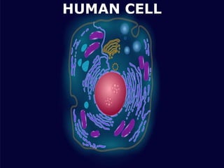
  • 6. PARTS OF THE HUMAN CELL The cell contains various structural components to allow it to maintain life which are known as organelles. All the organelles are suspended within a gelatinous matrix, the cytoplasm, which is contained within the cell membrane. One of the few cells in the human body that lacks almost all organelles are the red blood cells . The main organelles are as follows : Nucleus Cell membrane Endoplasmic reticulum Golgi apparatus Lysosomes Peroxisomes Mitochondria Microfilaments and microtubules. JNVU PHARMACY, JODHPUR
  • 8. JNVU PHARMACY, JODHPUR NUCLEUS The nucleus : is the master control of the cell. It contains genes, collections of DNA, which determines every aspect of human anatomy and physiology. The DNA which is arranged into chromosomes also contains the blueprint specific for each type of cell which allows for replication of the cell. Within the nucleus is an area known as the nucleolus. It is not enclosed by a membrane but is just an accumulation of RNA and proteins within the nucleus. The nucleolus is the site where the ribosomal RNA is transcribed from DNA and assembled.
  • 10. FUNCTIONS OF THE NUCLEUS The main function of the cell nucleus is to control gene expression and facilitate the replication of DNA during the cell cycle (which you will learn about in the next chapter). The nucleus controls the metabolic functions of the cell by producing mRNA which encodes for enzymes e.g. insulin. The nucleus controls the structure of the cell by transcribing DNA which encodes for structural proteins such as actin and keratin. The nucleus is the site of ribosomal RNA (rRNA) synthesis, which is important for the construction of ribosomes. Ribosomes are the site of protein translation (synthesis of proteins from amino acids). Characteristics are transmitted from parent to offspring through genetic material contained in the nucleus. JNVU PHARMACY, JODHPUR
  • 11. JNVU PHARMACY, JODHPUR CELL MEMBRANE The cell membrane is the outer coating of the cell and contains the cytoplasm, substances within it and the organelle. It is a double-layered membrane composed of proteins and lipids. The lipid molecules on the outer and inner part (lipid bilayer) allow it to selectively transport substances in and out of the cell. Endoplasmic Reticulum: The endoplasmic reticulum (ER) is a membranous structure that contains a network of tubules and vesicles. Its structure is such that substances can move through it and be kept in isolation from the rest of the cell until the manufacturing processes conducted within are completed. There are two types of endoplasmic reticulum – rough (granular) and smooth (a granular)
  • 13. Functions The cell membrane provides mechanical support that facilities the shape of the cell while enclosing the cell and its components from the external environment. It regulates what can be allowed to enter and exit the cell through channels, acting as a semi-permeable membrane, which facilities the exchange of essential compounds required for the survival of the cell. It generates and distributes signals in and outside of the cell for the proper functioning of the cell and all the organelles. It allows the interaction between cells required during tissue formation and cell fusion. JNVU PHARMACY, JODHPUR
  • 14. An additional non-living layer present outside the cell membrane in some cells that provides structure, protection, and filtering mechanism to the cell is the cell wall. Structure In a plant cell, the cell wall is made up of cellulose, hemicellulose, and proteins while in a fungal cell, it is composed of chitin. A cell wall is multilayered with a middle lamina, a primary cell wall, and a secondary cell wall. Middle lamina contains polysaccharides that provide adhesion and allows binding of the cells to one another. After middle lamina is the primary cell wall which is composed of cellulose. The last layer, which is not always present, is the secondary cell wall made of cellulose and hemicellulose. CELL WALL JNVU PHARMACY, JODHPUR
  • 15. Functions The critical function of the cell wall is protecting and maintaining the shape of the cell. It also helps the cell withstand the turgor pressure of the cell. It initiates cell division by providing signals to the cell and allows the passage of some molecules into the cell while blocking others. JNVU PHARMACY, JODHPUR
  • 16. JNVU PHARMACY, JODHPUR CYTOPLASM Cytoplasm The gel-like material within the cell membrane is referred to as the cytoplasm. It is a fluid matrix, the cytosol, which consists of 80% to 90% water, salts, organic molecules and many enzymes that catalyze reactions, along with dissolved substances such as proteins and nutrients. The cytoplasm plays an important role in a cell, serving as a "molecular soup" in which organelles are suspended and held together by a fatty membrane. Within the plasma membrane of a cell, the cytoplasm surrounds the nuclear envelope and the cytoplasmic organelles. It plays a mechanical role by moving around inside the membrane and pushing against the cell membrane helping to maintain the shape and consistency of the cell and again, to provide suspension to the organelles. It is also a storage space for chemical substances indispensable to life, which are involved in vital metabolic reactions, such as anaerobic glycolysis and protein synthesis. The cell membrane keeps the cytoplasm from leaking out. It contains many different organelles which are considered the insoluble constituents of the cytoplasm, such as the mitochondria, lysosomes, peroxysomes, ribosomes, several vacuoles and cytoskeletons, as well as complex cell membrane structures such as the endoplasmic reticulum and the Golgi apparatus that each have specific functions within the cell
  • 18. The cytoplasm is the jelly-like substance that fills the cell. It consists of up to 90%90% water. It also contains dissolved nutrients and waste products. Its main function is to hold together the organelles which make up the cytoplasm. It also nourishes the cell by supplying it with salts and sugars and provides a medium for metabolic reactions to occur. All the contents of prokaryotic cells are contained within the cytoplasm. In eukaryotic cells, all the organelles are contained within the cytoplasm except the nucleolus which is contained within the nucleus. JNVU PHARMACY, JODHPUR
  • 19. FUNCTIONS OF THE CYTOPLASM The cytoplasm provides mechanical support to the cell by exerting pressure against the cell's membrane which helps keep the shape of the cell. This pressure is known as turgor pressure. It is the site of most cellular activities including metabolism, cell division and protein synthesis. The cytoplasm contains ribosomes which assist in the synthesis of protein. The cytoplasm acts a storage area for small carbohydrate, lipid and protein molecules. The cytoplasm suspends and can transport organelles around the cell. JNVU PHARMACY, JODHPUR
  • 20. CENTRIOLES Centrioles are rod like structures composed of 9 bundles which contain three microtubules each. Two perpendicularly placed centrioles surrounded by proteins make up the centrosome. Centrioles are very important in cellular division, where they arrange the mitotic spindles that pull the chromosome apart. Centrioles and basal bodies act as microtubule organizing centers. A pair of centrioles (enclosed in a centrosome) located outside the nuclear envelope gives rise to the microtubules that make up the spindle apparatus used during cell division. Basal bodies are at the base of each flagellum and cilium and appear to organize their development. JNVU PHARMACY, JODHPUR
  • 21. JNVU PHARMACY, JODHPUR Functions During cell division, centrioles have a crucial role in forming spindle fibers which assist the movement of chromatids towards their respective sides. They are involved in the formation of cilia and flagella.
  • 22. CILIA AND FLAGELLA Cilia and Flagella are tiny hair-like projections from the cell made of microtubules and covered by the plasma membrane. Structure Cilia are hair-like projections that have a 9+2 arrangement of microtubules with a radial pattern of 9 outer microtubule doublet that surrounds two singlet microtubules. This arrangement is attached to the bottom with a basal body. Flagella is a filamentous organelle, the structure of which, is different in prokaryotes and eukaryotes. In prokaryotes, it is made up of the protein called flagellin wrapped around in a helical manner creating a hollow structure at the center throughout the length. In eukaryotes, however, the protein is absent and the structure is replaced with microtubules. JNVU PHARMACY, JODHPUR
  • 23. Functions The most critical role of cilia and flagella is movement. These are responsible for the movement of the organisms as well as for the movement of various particles present around the organisms. Some cilia present in some particular organs may have the function of sense. The cilium in the blood vessels, which helps in controlling the flow of blood is an example. JNVU PHARMACY, JODHPUR
  • 24. JNVU PHARMACY, JODHPUR GOLGI APPARATUS The Golgi apparatus is a stacked collection of flat vesicles. It is closely associated with the endoplasmic reticulum in that substances produced in the ER are transported as vesicles and fuses with the Golgi apparatus. In this way, the products from the ER are stored in the Golgi apparatus and converted into different substances that are necessary for the cell’s various functions
  • 26. FUNCTIONS OF THE GOLGI BODY It is important for proteins to be transported from where they are synthesised to where they are required in the cell. The organelle responsible for this is the Golgi Body. The Golgi body is the sorting organelle of the cell. Proteins are transported from the rough endoplasmic reticulum (RER) to the Golgi. In the Golgi, proteins are modified and packaged into vesicle. The Golgi body therefore receives proteins made in one location in the cell and transfers these to another location within the cell where they are required. For this reason the Golgi body can be considered to be the 'post office' of the cell. JNVU PHARMACY, JODHPUR
  • 27. LYSOSOMES Lysosomes: are vesicles that break off from the Golgi apparatus. It varies in size and function depending on the type of cell. Lysosomes contain enzymes that help with the digestion of nutrients in the cell and break down any cellular debris or invading microorganisms like bacteria. A structure that is similar to a lysosome is the secretory vesicle. It contains enzymes that are not used within the cell but emptied outside of the cell, for example the secretory vesicles of the pancreatic lacunar cell release digestive enzymes which help with the digestion of nutrients in the gut. JNVU PHARMACY, JODHPUR Lysosomes are known as suicide bags of the cell because they contain lytic enzymes capable of digesting cells and unwanted materials.
  • 28. Functions of Lysosomes Lysosomes serve two major functions: 1.Intracellular Digestion •To digest food, the lysosome membrane fuses with the membrane of food vacuole and squirts the enzymes inside. •The digested food then diffuses through the vacuole membrane and enters the cell to be used for energy and growth. 2.Autolytic Action •Cell organelles that need to be get ridden are covered by vesicles or vacuoles by the process of autophagy to form autophagosome. •The autophagosome is then destroyed by the action of lysosomal enzymes. Processes in which lysosomes play crucial roles include: a. Heterophagy The taking into the cell of exogenous material by phagocytosis or pinocytosis and the digestion of the ingested material after fusion of the newly formed vacuole with a lysosome. JNVU PHARMACY, JODHPUR
  • 29. b. Autophagy A normal physiological process that deals with the destruction of cells in the body. It is essential for maintaining homeostasis, for normal functioning by protein degradation, turnover of destroyed cell organelles for new cell formation c. Extracellular Digestion Primary lysosomes secrete hydrolases outside by exocytosis resulting in degradation of extracellular materials. Eg. Saprophytic fungi d. Autolysis It refers to the killing of an entire set of cells by the breakdown of the lysosomal membrane. It occurs during amphibian and insect metamorphosis. e. Fertilization The acrosome of the sperm head is a giant lysosome that ruptures and releases enzymes on the surface of the egg. This provides the way for sperm entry into the egg by digesting the egg membrane. f. As Janitors of the Cell Lysosomes remove ‘junk’ that may accumulate in the cell helping to prevent diseases JNVU PHARMACY, JODHPUR
  • 30. JNVU PHARMACY, JODHPUR PEROXISOMES These organelles are very similar to the lysosomes and contain enzymes that act together in the form of hydrogen peroxide to neutralize substances that may be toxic to the cell. Peroxisomes are formed directly from the endoplasmic reticulum rather than from the Golgi apparatus like lysosomes.
  • 31. Functions of Peroxisomes 1.Hydrogen Peroxide Metabolism: Enzymes present in the peroxisomes both lead to the production and elimination of H202 which is a reactive oxygen species. 2.Fatty acid oxidation: Oxidation of fatty acids, in animal cells, occurs in both peroxisomes and mitochondria, but in yeasts and plants, only limited to peroxisomes. Oxidation is accompanied by the production of H202 which is decomposed by catalase enzyme. This provides a major source of metabolic energy. 3.Lipid biosynthesis Synthesis of cholesterol and dolichol occurs in both ER and peroxisomes. Bile acid synthesis takes place from cholesterol in the liver. JNVU PHARMACY, JODHPUR
  • 32. 4.Germination of seeds Peroxisomes in seeds responsible for the conversion of stored fatty acids to carbohydrates, critical to providing energy and raw materials for the growth of germinating plants. 5.Photorespiration Peroxisomes in leaves particularly in the green ones carry out the photorespiration process along with chloroplasts. 6.Degradation of purines Carry out the catabolism of purines, polyamines and amino acids especially by uric acid oxidase 7.Bioluminescence Luciferase enzyme found in the peroxisomes of fireflies help in bioluminescence and thus aid the flies in finding a mate or its meal. Peroxisomes contain enzymes to synthesize plasmalogens, a family of phospholipids which are important membrane components of tissues of the heart and brain. JNVU PHARMACY, JODHPUR
  • 33. RIBOSOMES Ribosomes play an active role in the complex process of protein synthesis, where they serve as the structures that facilitate the joining of amino acids. Each ribosome is composed of a large and small subunit which are made up of ribosomal proteins and ribosomal RNAs. They can either be found in groups called polyribosomes within the cytoplasm or found alone. Occasionally they are attached to the endoplasmic reticulum JNVU PHARMACY, JODHPUR
  • 34. Function of ribosome The ribosome is a complex molecular machine, found within all living cells, that serves as the site of biological protein synthesis (translation). Ribosomes link amino acids together in the order specified by messenger RNA (mRNA) molecules. Ribosomes act as catalysts in two extremely important biological processes called peptidyl transfer and peptidyl hydrolysis. JNVU PHARMACY, JODHPUR
  • 35. JNVU PHARMACY, JODHPUR MITOCHONDRIA These are the powerhouses of the cell and break down nutrients to yield energy. Apart from producing its own energy, it also produces a high-energy compound called ATP (adenosine triphosphate) which can be used as a simple energy source elsewhere. Mitochondria are composed of two membranous layers – an outer membrane that surrounds the structure and an inner membrane that provides the physical sites of energy production. The inner membrane has many in folding that form shelves where enzymes attach and oxidize nutrients. The mitochondria also contain DNA which allows it to replicate where and when necessary.
  • 36. JNVU PHARMACY, JODHPUR MITOCHONDRIA Functions The primary function of mitochondria is the synthesis of energy in the form of ATP required for the proper functioning of all the cell organelles. Mitochondria also help in balancing the amount of Ca+ ions within the cell and assists the process of apoptosis. Different segments of hormones and components of blood are built within mitochondria. Mitochondria in the liver have the ability to detoxify ammonia.
  • 37. ENDOPLASMIC RETICULUM Endoplasmic Reticulum Endoplasmic means "within the plasm" and reticulum means "network". A complex three dimensional internal membrane system of flattened sheets, sacs and tubes, that play an important role in making proteins and shuttling cellular products; also involved in metabolisms of fats, and the production of various materials. In cross-section, they appear as a series of maze-like channels, often closely associated with the nucleus. When ribosomes are present, the rough ER attaches polysaccharide groups to the polypeptides as they are assembled by the ribosomes. Smooth ER, without ribosomes, is responsible for various activities, including the synthesis of lipids and hormones, especially in cells that produce these substances for export from the cell. JNVU PHARMACY, JODHPUR
  • 39. Rough endoplasmic reticulum has characteristic bumpy appearance due to the multitude of ribosomes coating it. It is the site where proteins not destined for the cytoplasm are synthesized. Smooth endoplasmic reticulum provides a variety of functions, including lipid synthesis and degradation, and calcium ion storage. In liver cells, the smooth ER is involved in the breakdown of toxins, drugs, and toxic byproducts from cellular reactions. JNVU PHARMACY, JODHPUR Functions ER contains many of the enzymes required for several metabolic processes, and the surface of the ER is essential for other operations like diffusion, osmosis, and active transport. One of the crucial functions of ER is the synthesis of lipids like cholesterol and steroids. Rough ER allows for the modification of polypeptides emerging out of the ribosomes to prepare secondary and tertiary structures of the protein. ER also synthesizes various membrane proteins and has a crucial role in preparing the nuclear envelope after cell division.
  • 40. JNVU PHARMACY, JODHPUR MICROFILAMENTS AND MICROTUBULES Microfilaments and microtubules are rigid protein substances that form the internal skeleton of the cell known as the cytoskeleton. Some of these microtubules also make up the centrioles and mitotic spindles within the cell which are responsible for the division of the cytoplasm when the cell divides. The microtubules are the central component of cilia, small hair-like projections that protrude from the surface of certain cells. It is also the central component of specialized cilia like the tail of the sperm cells which beats in a manner to allow the cell to move in a fluid medium.
  • 42. Functions of Microfilaments It generates the strength for the structure and movement of the cell in association with myosin protein. They help in cell division and are involved in the products of various cell surface projections Functions of Microtubules As a part of the cytoskeleton, they provide shape and movement to the cell. Microtubules facilitate the movement of other cell organelles within the cell through binding proteins. JNVU PHARMACY, JODHPUR
  • 43. VACUOLES Vacuoles Spaces in the cytoplasm that sometimes serve to carry materials to the cell membrane for discharge to the outside of the cell. Vacuoles are formed during endocytosis when portions of the cell membrane are pinched off. JNVU PHARMACY, JODHPUR
  • 44. FUNCTIONS OF THE VACUOLE The vacuole plays an important role in digestion and excretion of cellular waste and storage of water and organic and inorganic substances. The vacuole takes in and releases water by osmosis in response to changes in the cytoplasm, as well as in the environment around the cell. The vacuole is also responsible for maintaining the shape of plant cells. When the cell is full of water, the vacuole exerts pressure outwards, pushing the cell membrane against the cell wall. This pressure is called turgor pressure. If there is not sufficient water, pressure exerted by the vacuole is reduced and the cells become flaccid causing the plant to wilt. JNVU PHARMACY, JODHPUR
  • 45. JNVU PHARMACY, JODHPUR MOVEMENT ACROSS MEMBRANES Movement of substances across cell membranes is necessary as it allows cells to acquire oxygen and nutrients, excrete waste products and control the concentration of required substances in the cell (e.g oxygen, water, hormones, ions, etc). The key processes through which such movement occurs include diffusion, osmosis, facilitated diffusion and active transport. 1. DIFFUSION Diffusion is the movement of substances from a region of high concentration to low concentration. It is therefore said to occur down a concentration gradient. The diagram below shows the movement of dissolved particles within a liquid until eventually becoming randomly distributed.
  • 46. JNVU PHARMACY, JODHPUR Diffusion is a passive process which means it does not require any energy input. It can occur across a living or non-living membrane and can occur in a liquid or gas medium. Due to the fact that diffusion occurs across a concentration gradient it can result in the movement of substances into or out of the cell. Examples of substances moved by diffusion include carbon dioxide, oxygen, water and other small molecules that are able to dissolve within the lipid bilayer. Diffusion is the movement of molecules from a region of higher concentration to a lower concentration. It is a passive process (i.e. does not require input of energy).
  • 47. JNVU PHARMACY, JODHPUR 2. OSMOSIS When the concentration of solutes in solution is low, the water concentration is high, and we say there is a high water potential. Osmosis is the movement of water from a region of higher water potential to a region of lower water potential across a semi-permeable membrane that separates the two regions. Movement of water always occurs down a concentration gradient, i.e from higher water potential (dilute solution) to lower potential (concentrated solution). Osmosis is a passive process and does not require any input of energy. Cell membranes allow molecules of water to pass through, but they do not allow molecules of most dissolved substances, e.g. salt and sugar, to pass through. As water enters the cell via osmosis, it creates a pressure known as osmotic pressure.
  • 48. JNVU PHARMACY, JODHPUR Osmosis is the movement of water from an area of high water potential to an area of low water potential across a semi-permeable membrane.
  • 49. 3. FACILITATED DIFFUSION Facilitated diffusion is a special form of diffusion which allows rapid exchange of specific substances. Particles are taken up by carrier proteins which change their shape as a result. The change in shape causes the particles to be released on the other side of the membrane. Facilitated diffusion can only occur across living, biological membranes which contain the carrier proteins. A substance is transported via a carrier protein from a region of high concentration to a region of low concentration until it is randomly distributed. Therefore movement is down a concentration gradient. JNVU PHARMACY, JODHPUR Examples of substances moved via facilitated diffusion include all polar molecules such as glucose or amino acids.
  • 50. JNVU PHARMACY, JODHPUR 4. ACTIVE TRANSPORT Active transport is the movement of substances against a concentration gradient, from a region of low concentration to high concentration using an input of energy. In biological systems, the form in which this energy occurs is adenosine triphosphate (ATP). The process transports substances through a membrane protein. The movement of substances is selective via the carrier proteins and can occur into or out of the cell. Examples of substances moved include sodium and potassium ions
  • 51. JNVU PHARMACY, JODHPUR Endocytosis and exocytosis mechanisms are forms of Active Transport, both using energy to transport particles in and out of the cell. They both have different types similar in that they both transport materials across the cell membrane by forming vesicle pores.
  • 52. JNVU PHARMACY, JODHPUR EXOCYTOSIS As compared to endocytosis, exocytosis is a process that is used to transport materials from inside the cell to the external part of the cell by the use of energy. Therefore, it is a type of active transport mechanism and it is the opposite of endocytosis. Generally, in this mechanism of exocytosis, a special vesicle bound to the cell membrane, containing the cellular particles will expel the cell content to the external part of the cell. This mechanism has been used in the removal of waste materials from the cell, transport hormones, and proteins, for chemical signaling between cells and the construction of the cell membrane. The vesicles involved in exocytosis are formed by the Golgi bodies, endosomes, and the presynaptic neurons. These vesicles fuse with the cell membrane and they can either be complete or temporary vesicles, depending on their function. Normally after endocytic pinocytosis and phagocytosis, the cell membrane gets damaged, and therefore the exocytic process functions to repair the cell membrane by transporting proteins and lipids to the membrane for repair mechanisms. It is also the end-point mechanism for the transportation of protein complexes and packaging them in their destined sites and insertion to the cell membrane.
  • 53. JNVU PHARMACY, JODHPUR Exocytosis mechanism in summary involves: The transportation of the cell vesicle containing molecules from inside the cell to the cell membrane. The vesicle then attaches to the cell membrane. During vesicle fusion with the cell membrane, it allows the release of the contents in the vesicle to the outside of the cell.
  • 54. JNVU PHARMACY, JODHPUR TYPES OF EXOCYTOSIS There are three pathways involved in the exocytotic process. Constitutive exocytosis Regulated exocytosis Lysosome mediated exocytosis
  • 55. JNVU PHARMACY, JODHPUR Constitutive exocytosis This type of exocytosis involves the delivery of membrane proteins and lipids to the cell membrane and to also get rid of substances from the cell into the exterior. This is the most common pathway that is performed by all body cells. Regulated exocytosis This mechanism is common among the secretory cells whose function is to store hormones, digestive enzymes, and neurotransmitters. The secretion of these products must be triggered by extracellular signals to initiate the formation of secretory vesicles, which fuse with the cell membrane for a long time to allow the release of the cell contents out of the cell, into the exterior. After delivery, the vesicles are reformed and returned to the cytoplasm. Generally, this mechanism depends on extracellular signaling for the release of its content onto the outer part
  • 56. JNVU PHARMACY, JODHPUR Lysosome mediated exocytosis This process involves the fusion of cell vesicles with the cell lysosomes. Lysosomes contain digestive enzymes and hydrolase enzymes whose function involves the breakdown of cellular waste materials, microorganisms, and debris. The lysosome carries the elements that have been broken down onto the cell membrane where it fuses with the cell membrane releasing its elements into the extracellular cell matrix. EXAMPLE OF EXOCYTOSIS A good example of exocytosis in the transportation of glucagon from the pancreas in the Islets of Langerhans into the liver where they are broken down into glycogen which is further broken down to glucose which is simple for absorption. The glucose is then released into the bloodstream.
  • 57. JNVU PHARMACY, JODHPUR In the islets of Langerhans, the glucagon and insulin are stored in the secretory vesicles of the pancreas. When the glucose levels in the bloodstream are low, glucagon is secreted by the islets alpha cells, carried by the secretory vesicles into the liver cells where they are released by exocytosis for further processing and utilization. The pancreas also releases some digestive enzymes by exocytosis. Other exocytotic processes include the synaptic vesicle exocytosis where a synaptic vesicle filled with neurotransmitters in the pre-synaptic neuron fuses with the pre-synaptic membrane releasing neurotransmitters into the synaptic cleft (the gap between neurons). The neurotransmitters can then bind to receptors on the post- synaptic neuron.
  • 58. JNVU PHARMACY, JODHPUR Endocytosis is a cellular mechanism by which, a cell internalizes substances including proteins, fluids, electrolytes, microorganisms, and some macromolecules, from its external environment. These substances undergo certain processes of breaking down to smaller elements either for use by the cell or for elimination purposes. White blood cells, of the immune system, are the most common cells that use endocytosis mechanisms to eliminate microbial pathogens from the body. They entrap the pathogens, break them down and destroy them, for elimination from the body. Endocytosis was first described by Christian de Devu, A Belgium Cytologist and Biochemist who won several Nobel prizes for his role in discovering cellular elements such as lysosomes, peroxisomes, endosomes, and even exocytosis cellular mechanisms including the endocytosis mechanism. ENDOCYTOSIS
  • 59. There are three types of endocytosis mechanisms: Phagocytosis Pinocytosis Receptor-Mediated Endocytosis (Clathrin-Mediated Endocytosis JNVU PHARMACY, JODHPUR
  • 60. JNVU PHARMACY, JODHPUR PHAGOCYTOSIS Also known as cell eating This is the process whereby the cell membrane of a cell extends toward a particle, engulfing it and encloses it within this folded membrane forming a phagosome. The ingested material in the phagosome is later processed by cellular enzymes. Phagocytosis is a common mechanism in multicellular organisms by the White blood cells (macrophages, monocytes, neutrophils, Eosinophils, dendritic cells), in the elimination of pathogens from the system. Some protozoans such as Entamoeba spp use phagocytosis to acquire nutrients; Phagocytosis mechanism was first noted by Canadian physician William Osler (1876). Phagocytosis takes place in 5 steps: The phagocytic cells detect the molecule of interest or an antigen and moves towards it.
  • 62. JNVU PHARMACY, JODHPUR The phagocyte then attaches itself to the target molecule or antigen. Phagocytes have the ability to extend their membrane (pseudopodia) to the target particle and surround the particle of the pathogen. The pseudopodia extend toward each other while enclosing the particles. The particle is then enclosed within the vesicle formed from the extended pseudopodia that have fused. The vesicle with the enclosed particles is known as a phagosome. This is the vesicle that is digested by the phagocyte. The phagosome fuses with the lysosomes of the phagocyte forming a phagolysosome. The lysosomes have digestive enzymes that degrade or digest the materials contained in the vesicle. The degraded particles are then expelled from the phagocytic cell by exocytosis.
  • 63. JNVU PHARMACY, JODHPUR A typical example of phagocytosis is the mechanism of the immune cells such as the macrophages, dendritic cells, and neutrophils. Macrophages are the largest phagocytic cells in the immune system. They function by detecting, attaching, ingesting, digesting, and releasing digested particles from its cytoplasm by exocytosis. The antigens vary and they include bacteria, fungi, dust particles, dead cells, etc. Macrophages are the major phagocytic cells in the immune system. They have a pseudopodial membrane. On detection of an antigen, they move toward the antigen and extend their pseudopodia toward the antigen, and engulf it. On engulfment, the cell ingests the antigen forming a cell vesicle otherwise known as the phagosome. Within the macrophage, the vesicle encounters the lysosomes, forming a phagolysosome, which is digested my the lysosomic enzymes breaking the particle down which are then released from the cell by exocytosis.
  • 64. JNVU PHARMACY, JODHPUR Adherence of the particle with the phagocyte highly depends on the chemical nature of the particle. Some bacterial antigens bind directly and some need a protein component from blood, known as an opsonin (such as complements of antibodies), to form a film on the bacterial surface for it to adhere to the phagocyte, a process known as opsonization. So the phagocytes first will bind to the opsonin for phagocytosis to take place. Some bacteria with encapsulated cell walls are rather difficult to digest even with an opsonin. Therefore they must be bound to specific antibodies after the body responds to their presence. The antibody-bound encapsulated bacteria can then be acted upon by the phagocytes
  • 65. JNVU PHARMACY, JODHPUR Also known as cell drinking or fluid endocytosis; it is a form of endocytosis where small particles in extracellular fluids enter the cell through the cell membrane by invagination forming a small vesicle with suspended small molecules or particles within a cell. The pinocytic vesicle fuses with the cell endosome for the digestion of the particles. Its mechanisms are similar to the other endocytic processes, the major difference between pinocytosis and phagocytosis is that in pinocytosis particles ingested are contained within the cells extracellular fluids. The cell membrane invaginated together the vesicle containing the fluid particles, transporting them into the cell lysosomes. The vesicle and the lysosomes fuse, releasing digestive enzymes from the lysosomes. The enzymes degrade the vesicle, releasing its content into the cell cytoplasm, for utilization by the cell.. PINOCYTOSIS
  • 66. JNVU PHARMACY, JODHPUR Sometimes the vesicles do not interact with the lysosomes, instead, they move across the cell, fusing with the cell membrane causing a recycling effect of the membrane proteins and lipids. Pinocytosis takes place by two mechanisms: Micropinocytosis – This is the formation of small vesicles of about 0.1 um diameter; it takes place in the body cells forming tiny budding vesicles on the cell membrane known as caveolae. They are found in blood vessel endothelium. Macropinocytosis – This is the formation of larger vesicles of about 0.5-5um in diameter; they are found on the white blood cells; the large vesicles are formed by the cell membrane ruffles (villi), which are projections that extend to the extracellular fluids and have the ability to fold back by themselves. While folding, they shovel in some of the extracellular fluid forming a vesicle that pulls into the cell
  • 68. JNVU PHARMACY, JODHPUR Example of pinocytosis: The intake or absorption of nutrients in the small intestines RECEPTOR-MEDIATED ENDOCYTOSIS (CLATHRIN- MEDIATED ENDOCYTOSIS his is a type of endocytosis also known as clathrin- mediated endocytosis; It involves the internalization and recycling of receptors that are used in processes such as signal transduction (G-protein and tyrosine kinase receptors), nutrient uptake and synaptic vesicle reformation. This process is initiated by the accumulation of phosphatidylinositol-4,5-bisphosphate (PIP2) within the cell membrane. PIP2 accumulation is because of the catalyzation process of phosphoinositide within the plasma membrane.
  • 69. JNVU PHARMACY, JODHPUR PIP2 accumulates as a result of phosphoinositide by the lipid kinase and the hydrolyzation of phosphatases. The combination of adapter proteins (AP proteins) with the Phosphatidylinositol-4,5-bisphosphate (PIP2), leads to the attachment of a cytosol protein known as clathrin to the vesicle. This forms Clathrin-coated vesicles (CCV). The Clathrin-coated vesicles (CCV)must invaginate and mature to form the clathrin-coated pits. The Clathrin- coated vesicles bind to the cell membrane recruiting several proteins including Actin-binding proteins, Adapter proteins (AP) all of which play a major role in the maturation of the vesicle. The CCV, which contains several receptors bound with ligands and adapter proteins, then invaginated into the membrane and by the assistance of dynamin protein, it matures and scissions from the cell membrane, forming a clathrin-coated pit.
  • 70. JNVU PHARMACY, JODHPUR How are the Clathrin-coated pits formed? Clathrin- coated vesicles are found in most if not all cells, and therefore, after the detection of a signal, these vesicles recruit the adaptor proteins in the plasma membrane which accumulate on the lipid layer of the plasma membrane. The adapter proteins incorporate the Clathrin from the clathrin-coated vesicles into the cell membrane lipids along with the Actin-binding proteins. Due to the negative charge of the lipids layer, it gets attracted to the positive charge of the clathrin, forming a concave shape that is raised up from the membrane, thus forming pits all over the plasma membrane. The clathrin on the pits acts as a sensor for signals that activate endocytosis while the vesicle from the Clathrin Coated vesicles gets recycled to the cell membrane. The cycle between the clathrin-coated pits and clathrin-coated vesicles formation is continuous as long as there are signaling receptors and ligands that activate them.
  • 71. JNVU PHARMACY, JODHPUR The process of Receptor-mediated endocytosis has the following steps: The particles (ligands) that need to be synthesized are bound to the receptors on the cell membrane, The receptors with the ligands cluster forming the coated pits. The pits then undergo invagination with the help of the dynamin proteins forming a vesicle and the vesicle pinches-off within the cell membrane. The vesicles then lose the clathrin and the adaptor proteins. The uncoated vesicle then fuses with an early endosome to form the late endosome or the sorting vesicle. The late endosome segregates the particles within the vesicle i.e the receptors from the ligands recycling them into the cell membrane. The released particles interact with the lysosomes which contain digestive enzymes that hydrolyze the content in the vesicles. The digested particles are then released for utilization by the cell. This mechanism of receptor-mediated endocytosis (clathrin- coated Endocytosis) can best be used to bring macromolecules into the cell.
  • 72. JNVU PHARMACY, JODHPUR There are two classic examples of the Clathrin-Mediated Endocytosis which include iron-bound transferrin recycling receptor-mediated endocytosis is the uptake of cholesterol bound to low-density lipoprotein (LDL), a complex of phospholipid, protein, and cholesterol.
  • 73. JNVU PHARMACY, JODHPUR CHARACTERISTIC ENDOCYTOSIS EXOCYTOSIS DEFINITION Endocytosis is a cellular mechanism where a cell internalizes substances from the external environment. These substances undergo certain processes of breaking down to smaller elements either for use by the cell or for elimination purposes. Exocytosis is a process that is used to transport materials from inside the cell to the external part of the cell by the use of energy. The mechanism uses special vesicles fille with the particles of interest to transport Generally, in this mechanism of exocytosis, a special vesicle bound to the cell membrane, containing the cellular particles will expel the cell content to the external part of the cell. TYPES There are three types based on the mechanisms of particle. They include Phagocytosis Pinocytosis Receptor-mediated endocytosis (Clathrin- mediated endocytosis) There are also 3 types depending on the sequence of processes involved in transporting particles out of the cell. They include Constructive Exocytosis Regulated exocytosis Lysosome mediated Exocytosis ENERGY Being a form of active transport, they use some energy (ATP) during particle transportation. It is a type of active transport, using a lot of energy (ATP) for transporting particles out of the cell. FUNCTIONS Endocytic mechanisms are used to Breakdown and eliminate microbial antigens using phagocytic cells. Protozoans such as amoeba use phagocytosis to trap and digest nutrients (food). Uptake and absorption of nutrients through intestinal villi in the small intestines. Transportation of iron-transferrins in receptor-mediated endocytosis. To absorb cholesterol that is bound to ‘bad cholesterol’ (Low-density lipoproteins) in receptor-mediated endocytosis. Exocytosis is known for its functions in Transportation of proteins and lipids use to repair the cell membrane after endocytosis. Transportation of glucagon and insulin hormones from the pancreas to the liver for breakdown and utilization by the body depending on body sugar levels. Transportation of chemical signals between cells allowing cell communication. It is also important in synaptic transmission of information in the neurons.