2. What are Staphylococcus
2
Sir Alexander Ogston, a Scottish surgeon, first
showed in 1880 that a number of human pyogenic
diseases were associated with a cluster-forming
micro-organism.
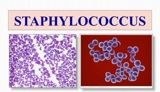
He introduced the name 'staphylococcus'
(Greek: staphyle = bunch of grapes; kokkos
= grain or berry), now used as the genus name
for a group of facultatively anaerobic, catalase-
positive, Gram-positive cocci.
4. Classification
4
Family
Genus
Species
Micrococcaceae
Micrococcus and Staphylococcus
S. aureus
S. saprophyticus
S. epidermidis
The genus Staphylococcus contains about
fourty species and subspecies today.
Only some of them are important as
human pathogens:
Staphylococcus aureus
Staphylococcus epidermidis
Staphylococcus saprophyticus
others
5. The genus Staphylococcus can be divided into two subgroups
(on the basis of its ability to clot blood plasma by enzyme coagulase):
5
• Coagulase-positive Staphylococcus
• Coagulase-negative Staphylococcus (CONS)
6. Morphology
6
Gram-positive cocci, non-motile, non-
sporing, approximately 1um in diameter.
Few strains may possess polysaccharide
capsules, in young cultures.
Cells occur in grape like clusters because
cells division occurs along different planes
and the daughter cells remain attached to one
another.
7. Antigenic structure : Staphylococcus aureus
7
1. Capsule: Facilitates adherence to catheters and other
synthetic material. Also inhibits phagocytosis.
2. Peptidoglycan: Major structural component of the cell
wall. It is important in the pathogenesis of staphylococcal
infections. Activates complement and evokes production
of inflammatory cytokines.
3. Teichoic acid. Facilitates adhesion to the host cell surface
and protects from opsonisation.
4. Protein A: The major protein component of the cell wall.
8. Protein A
8
The surface of most S. aureus strains (not CONS) is uniformly coated with protein A
responsible for antiphagocytic, anticomplementary effects & induces hypersensitivtiy.
This protein is linked to the peptidoglycan layer and has a unique affinity for binding to
the Fc receptor of immunoglobulin IgG. It is not an antigen-antibody specific reaction.
The presence of protein A has been exploited in passive agglutination tests, in which
protein A-coated S. aureus is used as a nonspecific carrier of antibodies directed against
other antigens in Coagglutination test.
Detection of protein A can be used as a specific identification test for S. aureus.
9. 9
Coagulase and other surface proteins
The outer surface of most strains of S. aureus contains clumping factor
(also called bound coagulase).
This protein binds fibrinogen, converts it to insoluble fibrin, causing the
staphylococci to clump or aggregate.
Detection of this protein is the primary test for identifying S. aureus.
Other surface proteins that appear to be important for adherence
to host tissues include:
Collagen-binding protein
Elastin-binding protein
Fibronectin-binding protein
10. Resistance:
10
S. aureus is rapidly killed by temperature above 60C (62C within 30 min).
Most strains can grow in presence of 10% NaCl.
S. aureus is susceptible to disinfectants and antiseptics commonly used.
S. aureus can survive and remain virulent long periods (2-3 months) of drying
especially in an environment with pus.
Killed by crystal violet.
12. Virulence Factors (contd....)
Cellwall asssociated
structures
Capsule
Adherance to host cell
Resist phagocytosis
Peptidoglycan Activates complement
Protein A
Binds to Fc moiety of IgG,
exerting antiopsonin /
antiphagocytic action
Clumping factor
(bound coagulase)
Cause organism to clump
in presence of plasma
13. Virulence Factors (contd....)
Extracellular toxins
Haemolysin (α,β,γ,δ)
Haemolytic dermo-necrotic and
leucocidal
Leucocidin (Panton-
Valentine factor)
Kills WBCS by producing holes in
their CM
Enterotoxin Act on ANS to cause illness
TSST (toxin shock syndrome
toxin)
Produce fever, skin
rashes,diarrhoea,conjunctivitis,an
d death to shock
Exfoliatin toxin
Breaks intracellular bridges in the
stratum granulosum of epidermis and
causes its separation from underlying
tissue, resulting in a blistering and
exfoliating disease of skin
14. Virulence Factors (....contd)
Extracellular Enzymes
Free coagulase
Clots plasma by acting along with CRF
present in plasma, binding to prothrombin
and converting fibrinogen to fibrin
Staphylokinase Degrades fibrin clots
Hyaluronidase
Hydrolyze the acidic
mucopolyysaccharides present
in matrix of connective tisues
Lipase, Phospholipase,
protease
Degrades lipid, phospholipid, and
protein respectively
16. Enzymes
16
1. Coagulase
Secreted free into the culture medium.
Triggers clotting of human and rabbit plasma.
Heat labile
Requires Coagulase reacting factor (CRF)
Converts fibrinogen to fibrin.
2. Clumping factor
Bound coagulase
Heat stable constituent of cell wall.
Can directly convert fibrinogen to insoluble fibrin and cause the staphylococci
to clump.
3. Hyaluronidase
Breaks down hyaluronic acid (mucopolysaccharide) of connective tissues,
enabling the bacteria to spread between cells.
* The role of coagulase in the
pathogenesis of disease is
speculative, but coagulase may
cause the formation of fibrin
layer around a staphylococcal
abscess, thus localizing the
infection and protecting the
organisms from phagocytosis.
17. 17
4. Staphylokinase (Fibrinolysin)
• Dissolves fibrin threads in blood clots, allowing Staphylococcus aureus to
free itself from clots.
5. Lipases
Digest lipids, allowing staphylococcus to grow on the skin’s surface and in
cutaneous oil glands.
6. -lactamase (Penicillinase)
Breaks down penicillin
Allows the bacteria to survive treatment with -lactam antimicrobial drugs.
7. Catalase
• All staphylococci produce catalase, which catalyzes the conversion of
toxic hydrogen peroxide to water and oxygen.
• Diagnostic value.
18. 18
Staphylococcal TOXINS
S. aureus produces many virulence factors, including at least five cytolytic or
membrane-damaging toxins:
1. Haemolysins
a) Alpha toxin
b) Beta toxin
c) Delta toxin
d) Gamma toxin
2. Leucocidin (Panton-Valentin toxin)
3. Two exfoliative toxins (A, B)
4. Eigth enterotoxins (A-E, G-I)
5. Toxic Shock Syndrome Toxin 1 (TSST-1)
19. Hemolysins: Exotoxins
19
1. Alpha toxin:
Protein in nature, inactivated at 60 C and regain activity when
further heated to 80 C - 100 C .
Produced only under aerobic conditions in cultures.
Lytic to rabbit RBC but less active against human and sheep
RBC.
It is leucocidal, cytotoxic, dermonecrotic, neurotoxic and lethal.
2. Beta toxin:
Hemolytic for sheep.
Can be produced aerobically and anaerobically.
Lysis initiated at 37 C but evident only on cold temperature. Property known as hot-cold
phenomenon.
3. Gamma toxin: Acts on human, sheep and rabbit RBCs.
4. Delta toxin: Lytic to human, sheep and rabbit RBCs.
20. Leucocidin:
20
Composed of 2 components; S (slow) and F (fast).
Damages polymorphs and macrophages.
Exfoliative toxin:
Epidermolytic toxin
Two types- A (heat stable) and B (heat labile) have been
described.
Causes epidermal splitting resulting in blistering diseases and
generalised desquamation leading to SSSS (staphylococcal
scalded skin syndrome).
Severe form of SSSS is known as Ritter’s disease in new born.
Milder forms are pemphigus neonatorum and bullous impetigo.
Stratum
corneum
DESMOSOME
22. Enterotoxin: Superantigen
22
Responsible for FOOD POISIONING.
Even microgram amount can cause illness.
Nausea, vomiting and diarrhoea within 2-6 hrs of intake.
Eigth serologically distinct staphylococcal enterotoxins (A, B,
C1-3, D, E and H). Type A responsible for most cases.
Enterotoxins C and D are found in contaminated milk products,
and enterotoxin B causes staphylococcal pseudomembranous
enterocolitis.
The enterotoxin are stable to heating at 100 °C for 30 minutes
and are resistant to hydrolysis by gastric and jejunal enzymes.
Believed to act directly on the autonomic nervous system.
23. TSST: Superantigen
23
Toxic Shock Syndrome characterised by
fever, hypotension, vomiting, diarrhoea and erythematous rash with desquamation
and hyperaemia of mucous membranes.
Belongs to bacteriophage group I.
TSST-1, formerly called pyrogenic exotoxin C & enterotoxin F, is a heat &
proteolysis resistant exotoxin.
The ability of TSST-1 to penetrate mucosal barriers is responsible for the systemic
effects of TSS.
Death in patients with TSS is due to hypovolemic shock leading to multiorgan
failure.
Was associated with use of tampons but is also known to be associated with
postoperative wound or soft tissue infections.
24. Staphylococcus aureus pathogenicity
24
S. aureus is pathogenic for human as well as for all domestic and free-living
warm-blooded animals.
Pyogenic organism and lesions are usually localised unlike that of streptococcal
lesion which are spreading in nature.
S. aureus causes disease through the production of toxin or through direct
invasion and destruction of tissue.
Causes cutaneous & deep infections; food poisoning; nosocomial infection; skin
exfoliative diseases and TSS.
25. Pathogenicity
25
1. Cutaneous Infections: Pustules, boils, carbuncles, abscesses, Styes, impetigo,
wound, burn infecion and pemphigus neonatorum.
2. Deep infections: Osteiomyelitis, tonsillitis, pharyngitis, sinusitis, penumonitis,
empyema, endocarditis, meningitis, septicemia and pyaemia. May also cause
UTI in association with local instrumentation, implants and diabetes.
3. Food poisioning
4. Nosocomial Infections
5. Skin exfoliative diseases: SSSS
6. Toxic Shock Syndrome
28. Antibiotic Sensitivity:
28
1. Beta-lactamase (Penicillinase):
Inactivates penicillin and other antibiotics
by splitting beta lactam ring.
2. Methicillin resistant Staphylococci (MRSA):
Due to reduction in affinity of penicillin binding proteins in cell wall for beta
lactam antibiotics.
Methicillin, nafcillin, oxacillin.
Cloxacillin used instead of Methicillin for testing.
Predominantly a hospital pathogen (nosocomial) but is becoming more common in
community.
Hospital staffs- chief source.
29. Antimicrobial susceptibility
29
MRSA can be due to
Production of penicillin-binding protein 2a (PBP2a)
encoded by mecA gene
Production of beta-lactamase
Resistance due to mecA can be detected via cefoxitin
disk diffusion or dilution methods.
Resistance due to beta-lactamase production can be
detected via the use of beta-lactamase inhibitor such
as clavulanic acid which would result in an increase
in zone size (disk diffusion method).
30. Bacteriophage Typing
30
Important for epidemiological studies; to find out the source of
outbreak.
Used to identify different strains of bacteria within a single
species.
Fixed dose of an internationally accepted set, of 23
bacteriophages is applied on the strain to be typed, grown on
nutrient agar (lawn culture).
Observed for lysis, after overnight incubation.
Phage type designated according to the phage that was capable
of lysis.
Eg: Strain of phage type 52/80/94 is the one that is lysed only
by phages 52, 80 and 94.
31. Person with lesions Airborne droplets
Asymptomatic carrier Cross-infection
Mode of
transmission
32. Lab Diagnosis
Specimen: Pus, sputum, blood, urine, CSF, synovial fluid etc.
A) Isolation: Inoculation on culture media.
B) Identification-Smear (Grams staining), Biochemical test,
Coagulase test, Novobiocin sensitivity test.
C) Microscopy: Smears of clinical materials are stained.
D) Phage typing – Not done routinely.
33. Staphylococcus aureus culture characteristics
33
Colonies on solid media are round, regular,
smooth, slightly convex and 1 to 3 mm in
diameter after 24h incubation.
Most strains show a -hemolysis
surrounding the colonies on blood agar.
S. aureus produce creamy, golden yellow or
orange pigment.
34. Culture
Aerobes and facultative anaerobes
Opt. Temp. For growth= 37°C
Opt. pH for growth= 7.5
On Nutrient agar
Golden yellow and opaque colonies with smooth
glistening surface, 1-2 mm in diameter (max.
pigment production at 22 °C)
On MacConkey agar
Smaller colonies than those on NA(0.1-0.5 mm) and
are pink coloured due to lactose fermentation
On Mannitol salt agar
S.aureus ferments mannitol and appear as yellow colonies
MSA is a useful selective medium for recovering S.aureus
from faecal specimens, when investigating food poisoning
35. Criteria for pathogenic strain of staph.
1. Golden yellow pigment
2. ß-Hemolysis on BA
3. Reduction of Potassium Tellurite -
Black colonies.
4. Coagulase positive
5. Catalse test: +ve
5. Mannitol fermentation +ve
6. Phosphatase production +ve
7. DNA-ase formation +ve
8. Liquiefy gelatin
36. 36
Catalase Test
Staphylococci
S. aureus
CONS
S. saprophyticus
S. xylosus
Streptococci
Gram
Positive Cocci
S. epidermidis
S. hemolyticus
S. hominis
S. lugdunesis
S. schleiferi
+
Coagulase and Protein A +
Coagulase and Protein A -
-
Novobiocin susceptibility +
Novobiocin susceptibility -
37. Biochemical Properties
Catalase positive
Oxidase negative
Ferment glucose, lactose, maltose,
sucrose and mannitol, with production
of acid but no gas.
Mannitol fermentation carries
diagnosis significance.
38. Biochemical Properties(....contd)
Indole test = negative
MR test = positive
VP test = positive
Urease test = positive
Hydrolyse gelatin
Reduces nitrate to nitrite
DNA-ase test = positive
Coagulase test = positive
39. Coagulase : (a) Slide test – Bound coagulase
(b) Tube test – Free coagulase.
a) Slide method:
Saline suspension of Staphylococcus.
1 drop undiluted human / rabbit plasma.
Mix
Look for clumping.
Detects coagulase bound to surface of
Staphylococcus
b) Tube method:
• 0.1ml overnight broth culture of
Staphylococcus
• 0.5ml of 1:5 dil. Human / Rabbit
plasma.
• Incubate at 37°c for 3-6 hrs.
• Clot formation in coagulase positive
reaction
40. Treatment
40
Antistaphylococcal antibiotics
of first choice:
– oxacillin (methicillin)
– cephalosporins of I. generation
(cefazolin)
Antistaphylococcal antibiotics
of the second choice:
– Lincosamides (e.g. clindamycin)
– Glycopeptides (vancomycin,
teicoplanin)
– Linezolid (for VRSA)
Benzyl penicillin in sensitive strains.
Cloxacillins for beta-lactamase producers.
Vancomycin, teichoplanin for MRSA
Topical applications of bacitracin or
chlorhexidine for superficial lesions and
for carriers.
Rifampicin along with oral antibiotic for
resistant superficial lesions and for carriers.
Drug tolerant strains: Higher doses given.
41. Coagulase-negative staphylococcal spp (CONS)
41
S. epidermidis – most frequently isolated Staphylococcal spp.
Has predilection for plastic material.
Associated with infection of IV lines, prosthetic heart valves, catheters etc.
Colonizes moist body areas such as axilla, inguinal and perianal areas,
anterior nares and toe webs.
Important cause of nosocomial infection.
Usually causes nosocomial infections in patients with predisposing
factors such as immunodeficiency/ immunocompromised or presence of
foreign bodies.
42. S. saprophyticus
42
S. saprophyticus frequently isolated in rectum
and genitourinary tract of young women.
Skin commensal.
Can be causative agent in UTI in young sexually
active young women.
2nd most common urinary pathogen (other than
E. coli) in young women.
Usually sensitive to wide range of antibiotics
43. Related microorganisms
43
The genus Micrococcus (two species)
– Micrococcus luteus
– Micrococcus lyla
Both species are found in nature and colonize humans, primarily on
the surface of the skin.
Although micrococci may be found in patients with opportunistic
infections, their isolation in clinical specimen usually represents
clinically insignificant contamination with skin flora.
44. Prevention
44
Hand antisepsis is the most
important measure in
preventing nosocomial
infections.
Also important is the proper
cleansing of wounds and
surgical openings, aseptic use
of catheters or indwelling
needles and appropriate use of
antiseptics.
Wash your hands Keep wounds covered Reduce tampon risks
Avoid sharing personal
care items
Cooking and storing
food properly