
Recommended
More Related Content
What's hot
What's hot (20)
Similar to Tocodynamometer, cardiotocogram
Similar to Tocodynamometer, cardiotocogram (20)
Recently uploaded
Recently uploaded (20)
Tocodynamometer, cardiotocogram
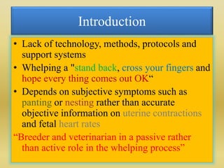
- 1. Introduction • Lack of technology, methods, protocols and support systems • Whelping a "stand back, cross your fingers and hope every thing comes out OK“ • Depends on subjective symptoms such as panting or nesting rather than accurate objective information on uterine contractions and fetal heart rates “Breeder and veterinarian in a passive rather than active role in the whelping process”
- 3. Purpose of Fetal Monitoring • To assess the influence of the intrauterine environment for fetal well-being – Identify the fetus at risk – Assess fetal well being • To assess progress of labor through measurement of uterine activity
- 4. Methods of Monitoring • Two methods of uterine activity monitoring – Palpation only – Electronic Monitoring • External with tocodynamometer • Internal with Intrauterine Pressure Catheter (IUPC)
- 5. Review • The presence of uterine contractions in an organized fashion in the canine was first documented by Vander et al., (1989) by surgically implanting electrodes in the canine myometrium. • Tocodynamometry is a novel approach to monitor parturition and obstetrical manipulations with the plan of reducing neonatal death (Davidson, 1998). • External monitoring devices using tocodynomometry and a hand held doppler to detect and record uterine activity and foetal heart rates is a novel approach to veterinary obstetrical monitoring (Davidson, 2001; Davidson, 2003).
- 6. Contd.. • The use of uterine and foetal monitors allows the veterinary clinician to observe and monitor labour, as well as manage labour medically or surgically with insight instead of guess work (Davidson et al., 2003). • Davidson et al., (2003) reported the overall still birth rate in female dogs declined from 9.2 per cent to 2.5 per cent with incorporation of uterine and foetal monitoring into the whelping process.
- 8. External Tocodynamometer • External tocography is currently the most extensively used technique to monitor the uterus during pregnancy and delivery. • A tocodynamometer, consists of a strain gauge transducer placed around the external surface of the abdomen. • The major benefit of a tocodynamometer is its noninvasiveness.
- 9. Instrumentation • Monitoring equipment – Graph paper – Display panel • External Monitoring – Tocodynamometer – Doppler
- 10. Graph Paper • Paper is heat sensitive • Two distinct sections or channels – FHR channel (on top) • Vertical – monitors the FHR on a 30-240 bpm per cm scale • Horizontal – each small box represents 20 sec, each dark line marks 1 minute – Uterine Activity “UA” (on bottom) • Vertical – measures the intensity of the contractions on a 0- 100 mmHg scale • Horizontal – each small box represents 20 sec, each dark line marks 1 minute
- 12. • The toco contains a flexible disk that responds to pressure. • When the uterus contracts, the fundus tightens and the change in pressure against the toco is amplified and conveyed to the electronic fetal monitor. • The monitor displays the uterine contraction as a pattern on graph paper.
- 13. • The lateral abdomen of the animal has to be clipped, and the animal has to be placed on animal examination table in the lateral recumbancy. • The uterine sensor should be placed and kept pressed on the abdominal skin and has secured with abdominal belts. • During the course of uterine monitoring, the bitches have to be kept quite avoiding any movements as far as possible. • The frequency, duration, amplitude and baseline shifts in uterine contraction have to be recorded for a minimum of thirty minutes.
- 14. • External monitoring provides a permanent recording of the frequency and duration of uterine contractions. However, it does not exactly record the intensity of the uterine contraction. • The signal provided by external tocography can be affected by many variables, such as the sensor position and the thickness of the subcutaneous fat tissue, body movements, gastric activity, and other nonlabor-induced stresses on the tocodynamometer can be erroneously interpreted for labor contractions.
- 15. • As a result, external recordings have an accuracy that is highly reliant on the examiner’s skills and are described by a low sensitivity. • Therefore, the use of external tocodynamometry can only provide information related to the frequency of the contractions while it does not measure the contraction amplitude and duration (Garfield et al., 1998).
- 17. WhelpwiseTM Veterinary Perinatal Specialties® • This system consists of a tocodynamometer (uterine sensor), a recorder, a modem, and a hand-held ecodoppler unit. • Tocodynamometry procedures can be used either in the home setting or at the veterinary clinic. • From the last week of pregnancy and throughout whelping and delivery, bitches will be monitored by tocodynamometry. • The uterine sensor has to be placed and to be kept pressed under the lightly clipped abdominal area, with the bitch in lateral recumbency. During uterine monitoring bitches have to be kept quiet, avoiding any movements throughout 20–30 min each session.
- 18. Contd.. • The recorder is worn in a small backpack placed over the caudal shoulder area. • During the monitoring sessions, bitches are at rest in the whelping box or in a crate or cage. • Following to each recording session, data is transferred from the recorder via a modem using standard telephones.
- 19. Fetal Doppler • Fetal doppler monitoring is performed bilaterally with a hand held unit with bitches in lateral recumbancy. • Directing the doppler perpendicularly over a fetus results in a distinctive amplification of the fetal heart sounds, distinct from maternal arterial or cardiac sounds, which enables determination of fetal heart rates (Davidson, 2003).
- 20. Procedure • WhelpWise begins 4-5 days before the expected due date and continues until whelping is complete. • Information on uterine contractions is gathered twice daily for one hour, and fetal heart rates are checked at least once daily. • Early detection of distressed pups allows the breeder to make informed decisions about interventions. • The fetal heart rate Doppler can also be used around 6-7 weeks to confirm pregnancy.
- 21. Contd.. • Recordings are made twice daily, then intermittently on bitches at home. • During active labor shorter periods of time, when patients are being evaluated for suspected dystocia minimally 20 minutes (Davidson, 2001 and 2003). • Pre-labour: 2–8 mm Hg above the baseline uterine pressure and frequency of 0 –3 contractions per hour, lasting 2–5 min in duration. • Labour: 10 mm Hg in strength or more and frequency ranged from 4 to 12 contractions per hour, each one of 2–5 min duration.
- 22. Benefits • Providing the breeder with accurate information on presence or absence of labor, and assessments of fetal well being as detected by the monitoring equipment • Uterine contraction and fetal heart rate monitoring will allow to know when labor has begun, and directly monitor the heart rates of the puppies.
- 23. • Common problems such as inertia can be identified and treated • All medication doses are determined by the uterine contraction pattern • Instances such as a stuck pup where strong contractions exist and medication should not be given can also be identified, helping to prevent problems such as uterine rupture.
- 24. Active labor • 0–3 mild (2–8 mmHg) contractions per 30 min in pre-labour bitches, monitored every 12 h. • 3–7 strength (10 mm of Hg) contractions per 30 min in bitches delivering puppies in a interval comprised between 30 min and 3h. Groppetti et al., (2010)
- 25. Inertia • After the onset of labour, the intra-partum detection of 0–3 mild contractions per 30 min without parturition, constituted recommendation for medical management of uterine inertia as long as no fetal distress was noted. • Bitches showing 3 strong intra-partum contractions not followed by parturition within 3h were submitted to emergency C-section for dystocia. Groppetti et al., (2010)
- 26. Spontaneous whelping (Jayakumar, 2015) • Escalating and regular type of active labor pattern of uterine contractions followed and coupled with abdominal pressure spikes. • Frequent and regular uterine contractions with duration of 2 to 5 minutes • Uterine contractile tone of 30 to 40% followed and coupled with abdominal pressure increase in the form of spikes of 80 to 99 % at expulsive phase. • The number of spikes representing abdominal activity along with uterine contractions at physiological births varied between 2 to 4 in a period of thirty minutes.
- 27. Complete primary uterine inertia • Baseline contraction strength of only 10% without augmentation from the baseline. (Jayakumar, 2015)
- 28. Partial primary uterine inertia • Baseline contraction strength of only 10% with marginal augmentation from the baseline. • No consistent pattern either in frequency or the duration of uterine contraction and abdominal pressure spikes increased throughout the monitoring period. (Jayakumar, 2015)
- 29. Successful medical management in primary uterine inertia (Jayakumar, 2015) • Response to medical management evinced as increase in strength and overall frequency as well as duration of contraction. • Augmentation of uterine contraction strength to 30% to 50% followed and coupled with abdominal pressure increase spikes upto 70 to 95% during the expulsion phase. • Frequency of 2 to 5 close coupled uterine contractions per 30 minutes, each with duration of 2 to 3 minutes followed and coupled with abdominal pressure increase spikes of 3 to 4 noticed.
- 30. Unsuccessful medical management in primary uterine inertia • In unresponsive dogs to medical management, no improvement in the contractility pattern was noticed even after treatment. • In few dogs, close coupled tetanic contractions without fetal expulsion was noticed. (Jayakumar, 2015)
- 31. Dystocia from obstructive cause • Uterine hyperstimulation indicated by abnormally high uterine resting tone (>20 to 30%) for an extended period. • Increase in abdominal pressure spikes of prolonged duration without fetal expulsion. (Jayakumar, 2015)
- 32. Interpretation (Copley, 2002) • The occurrence of 1-3 contractions an hour a normal “baseline” uterine contraction pattern after 53 days post LH surge. • Further it was reported that uterine contractions occurring before day 53, especially with the presence of irritability (contractions that are less than a minute in length) have a high incidence of premature delivery and/or premature placental separation. • Studies in bitches after uterine and fetal monitoring have shown significantly lower stillbirth rates (2.5% to 3.7%) than previously reported.
- 33. Conclusion • Medical therapy for dystocia, based on the administration of oxytocin and calcium gluconate, can be customized based on the results of monitoring. • Cesarean section can be indicated if aberrant contractile patterns are noticed during uterine monitoring. • The overall stillbirth rate declined from 9.2 per cent to 2.5 per cent with incorporation of uterine and fetal monitoring into the whelping process (Davidson, 2010).
- 34. Cardiotocography • Uterine contractions can affect the fetal heart rate (FHR) by subjecting the fetus to an intermittent hyperbaric state. • They also compress the myometrial vessels, may influence the cerebral blood flow and depending on the umbilical cord location, they may cause occlusion of the umbilical cord with a resultant decrease in fetal oxygenation. • These situations are usually reflected in a deceleration of the FHR. • Scrutinizing the FHR in combination with the uterine activity is referred to as cardiotocography (CTG).
- 35. Features of a CTG • Baseline Heart Rate • Short term variability • Accelerations • Decelerations • Response to stimuli – Contractions – Fetal movements – Others eg drugs
- 36. Baseline Fetal Heart Rate • Normal rate 110 to 150 bpm at term • Faster in early pregnancy • Below 100 = baseline bradycardia • Below 80 = severe bradycardia • Tachycardia > 160 bpm • Tachycardia if mother has fever
- 37. Hypoxia Chorioamnionitis Maternal fever Drugs Fetal anaemia,sepsis,heart failure, arrhythmias 37 TACHYCARDIA
- 38. Short Term Variability or Beat to Beat Variability • Should be 10 to 25 beats • The most important feature of any CTG • Reflection of competing acceleratory and decelerating CNS influences on the fetal heart • Represents the best measure of CNS oxygenation • Will be affected by drugs • Will be reduced in the pre term fetus
- 40. 40 Hypoxia Drugs Extreme prematurity Sleep CNS abnormality REDUCED VARIABILITY
- 41. Sinusoidal pattern • A regular oscillation of the baseline long-term variability resembling a sine wave. This smooth, undulating pattern, lasting at least 10 minutes, has a relatively fixed period of 3–5 cycles per minute and an amplitude of 5–15 bpm above and below the baseline. • Associated with - Severe chronic fetal anaemia Severe hypoxia & acidosis 41
- 42. Accelerations • Must be >15 bpm and >15 sec above baseline • Should be >2 per 15 min period • Always reassuring when present • May not occur when fetus is “sleeping” • Should occur in response to fetal movements or fetal stimulation • Non reactive periods usually do not exceed 45 min – >90 min and no accelerations is worrying
- 43. DECCELERATIONS • EARLY : Head compression • LATE : Utero placental insufficiency • VARIABLE : Cord compression Primary CNS dysfunction 43
- 44. Early decelerations • Begin with head compression. • This reduction of cerebral blood flow leads to hypoxia and hypercapnia • Hypercapnia leads to hypertension with triggering of baroreceptors • Results in bradycardia mediated by parasympathetic nervous system (via the vagal nerve) • Fall in FHR is matched to rise in contraction strength • Not indicative of fetal compromise
- 45. Late Decelerations • Repetitive from one contraction to the next (3 or more) • Recovery to baseline is late, well after the end of the contraction • More ominous when associated with minimal variability & baseline • Reflects a change in placental ability to adequately meet fetal needs • May indicate the presence of fetal hypoxia and acidosis • Often signifies fetal decompensation.
- 46. Variable Decelerations • Repetitive or intermittent. • Often mimic letters of the alphabet U V W M • Rapid sudden fall in FHR. • Often rapid recovery. • Reflect some degree of umbilical cord impingement • Often seen when liquor volume is .