
Recommended
More Related Content
What's hot
What's hot (20)
Similar to BREAST - ANATOMY AND PHYSIOLOGY.pptx
Similar to BREAST - ANATOMY AND PHYSIOLOGY.pptx (20)
Recently uploaded
Recently uploaded (20)
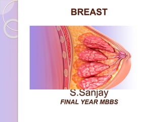
BREAST - ANATOMY AND PHYSIOLOGY.pptx
- 2. ANATOMY • Breast is a modified sweat gland derived from ectoderm, as branching epithelial cords which form lactiferous ducts. • It lies within the superficial fascia(sub- cutaneous). • It is present in both the sexes. • Rudimentary in male and well developed in female after puberty. • EXTENT • Vertical - 2nd to 6th ribs. • Horizontal - Lateral border of sternum to anterior axillary line. • 2/3rd of breast rests upon pectoralis major. • 1/3rd rests upon serratus anterior.
- 3. AXILLARY TAIL OF SPENCE It is the prolongation of Superolateral quadrant to the axilla along the lower border of pectoralis major through an opening in the deep fascia called Foramen of Langer. It is sometimes mistaken for an enlarged lymph node or a lipoma.
- 4. STRUCTURES DEEP TO BREAST Retromammary space Pectoral deep fascia Pectoralis major Serratus anterior External oblique Chest wall
- 5. AREOLA A circular pigmented area around the nipple. It contains involuntary muscle, sweat glands and sebaceous glands which enlarges during pregnancy and helps in lubrication during lactation (Montgomery tubercles). NIPPLE It is located at the level of 4th intercostal space. Covered by thick skin with corrugations. Near its apex lie the orifices of the lactiferous ducts. Erectile structure – smooth muscle fibres arranged concentrically and longitudinally- resemble panniculus carnosus.
- 6. SUSPENSORY LIGAMENT OF COOPER They are hollow conical projections of fibrous tissue. Anchors breast to underlying superficial pectoral fascia and to the overlying skin . These ligaments account for the dimpling of the skin overlying a carcinoma.
- 7. LOBES 15 to 20 in number. Composed of glandular structures called lobules which empty via ductules into lactiferous duct. LACTIFEROUS DUCTS It drains each lobes of the breast pass through the nipple and open on its tip as 15 to 20 orifices. Contains ampulla near its end (Reservoir for milk or abnormal discharge)
- 8. Histology of ducts and acini
- 9. Cont…
- 10. Histology of breast Lined by a layer of cuboidal epithelium(single layer thick in smaller ducts, two cells thick in the larger ducts) They lie on the myo-epithelial cells(ectoderm) and basement membrane. The morphology varies with age, menstrual cycle, pregnancy and lactation. Close to the opening of lactiferous duct they become from columnar to keratinizing stratified squamous
- 12. Cont… 5-6th week, two ventral bands of epithelium forms from axilla to groin region(Mil k line of schlutz) At day 49, thoracic bud only invaginat es and rest vanish The ectoderm ingrowth forms lactiferous duct and acini, surrounding perisomatop leuric mesoderm forms the fat and fascia. At day 56, the nipple forms At day 84, Primitive mammar y sprouts At 15th day, canalization occurs The ducts open into the mammary pits
- 13. Applied: Witch’s milk : ◦ Milk secreted from the new born infants. ◦ Due to combination of fetal prolactin and maternal oestrogens Polythelia ◦ Presence of extra nipples in the body. ◦ They’ll usually develop along the milk line. Athelia : ◦ Absence of nipple devolpement.
- 14. BLOOD SUPPLY OF BREAST Internal thoracic artery- 60% Perforating cutaneous branches to 2nd 3rd and 4th intercostal spaces Axillary artery- 30% lateral thoracic artery (2nd branch) pectoral branch of acromiothoracic artery superior thoracic artery Lateral branch of 2nd 3rd and 4th intercostal arteries.
- 15. VENOUS DRAINAGE OF BREAST Initially they converge around the nipple to form a circular venous plexus. SUPERFICIAL VEINS – drain into axillary, internal mammary and intercostal vessels. Through posterior intercostal veins they communicate with paravertebral venous plexus (Batson’s plexus) - CA Breast metastasis to vertebrae.
- 16. NERVE SUPPLY OF BREAST Anterior and lateral cutaneous branches of 4th to 6th intercostal nerves.
- 17. LYMPHATIC DRAINAGE OF BREAST AXILLARY LYMPH NODES – 75% Lateral - along the axillary vein . Anterior - along the lateral thoracic vessels. Posterior - along the subscapular vessels. Central - embedded in fat in the centre of the axilla. Interpectoral - also called Rotter’s node lies between the pectoralis major and minor muscles. Apical - also called subclavicular or Halsted nodes. lies most superior and deep to pectoralis minor medial to axillary vessels.
- 19. BERG’S LEVEL OF AXILLARY LYMPH NODE LEVEL I - Below and lateral to pectoralis minor. Anterior, posterior and lateral LEVEL II - Behind the pectoralis minor. Central and interpectoral LEVEL III - Above and medial to pectoralis minor. Apical
- 20. 25% drains mainly from medial half of the breast into 2nd 3rd 4th intercostal space internal mammary lymph nodes. 1 to 2 cm lateral to sternal margin. Drainage into contralateral axilla and opposite lymph nodes. Sentinel lymph node – first node in the regional lymphatic basin that receives the lymph flow from the primary tumor.
- 21. PHYSIOLOGY OF LACTATION Mammogenesis - Development of breast to a functional state. Lactogenesis - Synthesis and secretion of milk from the alveoli. Galactokinesis - Ejection of milk outside the breast. Galactopoiesis - Maintenance of lactation. Involution- Regression and atrophy post lactation
- 22. Estrogen stimulates growth of the ductal system. Progesterone is required for full development of lobe – alveolar system. Prolactin promotes lactation. Milk ejection reflex:
- 23. KEY POINTS Breast is derived from ectoderm. Extents from 2nd to 6th rib. 2/3rd rests on pectoralis major and 1/3rd on serratus anterior. Montgomery tubercle is anatomical. Axillary tail of Spence extends through foramen of Langer. Infiltration of Cooper’s ligament causes dimpling. Internal thoracic artery – 60% Batson’s plexus – Metastasis to vertebrae. Rotter’s node – Interpectoral node. Halsted nodes – Apical lymph node
- 24. THANK YOU