SlideShare a Scribd company logo
1 of 31
Dual-Energy imaging and Digital Tomosynthesis:
Innovative X-ray based imaging technologies
S Sajja, F Ursani, A Ursani, and N S Paul
TRIIO Core Laboratory, Departments of Medical Imaging,
University Health Network, University of Toronto
S Richard, N Packard, X Wang, and L Vogelsang
Carestream Health, Rochester
2
Outline
•Chest Radiography – Limitations of 2D imaging
•Anatomical clutter
•Dual-Energy (DE) X-ray
•Anatomical clutter reduction via tissue discrimination
•Factors that impact DE imaging performance
•Clinical examples
•Digital Tomosynthesis (DT) X-ray
•Anatomical clutter reduction via spatial discrimination
•Factors that impact DT imaging performance
•Clinical examples
3
Chest Radiography (CXR)
• Chest Radiography is the most common technique for 2D chest imaging
•Limitation
• A chest radiograph is a 2-dimensional projection of a complex 3-
dimensional volume in which several tissues overlay each other.
• Overlapping anatomy – ribs, lungs and vessels are background
structures which may confound detection of a lung nodule on a single
projection.
Conspicuity (detectability) = Contrast of the object
Complexity of the background
4
Limitation of Chest Radiography (CXR)
3-d object
2-d projection of
the objectsCylinders made of material 1
Different objects made of
material 2
Overcoming anatomical noise via tissue discrimination
•Removing overlapping ribs via tissue removal using dual-energy imaging.
Material 1 only image2-d projection
Tissue discrimination using x-ray
imaging results in reduced
background anatomical noise
and improved feature
conspicuity.
DR - Digital Radiography
DE - Dual Energy X-ray
Tissue Discrimination – Dual-Energy (DE) imaging
6
High energy image
Low energy image
Bone image
WbWs
Soft-tissue image
X-ray tube
Anti-scatter bucky grid
flat-panel
digital detector
 Energy selection: Choice of
high and low energy values
 Dose allocation:
 Filtration:
 Delay between two
acquisitions:
7
Factors that affect DE imaging performance
• DE soft tissue images of polyethylene nodule
• High energy = 130 kVp (fixed)
• Low energy = 60-90 kVp
Patient (cm) equivalent phantom [acrylic] thicknesses:
“slim patient” (18 cm) [7.5cm]
“average patient” (24 cm) [10cm]
“large patient” (30 cm) [12cm]
Nodule contrast is highest at low kVp (60 kVp) and in a
slim patient
Nick Schumat, Masters Thesis, University of Toronto, 2008
 Energy selection:
 Dose allocation: Ratio of dose
between high and low energies
 Filtration:
 Delay between two
acquisitions:
8
Factors that affect DE imaging performance
Aε = 0.06 Aε = 0.29
Aε = 0.72 Aε = 0.91
• DE bone-only images at different dose
allocations (AƐ)
• Noticeable increase in image noise at very
low and very high allocations
Nick Schumat, Masters Thesis, University of Toronto, 2008
9
Factors that affect DE imaging performance
 Energy selection:
 Dose allocation:
 Filtration: helps in increasing the
energy separation between the high
and low energy images
Fixed filtration: Same filtration for both
high and low energy acquisitions.
 Delay between two
acquisitions:
4
0 50 100 150
0
2
4
6
8
10
12
x 10
Intensity
4
keV
0 50 100 150
0
2
4
6
8
10
12
x 10
Intensity
60 kVp
60 kVp
1 mm Cu
keV
No Filtration – high unused radiation
Fixed Filtration – lower unused radiation
10
Factors that affect DE imaging performance
Differential Filtration – very low unused
radiation
 Energy selection:
 Dose allocation:
 Filtration: helps in increasing the
energy separation between the high
and low energy images
Differential filtration: Filtration is
changed between high and low
images to separate spectra.
 Delay between two
acquisitions:
4
keV
0 50 100 150
0
2
4
6
8
10
12
x 10
Intensity
4
keV
0 50 100 150
0
2
4
6
8
10
12
x 10
Intensity
60 kVp
1 mm Cu
120 kVp
1mm Cu
70 kVp
0.1 mm Cu
120 kVp
0.5 Ag
Fixed Filtration – lower unused radiation
 Energy selection:
 Dose allocation:
 Filtration:
 Delay between two
acquisitions: This has an impact
on the motion artifacts observed in
resultant DE image.
11
Factors that affect DE imaging performance
ECG gating can be used to reduce motion artifacts.
Timing diagram displaying the (ECG) trace,
plethysmogram, and digital trigger.
DE “soft-tissue” images acquired (a) with (b) without
cardiac motion – custom built insert for motion
simulation. Some motion blur is observed.
Nick Schumat, Masters Thesis, University of Toronto, 2008
12
Modelling nodule conspicuity
DR
Objective function for system optimization - combines:
MTF – System resolution
GNPS- Noise and anatomical clutter
Wtask- description of task i.e., nodule detection
d‘ – Detectability index – surrogate for nodule conspicuity
13
Nodule detectability – fixed versus differential filtration
DR
DR DE fixed filtration
DE differential
filtration
• Figure shows the detectability index values for
DE fixed and differential filtration.
• The results are normalized such that d’ = 1 for
DR.
• d’ = 1.2 and 1.3 for fixed and differential filtration
respectively.
• d’norm refers to detectability normalized by dose.
• d’norm = 0.7 and 1.1 for fixed and differential
filtration indicating fixed filtration less dose
efficient than differential filtration.
Richard S. et al., Diagnostic Imaging, 2015
14
DE Clinical patient imaging
CXR of a patient with lung nodules:
limited conspicuity due to
overlapping structures
DE-soft tissue of a patient with lung
nodules: Improved conspicuity due
to subtracted ribs.
15
DE – Cadaver Images
Cadaveric chest x-rays were performed using conventional Digital
Radiography (DR) (a) followed by DE projections decomposed into Bone
only (b) and Soft tissue only (c) images to demonstrate improved
conspicuity of bone and soft tissue details from DE projections compared
to DR chest x-ray.
How to overcome anatomical clutter via spatial
discrimination
•Separation of overlapping structures. Commonly used techniques –
Computed tomography (CT) and Digital Tomosynthesis (DT) .
Illustration of CT
Illustration of DT
Illustration of spatial discrimination in x-ray medical
imaging resulting in reduced overlying clutter and
improved feature conspicuity.
17
DT (Spatial discrimination) – Imaging
Detector
Detector
X-ray
tube
Table top
Wall Stand
(a) Working of the tomosynthesis in wallstand position (b) Working of the tomosynthesis in table top
position
Phantom
Phantom
Factors that affect DT imaging performance
 Angular range (Ɵ), number of
projections (N) : A smaller range for
image acquisition results in a lower tomo
angle and reduced x-ray dose; but the
depth resolution is also reduced.
 Detector binning: The process of
selecting regions of pixel and finding the
mean value. Improves signal-to-noise
ratio, speeds but results in loss of spatial
resolution.
 Scan time: The time taken to acquire
the set of projections. This has an impact
on motion artifacts due to cardiac and
respiratory motion.
(a) N=15, Ɵ=30o (b) N=60, Ɵ=30o
(a) N=15, Ɵ=7.5o (b) N=60, Ɵ=30o
Söderman C. et al., Medical Physics, 2015
Factors that affect DT imaging performance
 Angular range (Ɵ), number of
projections: A smaller range for
image acquisition results in a lower tomo
angle and reduced x-ray dose; but the
depth resolution is also reduced.
 Detector binning: The process of
selecting regions of pixel (NxN, N=
number of pixels) and finding the mean
value. Improves signal-to-noise ratio,
speeds but results in loss of spatial
resolution.
 Scan time: The time taken to acquire
the set of projections. This has an impact
on motion artifacts due to cardiac and
respiratory motion.
Pacemaker generator: Greater detail is seen
with 1X1 binning
Factors that affect DT imaging performance
 Angular range (Ɵ), number of
projections: A smaller range for
image acquisition results in a lower tomo
angle and reduced x-ray dose; but the
depth resolution is also reduced.
 Detector binning: The process of
selecting regions of pixel (NxN, N=
number of pixels) and finding the mean
value. Improves signal-to-noise ratio,
speeds but results in loss of spatial
resolution. .
 Scan time: The time taken to acquire
the set of projections. This has an impact
on motion artifacts due to cardiac and
respiratory motion.
DT phantom images acquired
(a) without motion
(b) with respiratory (breathing) motion
(c) with cardiac motion using a custom built
insert for motion simulation.
21
Nodule detectability – Impact of dose in IQ
DT 100% DT 50% DT 30%
• Figure shows the detectability index values for
DT at different doses (30%, 50% and 100%).
• The results are normalized such that d’ = 1 for
DR.
• d’ = 11.8, 14.6 and 18.1 for 30%, 50% and
100% of the nominal doses respectively.
• d’norm refers to detectability normalized by dose.
• d’norm = 7.8, 7.7 and 5.9 for 30%, 50% and 100%
respectively since DT becomes anatomical noise
limited as opposed to quantum noise limited.Richard S. et al., Diagnostic Imaging, 2015
22
DT – Patient imaging
Reconstructed DT patient image Corresponding coronal CT patient
23
DT – Cadaver Images
Cadaveric DT through
the pulmonary hila
demonstrate high
resolution images of
the bifurcating left
main bronchus (inset)
using:
Slice thickness = 5 mm
Slice interval = 3 mm
Dose = full
Binning=1x1
24
DT – Cadaver Images
Cadaveric DT
through the
pulmonary hila
demonstrate high
resolution images of
the branch
pulmonary arteries
and veins (inset)
using:
Slice thickness = 5 mm
Slice interval = 3 mm
Dose = full
Binning =1x1
25
DT – Limitations
A nodule in a vessel branching point may be mistaken for
an enlarged vessel in tomosynthesis – DT (left), CT (right)
(Asplund S. , Acta Radiologica, 2011)
•DT involves a limited angle of
acquisition compared to CT.
•This results in limited sampling of
the signal in the frequency domain.
• Depth resolution is poorer in DT
than CT.
• In some cases this may result in
misinterpretation of structures as
seen in the image here.
•This can be alleviated by relating
the location of the ribs as compared
to the structure in question.
•Also for this particular example, the
potential use of contrast medium can
be explored.
26
DT – Limitations
Nodule close to
pleura border –
With X-ray beam
tangential to ribs
Nodule close to
pleura border –
With X-ray beam
not tangential to
ribs
Nodule close to
pleura border –
With X-ray
beam not
tangential to
ribs
Nodule on vessel
branching point –
may be mistaken
for an enlarged
vessel on DT
DT may provide a false anatomical location of a lung nodule when it is located close to the
pleura; the effect depends on the angle of the incident X-ray beam relative to the nodule.
Asplund S. , Acta Radiologica, 2011
27
DE-DT – Comparison Study – Low density objects
• Low density objects (cotton spheres) were inserted into the anthropomorphic phantom:
1 dry cotton sphere (green arrow) and 2 cotton spheres dipped in distilled water (blue
arrows)
• The phantom was imaged using (a) DR (b) DE (c) and DT and low dose CT (1mSv).
• CT images served as a reference standard (slide 28).
• DR and soft tissue DE images demonstrate the wet cotton spheres with a faint
projection of the dry cotton sphere. The DT images clearly demonstrate all of the
spheres.
• Lung pathologies (tumors) vary in their water content
• DE-DT may improve lesion characterization
Digital Radiograph Dual Energy Digital Tomosynthesis
28
DE-DT – Comparison Study – Low density object
Coronal low dose CT images
Digital Radiograph Dual Energy Digital Tomosynthesis
29
What is the future of DE and DT?
Future innovation: Qualitative to quantitative
Increased detector spatial resolution will facilitate extraction of quantitative image
data to provide more accurate diagnosis.
Application study: Volume estimation of thoracic water content
• In critically ill patients, clinical assessment of change
in thoracic fluid volume is necessary.
• A method based on temporal subtraction of CXR is
proposed to quantify the change in fluid volume
• Proof of concept testing was done using a chest
anthropomorphic phantom and solid water blocks.
• The estimated volume based on the technique was
compared with the actual volume.
•Dual energy (DE)- soft tissue only images had the
highest accuracy and correlated closely with the actual
volume with a root mean square (RMS)=.4.74 ml
Sajja et al., World Congress of Med. Phys. and Biomed. Eng. 2015
30
What is the future of DE and DT?
DE- DT
An integrated DE-DT system would be beneficial for increasing the scope of
thoracic diseases which can be potentially diagnosed using chest radiography.
Illustration of DE-DT – material 1 Illustration of DE-DT – material 2
31
Summary
• For DR CXR, overlapping of structures confounds the detection of
nodules. This results in anatomical noise.
• Anatomical noise can be overcome either via tissue discrimination or
spatial discrimination.
• Tissue discrimination is achieved via Dual-Energy (DE) which involves
acquisition of paired radiographs at 2 energies.
• Spatial discrimination is achieved via Digital Tomosynthesis (DT) which
involves acquisition of radiographs at different angles.
• It would be beneficial to combine the spatial and tissue discrimination
through an integrated DE-DT system.

More Related Content

What's hot

Magnetic Resonance Perfusion
Magnetic Resonance PerfusionMagnetic Resonance Perfusion
Magnetic Resonance PerfusionRahman Ud Din
 
Current applications of interventional radiology 97
Current applications of interventional radiology 97Current applications of interventional radiology 97
Current applications of interventional radiology 97Arun Jagannathan
 
Image guided radiation therapy (IGRT)
Image guided radiation therapy (IGRT)Image guided radiation therapy (IGRT)
Image guided radiation therapy (IGRT)Anees Muhammed
 
Presentation1.pptx, diffusion tensor imaging of white matter tract in cerebra...
Presentation1.pptx, diffusion tensor imaging of white matter tract in cerebra...Presentation1.pptx, diffusion tensor imaging of white matter tract in cerebra...
Presentation1.pptx, diffusion tensor imaging of white matter tract in cerebra...Abdellah Nazeer
 
Radio Frequency Ablation (RFA Treatment ) -Modern Technology for management l...
Radio Frequency Ablation (RFA Treatment ) -Modern Technology for management l...Radio Frequency Ablation (RFA Treatment ) -Modern Technology for management l...
Radio Frequency Ablation (RFA Treatment ) -Modern Technology for management l...SafeMedTrip
 
MANUAL ISODOSE PLOTTING.pptx
MANUAL ISODOSE PLOTTING.pptxMANUAL ISODOSE PLOTTING.pptx
MANUAL ISODOSE PLOTTING.pptxPIJUSH PAUL
 
magnetic resonance in angiography
magnetic resonance in  angiography magnetic resonance in  angiography
magnetic resonance in angiography BISHAL KHANAL
 
Portable ct mobile ct
Portable ct mobile ctPortable ct mobile ct
Portable ct mobile ctanilayyakutty
 
Image guided radiation therapy
Image guided radiation therapyImage guided radiation therapy
Image guided radiation therapySwarnita Sahu
 
Image Quality, Artifacts and it's Remedies in CT-Avinesh Shrestha
Image Quality, Artifacts and it's Remedies in CT-Avinesh ShresthaImage Quality, Artifacts and it's Remedies in CT-Avinesh Shrestha
Image Quality, Artifacts and it's Remedies in CT-Avinesh ShresthaAvinesh Shrestha
 
Diffusion weighted imaging
Diffusion  weighted imagingDiffusion  weighted imaging
Diffusion weighted imagingSharath Raj
 
Dose volume histogram
Dose volume histogramDose volume histogram
Dose volume histogramNilesh Kucha
 
CEUS final.pptx
CEUS final.pptxCEUS final.pptx
CEUS final.pptxjim kuok
 
Medulloblatoma - Field Matching In RT Planning - CSI
Medulloblatoma - Field Matching In RT Planning - CSIMedulloblatoma - Field Matching In RT Planning - CSI
Medulloblatoma - Field Matching In RT Planning - CSISubhash Thakur
 
Perfusion MRI (DSC and DCE perfusion techniques) for radiology residents
Perfusion MRI (DSC and DCE perfusion techniques) for radiology residentsPerfusion MRI (DSC and DCE perfusion techniques) for radiology residents
Perfusion MRI (DSC and DCE perfusion techniques) for radiology residentsRiham Dessouky
 
EWINGS SARCOMA & RADIOTHERAPY
EWINGS SARCOMA & RADIOTHERAPYEWINGS SARCOMA & RADIOTHERAPY
EWINGS SARCOMA & RADIOTHERAPYPaul George
 

What's hot (20)

Magnetic Resonance Perfusion
Magnetic Resonance PerfusionMagnetic Resonance Perfusion
Magnetic Resonance Perfusion
 
Ctcontrast
Ctcontrast Ctcontrast
Ctcontrast
 
Current applications of interventional radiology 97
Current applications of interventional radiology 97Current applications of interventional radiology 97
Current applications of interventional radiology 97
 
CT fluoroscopy
CT fluoroscopyCT fluoroscopy
CT fluoroscopy
 
MRI in Cirrhotic Liver
MRI in Cirrhotic LiverMRI in Cirrhotic Liver
MRI in Cirrhotic Liver
 
Image guided radiation therapy (IGRT)
Image guided radiation therapy (IGRT)Image guided radiation therapy (IGRT)
Image guided radiation therapy (IGRT)
 
Modern Radiotherapy
Modern RadiotherapyModern Radiotherapy
Modern Radiotherapy
 
Presentation1.pptx, diffusion tensor imaging of white matter tract in cerebra...
Presentation1.pptx, diffusion tensor imaging of white matter tract in cerebra...Presentation1.pptx, diffusion tensor imaging of white matter tract in cerebra...
Presentation1.pptx, diffusion tensor imaging of white matter tract in cerebra...
 
Radio Frequency Ablation (RFA Treatment ) -Modern Technology for management l...
Radio Frequency Ablation (RFA Treatment ) -Modern Technology for management l...Radio Frequency Ablation (RFA Treatment ) -Modern Technology for management l...
Radio Frequency Ablation (RFA Treatment ) -Modern Technology for management l...
 
MANUAL ISODOSE PLOTTING.pptx
MANUAL ISODOSE PLOTTING.pptxMANUAL ISODOSE PLOTTING.pptx
MANUAL ISODOSE PLOTTING.pptx
 
magnetic resonance in angiography
magnetic resonance in  angiography magnetic resonance in  angiography
magnetic resonance in angiography
 
Portable ct mobile ct
Portable ct mobile ctPortable ct mobile ct
Portable ct mobile ct
 
Image guided radiation therapy
Image guided radiation therapyImage guided radiation therapy
Image guided radiation therapy
 
Image Quality, Artifacts and it's Remedies in CT-Avinesh Shrestha
Image Quality, Artifacts and it's Remedies in CT-Avinesh ShresthaImage Quality, Artifacts and it's Remedies in CT-Avinesh Shrestha
Image Quality, Artifacts and it's Remedies in CT-Avinesh Shrestha
 
Diffusion weighted imaging
Diffusion  weighted imagingDiffusion  weighted imaging
Diffusion weighted imaging
 
Dose volume histogram
Dose volume histogramDose volume histogram
Dose volume histogram
 
CEUS final.pptx
CEUS final.pptxCEUS final.pptx
CEUS final.pptx
 
Medulloblatoma - Field Matching In RT Planning - CSI
Medulloblatoma - Field Matching In RT Planning - CSIMedulloblatoma - Field Matching In RT Planning - CSI
Medulloblatoma - Field Matching In RT Planning - CSI
 
Perfusion MRI (DSC and DCE perfusion techniques) for radiology residents
Perfusion MRI (DSC and DCE perfusion techniques) for radiology residentsPerfusion MRI (DSC and DCE perfusion techniques) for radiology residents
Perfusion MRI (DSC and DCE perfusion techniques) for radiology residents
 
EWINGS SARCOMA & RADIOTHERAPY
EWINGS SARCOMA & RADIOTHERAPYEWINGS SARCOMA & RADIOTHERAPY
EWINGS SARCOMA & RADIOTHERAPY
 

Similar to Dual energy imaging and digital tomosynthesis: Innovative X-ray based imaging technologies

medical equipment lec 6 CT scan
medical equipment lec 6 CT scanmedical equipment lec 6 CT scan
medical equipment lec 6 CT scancairo university
 
CT SCAN - 3 Marks - QUESTION AND ANSWERS
CT SCAN - 3 Marks - QUESTION AND ANSWERSCT SCAN - 3 Marks - QUESTION AND ANSWERS
CT SCAN - 3 Marks - QUESTION AND ANSWERSGanesan Yogananthem
 
Dose reduction in MDCT . Daniel J.P , Khorfakhan hospital . UAE
Dose reduction in MDCT . Daniel J.P , Khorfakhan hospital . UAEDose reduction in MDCT . Daniel J.P , Khorfakhan hospital . UAE
Dose reduction in MDCT . Daniel J.P , Khorfakhan hospital . UAEhaijaypee_dan
 
On Dose Reduction and View Number
On Dose Reduction and View NumberOn Dose Reduction and View Number
On Dose Reduction and View NumberKaijie Lu
 
CT ITS BASIC PHYSICS
CT ITS BASIC PHYSICSCT ITS BASIC PHYSICS
CT ITS BASIC PHYSICSDEEPAK
 
Image Quality And Artifacts in Computed Tomography.pptx
Image Quality And Artifacts in Computed Tomography.pptxImage Quality And Artifacts in Computed Tomography.pptx
Image Quality And Artifacts in Computed Tomography.pptxivanKeshari
 
The Use Of High Energy Protons
The Use Of High Energy ProtonsThe Use Of High Energy Protons
The Use Of High Energy Protonsfondas vakalis
 
CT Dose Issues.pptx on the factors to be considered on radiation protection
CT Dose Issues.pptx on the factors to be considered on radiation protectionCT Dose Issues.pptx on the factors to be considered on radiation protection
CT Dose Issues.pptx on the factors to be considered on radiation protectionsanyengere
 
principals of computed tomography / dental implant courses
principals of computed tomography / dental implant coursesprincipals of computed tomography / dental implant courses
principals of computed tomography / dental implant coursesIndian dental academy
 
MDCT Principles and Applications- Avinesh Shrestha
MDCT Principles and Applications- Avinesh ShresthaMDCT Principles and Applications- Avinesh Shrestha
MDCT Principles and Applications- Avinesh ShresthaAvinesh Shrestha
 
Patient data acquisition and treatment verification.pptx
Patient data acquisition and treatment verification.pptxPatient data acquisition and treatment verification.pptx
Patient data acquisition and treatment verification.pptxJuliyet K Joy
 
COMPUTED TOMOGRAPHY SCAN
COMPUTED TOMOGRAPHY SCANCOMPUTED TOMOGRAPHY SCAN
COMPUTED TOMOGRAPHY SCANShounak Nandi
 
Ct radiation dose & safety lecture 1
Ct radiation dose & safety lecture 1Ct radiation dose & safety lecture 1
Ct radiation dose & safety lecture 1Gamal Mahdaly
 
Comparison of ct and cbct
Comparison of ct and cbct Comparison of ct and cbct
Comparison of ct and cbct Amritha James
 

Similar to Dual energy imaging and digital tomosynthesis: Innovative X-ray based imaging technologies (20)

medical equipment lec 6 CT scan
medical equipment lec 6 CT scanmedical equipment lec 6 CT scan
medical equipment lec 6 CT scan
 
CT SCAN - 3 Marks - QUESTION AND ANSWERS
CT SCAN - 3 Marks - QUESTION AND ANSWERSCT SCAN - 3 Marks - QUESTION AND ANSWERS
CT SCAN - 3 Marks - QUESTION AND ANSWERS
 
3.ppt
3.ppt3.ppt
3.ppt
 
Dose reduction in MDCT . Daniel J.P , Khorfakhan hospital . UAE
Dose reduction in MDCT . Daniel J.P , Khorfakhan hospital . UAEDose reduction in MDCT . Daniel J.P , Khorfakhan hospital . UAE
Dose reduction in MDCT . Daniel J.P , Khorfakhan hospital . UAE
 
On Dose Reduction and View Number
On Dose Reduction and View NumberOn Dose Reduction and View Number
On Dose Reduction and View Number
 
Dose reduction
Dose reductionDose reduction
Dose reduction
 
CT ITS BASIC PHYSICS
CT ITS BASIC PHYSICSCT ITS BASIC PHYSICS
CT ITS BASIC PHYSICS
 
Image Quality And Artifacts in Computed Tomography.pptx
Image Quality And Artifacts in Computed Tomography.pptxImage Quality And Artifacts in Computed Tomography.pptx
Image Quality And Artifacts in Computed Tomography.pptx
 
Ct brain presentation
Ct brain presentationCt brain presentation
Ct brain presentation
 
CT dose reduction
CT dose reductionCT dose reduction
CT dose reduction
 
Ct i patient_whitepaper
Ct i patient_whitepaperCt i patient_whitepaper
Ct i patient_whitepaper
 
The Use Of High Energy Protons
The Use Of High Energy ProtonsThe Use Of High Energy Protons
The Use Of High Energy Protons
 
CT Dose Issues.pptx on the factors to be considered on radiation protection
CT Dose Issues.pptx on the factors to be considered on radiation protectionCT Dose Issues.pptx on the factors to be considered on radiation protection
CT Dose Issues.pptx on the factors to be considered on radiation protection
 
principals of computed tomography / dental implant courses
principals of computed tomography / dental implant coursesprincipals of computed tomography / dental implant courses
principals of computed tomography / dental implant courses
 
MDCT Principles and Applications- Avinesh Shrestha
MDCT Principles and Applications- Avinesh ShresthaMDCT Principles and Applications- Avinesh Shrestha
MDCT Principles and Applications- Avinesh Shrestha
 
Patient data acquisition and treatment verification.pptx
Patient data acquisition and treatment verification.pptxPatient data acquisition and treatment verification.pptx
Patient data acquisition and treatment verification.pptx
 
COMPUTED TOMOGRAPHY SCAN
COMPUTED TOMOGRAPHY SCANCOMPUTED TOMOGRAPHY SCAN
COMPUTED TOMOGRAPHY SCAN
 
X ray tomography
X ray tomographyX ray tomography
X ray tomography
 
Ct radiation dose & safety lecture 1
Ct radiation dose & safety lecture 1Ct radiation dose & safety lecture 1
Ct radiation dose & safety lecture 1
 
Comparison of ct and cbct
Comparison of ct and cbct Comparison of ct and cbct
Comparison of ct and cbct
 

More from Carestream

Affordable Digital Upgrade for Medical Imaging - the Benefits and the Return ...
Affordable Digital Upgrade for Medical Imaging - the Benefits and the Return ...Affordable Digital Upgrade for Medical Imaging - the Benefits and the Return ...
Affordable Digital Upgrade for Medical Imaging - the Benefits and the Return ...Carestream
 
Digital Tomosynthesis: Theory of Operation
Digital Tomosynthesis: Theory of OperationDigital Tomosynthesis: Theory of Operation
Digital Tomosynthesis: Theory of OperationCarestream
 
Tube and Line and Pneumothorax Visualization Software
Tube and Line and Pneumothorax Visualization SoftwareTube and Line and Pneumothorax Visualization Software
Tube and Line and Pneumothorax Visualization SoftwareCarestream
 
EVP Plus Software delivers state-of-the-art image processing for CR and DR sy...
EVP Plus Software delivers state-of-the-art image processing for CR and DR sy...EVP Plus Software delivers state-of-the-art image processing for CR and DR sy...
EVP Plus Software delivers state-of-the-art image processing for CR and DR sy...Carestream
 
Dose Efficient Dual Energy Subtraction Radiography - Theory of Operations
Dose Efficient Dual Energy Subtraction Radiography - Theory of OperationsDose Efficient Dual Energy Subtraction Radiography - Theory of Operations
Dose Efficient Dual Energy Subtraction Radiography - Theory of OperationsCarestream
 
Smart Noise Cancellation Processing: New Level of Clarity in Digital Radiography
Smart Noise Cancellation Processing: New Level of Clarity in Digital RadiographySmart Noise Cancellation Processing: New Level of Clarity in Digital Radiography
Smart Noise Cancellation Processing: New Level of Clarity in Digital RadiographyCarestream
 
Special Report: Challenges and Solutions in Pediatric X-ray
Special Report: Challenges and Solutions in Pediatric X-raySpecial Report: Challenges and Solutions in Pediatric X-ray
Special Report: Challenges and Solutions in Pediatric X-rayCarestream
 
Special Report: Getting the Optimal Return on X-ray Equipment
Special Report: Getting the Optimal Return on X-ray EquipmentSpecial Report: Getting the Optimal Return on X-ray Equipment
Special Report: Getting the Optimal Return on X-ray EquipmentCarestream
 
The Pursuit of Excellence in Image Quality
The Pursuit of Excellence in Image QualityThe Pursuit of Excellence in Image Quality
The Pursuit of Excellence in Image QualityCarestream
 
Whitepaper: Healthcare Data Migration - Top 10 Questions
Whitepaper: Healthcare Data Migration - Top 10 Questions Whitepaper: Healthcare Data Migration - Top 10 Questions
Whitepaper: Healthcare Data Migration - Top 10 Questions Carestream
 
Whitepaper: Image Quality Impact of SmartGrid Processing in Bedside Chest Ima...
Whitepaper: Image Quality Impact of SmartGrid Processing in Bedside Chest Ima...Whitepaper: Image Quality Impact of SmartGrid Processing in Bedside Chest Ima...
Whitepaper: Image Quality Impact of SmartGrid Processing in Bedside Chest Ima...Carestream
 
Using Carbon Nano-Tube Field Emitters to Miniaturize X-Ray Tubes
Using Carbon Nano-Tube Field Emitters to Miniaturize X-Ray TubesUsing Carbon Nano-Tube Field Emitters to Miniaturize X-Ray Tubes
Using Carbon Nano-Tube Field Emitters to Miniaturize X-Ray TubesCarestream
 
Guia obsolescencia seram
Guia obsolescencia seramGuia obsolescencia seram
Guia obsolescencia seramCarestream
 
Cloud Security
Cloud Security Cloud Security
Cloud Security Carestream
 
Sunway Medical Centre Installs CARESTREAM Vue PACS to Streamline Imaging Proc...
Sunway Medical Centre Installs CARESTREAM Vue PACS to Streamline Imaging Proc...Sunway Medical Centre Installs CARESTREAM Vue PACS to Streamline Imaging Proc...
Sunway Medical Centre Installs CARESTREAM Vue PACS to Streamline Imaging Proc...Carestream
 
White Paper: The Benefits of Mobile X-rays in Thoracic and Cardiac Care
White Paper: The Benefits of Mobile X-rays in Thoracic and Cardiac CareWhite Paper: The Benefits of Mobile X-rays in Thoracic and Cardiac Care
White Paper: The Benefits of Mobile X-rays in Thoracic and Cardiac CareCarestream
 
Study: University Clinic, Regensburg, Evaluates Smart Flow in Ultrasound
Study: University Clinic, Regensburg, Evaluates Smart Flow in UltrasoundStudy: University Clinic, Regensburg, Evaluates Smart Flow in Ultrasound
Study: University Clinic, Regensburg, Evaluates Smart Flow in UltrasoundCarestream
 
Financial Implications for Integrating Carestream OnSight 3D Extremity System...
Financial Implications for Integrating Carestream OnSight 3D Extremity System...Financial Implications for Integrating Carestream OnSight 3D Extremity System...
Financial Implications for Integrating Carestream OnSight 3D Extremity System...Carestream
 
Whitepaper: Enterprise Access Viewer Connecting technologies, physicians, and...
Whitepaper: Enterprise Access Viewer Connecting technologies, physicians, and...Whitepaper: Enterprise Access Viewer Connecting technologies, physicians, and...
Whitepaper: Enterprise Access Viewer Connecting technologies, physicians, and...Carestream
 
Evaluating Enterprise Clinical Data-Management Systems at RSNA 2016
Evaluating Enterprise Clinical Data-Management Systems at RSNA 2016Evaluating Enterprise Clinical Data-Management Systems at RSNA 2016
Evaluating Enterprise Clinical Data-Management Systems at RSNA 2016Carestream
 

More from Carestream (20)

Affordable Digital Upgrade for Medical Imaging - the Benefits and the Return ...
Affordable Digital Upgrade for Medical Imaging - the Benefits and the Return ...Affordable Digital Upgrade for Medical Imaging - the Benefits and the Return ...
Affordable Digital Upgrade for Medical Imaging - the Benefits and the Return ...
 
Digital Tomosynthesis: Theory of Operation
Digital Tomosynthesis: Theory of OperationDigital Tomosynthesis: Theory of Operation
Digital Tomosynthesis: Theory of Operation
 
Tube and Line and Pneumothorax Visualization Software
Tube and Line and Pneumothorax Visualization SoftwareTube and Line and Pneumothorax Visualization Software
Tube and Line and Pneumothorax Visualization Software
 
EVP Plus Software delivers state-of-the-art image processing for CR and DR sy...
EVP Plus Software delivers state-of-the-art image processing for CR and DR sy...EVP Plus Software delivers state-of-the-art image processing for CR and DR sy...
EVP Plus Software delivers state-of-the-art image processing for CR and DR sy...
 
Dose Efficient Dual Energy Subtraction Radiography - Theory of Operations
Dose Efficient Dual Energy Subtraction Radiography - Theory of OperationsDose Efficient Dual Energy Subtraction Radiography - Theory of Operations
Dose Efficient Dual Energy Subtraction Radiography - Theory of Operations
 
Smart Noise Cancellation Processing: New Level of Clarity in Digital Radiography
Smart Noise Cancellation Processing: New Level of Clarity in Digital RadiographySmart Noise Cancellation Processing: New Level of Clarity in Digital Radiography
Smart Noise Cancellation Processing: New Level of Clarity in Digital Radiography
 
Special Report: Challenges and Solutions in Pediatric X-ray
Special Report: Challenges and Solutions in Pediatric X-raySpecial Report: Challenges and Solutions in Pediatric X-ray
Special Report: Challenges and Solutions in Pediatric X-ray
 
Special Report: Getting the Optimal Return on X-ray Equipment
Special Report: Getting the Optimal Return on X-ray EquipmentSpecial Report: Getting the Optimal Return on X-ray Equipment
Special Report: Getting the Optimal Return on X-ray Equipment
 
The Pursuit of Excellence in Image Quality
The Pursuit of Excellence in Image QualityThe Pursuit of Excellence in Image Quality
The Pursuit of Excellence in Image Quality
 
Whitepaper: Healthcare Data Migration - Top 10 Questions
Whitepaper: Healthcare Data Migration - Top 10 Questions Whitepaper: Healthcare Data Migration - Top 10 Questions
Whitepaper: Healthcare Data Migration - Top 10 Questions
 
Whitepaper: Image Quality Impact of SmartGrid Processing in Bedside Chest Ima...
Whitepaper: Image Quality Impact of SmartGrid Processing in Bedside Chest Ima...Whitepaper: Image Quality Impact of SmartGrid Processing in Bedside Chest Ima...
Whitepaper: Image Quality Impact of SmartGrid Processing in Bedside Chest Ima...
 
Using Carbon Nano-Tube Field Emitters to Miniaturize X-Ray Tubes
Using Carbon Nano-Tube Field Emitters to Miniaturize X-Ray TubesUsing Carbon Nano-Tube Field Emitters to Miniaturize X-Ray Tubes
Using Carbon Nano-Tube Field Emitters to Miniaturize X-Ray Tubes
 
Guia obsolescencia seram
Guia obsolescencia seramGuia obsolescencia seram
Guia obsolescencia seram
 
Cloud Security
Cloud Security Cloud Security
Cloud Security
 
Sunway Medical Centre Installs CARESTREAM Vue PACS to Streamline Imaging Proc...
Sunway Medical Centre Installs CARESTREAM Vue PACS to Streamline Imaging Proc...Sunway Medical Centre Installs CARESTREAM Vue PACS to Streamline Imaging Proc...
Sunway Medical Centre Installs CARESTREAM Vue PACS to Streamline Imaging Proc...
 
White Paper: The Benefits of Mobile X-rays in Thoracic and Cardiac Care
White Paper: The Benefits of Mobile X-rays in Thoracic and Cardiac CareWhite Paper: The Benefits of Mobile X-rays in Thoracic and Cardiac Care
White Paper: The Benefits of Mobile X-rays in Thoracic and Cardiac Care
 
Study: University Clinic, Regensburg, Evaluates Smart Flow in Ultrasound
Study: University Clinic, Regensburg, Evaluates Smart Flow in UltrasoundStudy: University Clinic, Regensburg, Evaluates Smart Flow in Ultrasound
Study: University Clinic, Regensburg, Evaluates Smart Flow in Ultrasound
 
Financial Implications for Integrating Carestream OnSight 3D Extremity System...
Financial Implications for Integrating Carestream OnSight 3D Extremity System...Financial Implications for Integrating Carestream OnSight 3D Extremity System...
Financial Implications for Integrating Carestream OnSight 3D Extremity System...
 
Whitepaper: Enterprise Access Viewer Connecting technologies, physicians, and...
Whitepaper: Enterprise Access Viewer Connecting technologies, physicians, and...Whitepaper: Enterprise Access Viewer Connecting technologies, physicians, and...
Whitepaper: Enterprise Access Viewer Connecting technologies, physicians, and...
 
Evaluating Enterprise Clinical Data-Management Systems at RSNA 2016
Evaluating Enterprise Clinical Data-Management Systems at RSNA 2016Evaluating Enterprise Clinical Data-Management Systems at RSNA 2016
Evaluating Enterprise Clinical Data-Management Systems at RSNA 2016
 

Recently uploaded

Vip Call Girls Makarba 👙 6367187148 👙 Genuine WhatsApp Number for Real Meet
Vip Call Girls Makarba 👙 6367187148 👙 Genuine WhatsApp Number for Real MeetVip Call Girls Makarba 👙 6367187148 👙 Genuine WhatsApp Number for Real Meet
Vip Call Girls Makarba 👙 6367187148 👙 Genuine WhatsApp Number for Real MeetAhmedabad Call Girls
 
(Deeksha) 💓 9920725232 💓High Profile Call Girls Navi Mumbai You Can Get The S...
(Deeksha) 💓 9920725232 💓High Profile Call Girls Navi Mumbai You Can Get The S...(Deeksha) 💓 9920725232 💓High Profile Call Girls Navi Mumbai You Can Get The S...
(Deeksha) 💓 9920725232 💓High Profile Call Girls Navi Mumbai You Can Get The S...Ahmedabad Call Girls
 
Call Girls in Udaipur Girija Udaipur Call Girl ✔ VQRWTO ❤️ 100% offer with...
Call Girls in Udaipur  Girija  Udaipur Call Girl  ✔ VQRWTO ❤️ 100% offer with...Call Girls in Udaipur  Girija  Udaipur Call Girl  ✔ VQRWTO ❤️ 100% offer with...
Call Girls in Udaipur Girija Udaipur Call Girl ✔ VQRWTO ❤️ 100% offer with...mahaiklolahd
 
Call Girls Patiala Just Call 8250077686 Top Class Call Girl Service Available
Call Girls Patiala Just Call 8250077686 Top Class Call Girl Service AvailableCall Girls Patiala Just Call 8250077686 Top Class Call Girl Service Available
Call Girls Patiala Just Call 8250077686 Top Class Call Girl Service AvailableDipal Arora
 
Patna Call Girls 👙 6297143586 👙 Genuine WhatsApp Number for Real Meet
Patna Call Girls 👙 6297143586 👙 Genuine WhatsApp Number for Real MeetPatna Call Girls 👙 6297143586 👙 Genuine WhatsApp Number for Real Meet
Patna Call Girls 👙 6297143586 👙 Genuine WhatsApp Number for Real MeetCall Girls Service
 
Muzaffarpur Call Girls 👙 6297143586 👙 Genuine WhatsApp Number for Real Meet
Muzaffarpur Call Girls 👙 6297143586 👙 Genuine WhatsApp Number for Real MeetMuzaffarpur Call Girls 👙 6297143586 👙 Genuine WhatsApp Number for Real Meet
Muzaffarpur Call Girls 👙 6297143586 👙 Genuine WhatsApp Number for Real MeetCall Girls Service
 
Tirupati Call Girls 👙 6297143586 👙 Genuine WhatsApp Number for Real Meet
Tirupati Call Girls 👙 6297143586 👙 Genuine WhatsApp Number for Real MeetTirupati Call Girls 👙 6297143586 👙 Genuine WhatsApp Number for Real Meet
Tirupati Call Girls 👙 6297143586 👙 Genuine WhatsApp Number for Real MeetCall Girls Service
 
VIP Call Girls Noida Jhanvi 9711199171 Best VIP Call Girls Near Me
VIP Call Girls Noida Jhanvi 9711199171 Best VIP Call Girls Near MeVIP Call Girls Noida Jhanvi 9711199171 Best VIP Call Girls Near Me
VIP Call Girls Noida Jhanvi 9711199171 Best VIP Call Girls Near Memriyagarg453
 
coimbatore Call Girls 👙 6297143586 👙 Genuine WhatsApp Number for Real Meet
coimbatore Call Girls 👙 6297143586 👙 Genuine WhatsApp Number for Real Meetcoimbatore Call Girls 👙 6297143586 👙 Genuine WhatsApp Number for Real Meet
coimbatore Call Girls 👙 6297143586 👙 Genuine WhatsApp Number for Real MeetCall Girls Service
 
Kochi call girls Mallu escort girls available 7877702510
Kochi call girls Mallu escort girls available 7877702510Kochi call girls Mallu escort girls available 7877702510
Kochi call girls Mallu escort girls available 7877702510Vipesco
 
Hubli Call Girls 👙 6297143586 👙 Genuine WhatsApp Number for Real Meet
Hubli Call Girls 👙 6297143586 👙 Genuine WhatsApp Number for Real MeetHubli Call Girls 👙 6297143586 👙 Genuine WhatsApp Number for Real Meet
Hubli Call Girls 👙 6297143586 👙 Genuine WhatsApp Number for Real MeetCall Girls Service
 
Kolkata Call Girls Miss Inaaya ❤️ at @30% discount Everyday Call girl
Kolkata Call Girls Miss Inaaya ❤️ at @30% discount Everyday Call girlKolkata Call Girls Miss Inaaya ❤️ at @30% discount Everyday Call girl
Kolkata Call Girls Miss Inaaya ❤️ at @30% discount Everyday Call girlonly4webmaster01
 
raisen Call Girls 👙 6297143586 👙 Genuine WhatsApp Number for Real Meet
raisen Call Girls 👙 6297143586 👙 Genuine WhatsApp Number for Real Meetraisen Call Girls 👙 6297143586 👙 Genuine WhatsApp Number for Real Meet
raisen Call Girls 👙 6297143586 👙 Genuine WhatsApp Number for Real MeetCall Girls Service
 
Escorts Service Ahmedabad🌹6367187148 🌹 No Need For Advance Payments
Escorts Service Ahmedabad🌹6367187148 🌹 No Need For Advance PaymentsEscorts Service Ahmedabad🌹6367187148 🌹 No Need For Advance Payments
Escorts Service Ahmedabad🌹6367187148 🌹 No Need For Advance PaymentsAhmedabad Call Girls
 
Jaipur Call Girls 9257276172 Call Girl in Jaipur Rajasthan
Jaipur Call Girls 9257276172 Call Girl in Jaipur RajasthanJaipur Call Girls 9257276172 Call Girl in Jaipur Rajasthan
Jaipur Call Girls 9257276172 Call Girl in Jaipur Rajasthanindiancallgirl4rent
 
💚 Punjabi Call Girls In Chandigarh 💯Lucky 🔝8868886958🔝Call Girl In Chandigarh
💚 Punjabi Call Girls In Chandigarh 💯Lucky 🔝8868886958🔝Call Girl In Chandigarh💚 Punjabi Call Girls In Chandigarh 💯Lucky 🔝8868886958🔝Call Girl In Chandigarh
💚 Punjabi Call Girls In Chandigarh 💯Lucky 🔝8868886958🔝Call Girl In ChandigarhSheetaleventcompany
 
Krishnagiri call girls Tamil aunty 7877702510
Krishnagiri call girls Tamil aunty 7877702510Krishnagiri call girls Tamil aunty 7877702510
Krishnagiri call girls Tamil aunty 7877702510Vipesco
 
Best Lahore Escorts 😮‍💨03250114445 || VIP escorts in Lahore
Best Lahore Escorts 😮‍💨03250114445 || VIP escorts in LahoreBest Lahore Escorts 😮‍💨03250114445 || VIP escorts in Lahore
Best Lahore Escorts 😮‍💨03250114445 || VIP escorts in LahoreDeny Daniel
 
Call Girls Hyderabad Just Call 9907093804 Top Class Call Girl Service Available
Call Girls Hyderabad Just Call 9907093804 Top Class Call Girl Service AvailableCall Girls Hyderabad Just Call 9907093804 Top Class Call Girl Service Available
Call Girls Hyderabad Just Call 9907093804 Top Class Call Girl Service AvailableDipal Arora
 
Ernakulam Call Girls 👙 6297143586 👙 Genuine WhatsApp Number for Real Meet
Ernakulam Call Girls 👙 6297143586 👙 Genuine WhatsApp Number for Real MeetErnakulam Call Girls 👙 6297143586 👙 Genuine WhatsApp Number for Real Meet
Ernakulam Call Girls 👙 6297143586 👙 Genuine WhatsApp Number for Real MeetCall Girls Chandigarh
 

Recently uploaded (20)

Vip Call Girls Makarba 👙 6367187148 👙 Genuine WhatsApp Number for Real Meet
Vip Call Girls Makarba 👙 6367187148 👙 Genuine WhatsApp Number for Real MeetVip Call Girls Makarba 👙 6367187148 👙 Genuine WhatsApp Number for Real Meet
Vip Call Girls Makarba 👙 6367187148 👙 Genuine WhatsApp Number for Real Meet
 
(Deeksha) 💓 9920725232 💓High Profile Call Girls Navi Mumbai You Can Get The S...
(Deeksha) 💓 9920725232 💓High Profile Call Girls Navi Mumbai You Can Get The S...(Deeksha) 💓 9920725232 💓High Profile Call Girls Navi Mumbai You Can Get The S...
(Deeksha) 💓 9920725232 💓High Profile Call Girls Navi Mumbai You Can Get The S...
 
Call Girls in Udaipur Girija Udaipur Call Girl ✔ VQRWTO ❤️ 100% offer with...
Call Girls in Udaipur  Girija  Udaipur Call Girl  ✔ VQRWTO ❤️ 100% offer with...Call Girls in Udaipur  Girija  Udaipur Call Girl  ✔ VQRWTO ❤️ 100% offer with...
Call Girls in Udaipur Girija Udaipur Call Girl ✔ VQRWTO ❤️ 100% offer with...
 
Call Girls Patiala Just Call 8250077686 Top Class Call Girl Service Available
Call Girls Patiala Just Call 8250077686 Top Class Call Girl Service AvailableCall Girls Patiala Just Call 8250077686 Top Class Call Girl Service Available
Call Girls Patiala Just Call 8250077686 Top Class Call Girl Service Available
 
Patna Call Girls 👙 6297143586 👙 Genuine WhatsApp Number for Real Meet
Patna Call Girls 👙 6297143586 👙 Genuine WhatsApp Number for Real MeetPatna Call Girls 👙 6297143586 👙 Genuine WhatsApp Number for Real Meet
Patna Call Girls 👙 6297143586 👙 Genuine WhatsApp Number for Real Meet
 
Muzaffarpur Call Girls 👙 6297143586 👙 Genuine WhatsApp Number for Real Meet
Muzaffarpur Call Girls 👙 6297143586 👙 Genuine WhatsApp Number for Real MeetMuzaffarpur Call Girls 👙 6297143586 👙 Genuine WhatsApp Number for Real Meet
Muzaffarpur Call Girls 👙 6297143586 👙 Genuine WhatsApp Number for Real Meet
 
Tirupati Call Girls 👙 6297143586 👙 Genuine WhatsApp Number for Real Meet
Tirupati Call Girls 👙 6297143586 👙 Genuine WhatsApp Number for Real MeetTirupati Call Girls 👙 6297143586 👙 Genuine WhatsApp Number for Real Meet
Tirupati Call Girls 👙 6297143586 👙 Genuine WhatsApp Number for Real Meet
 
VIP Call Girls Noida Jhanvi 9711199171 Best VIP Call Girls Near Me
VIP Call Girls Noida Jhanvi 9711199171 Best VIP Call Girls Near MeVIP Call Girls Noida Jhanvi 9711199171 Best VIP Call Girls Near Me
VIP Call Girls Noida Jhanvi 9711199171 Best VIP Call Girls Near Me
 
coimbatore Call Girls 👙 6297143586 👙 Genuine WhatsApp Number for Real Meet
coimbatore Call Girls 👙 6297143586 👙 Genuine WhatsApp Number for Real Meetcoimbatore Call Girls 👙 6297143586 👙 Genuine WhatsApp Number for Real Meet
coimbatore Call Girls 👙 6297143586 👙 Genuine WhatsApp Number for Real Meet
 
Kochi call girls Mallu escort girls available 7877702510
Kochi call girls Mallu escort girls available 7877702510Kochi call girls Mallu escort girls available 7877702510
Kochi call girls Mallu escort girls available 7877702510
 
Hubli Call Girls 👙 6297143586 👙 Genuine WhatsApp Number for Real Meet
Hubli Call Girls 👙 6297143586 👙 Genuine WhatsApp Number for Real MeetHubli Call Girls 👙 6297143586 👙 Genuine WhatsApp Number for Real Meet
Hubli Call Girls 👙 6297143586 👙 Genuine WhatsApp Number for Real Meet
 
Kolkata Call Girls Miss Inaaya ❤️ at @30% discount Everyday Call girl
Kolkata Call Girls Miss Inaaya ❤️ at @30% discount Everyday Call girlKolkata Call Girls Miss Inaaya ❤️ at @30% discount Everyday Call girl
Kolkata Call Girls Miss Inaaya ❤️ at @30% discount Everyday Call girl
 
raisen Call Girls 👙 6297143586 👙 Genuine WhatsApp Number for Real Meet
raisen Call Girls 👙 6297143586 👙 Genuine WhatsApp Number for Real Meetraisen Call Girls 👙 6297143586 👙 Genuine WhatsApp Number for Real Meet
raisen Call Girls 👙 6297143586 👙 Genuine WhatsApp Number for Real Meet
 
Escorts Service Ahmedabad🌹6367187148 🌹 No Need For Advance Payments
Escorts Service Ahmedabad🌹6367187148 🌹 No Need For Advance PaymentsEscorts Service Ahmedabad🌹6367187148 🌹 No Need For Advance Payments
Escorts Service Ahmedabad🌹6367187148 🌹 No Need For Advance Payments
 
Jaipur Call Girls 9257276172 Call Girl in Jaipur Rajasthan
Jaipur Call Girls 9257276172 Call Girl in Jaipur RajasthanJaipur Call Girls 9257276172 Call Girl in Jaipur Rajasthan
Jaipur Call Girls 9257276172 Call Girl in Jaipur Rajasthan
 
💚 Punjabi Call Girls In Chandigarh 💯Lucky 🔝8868886958🔝Call Girl In Chandigarh
💚 Punjabi Call Girls In Chandigarh 💯Lucky 🔝8868886958🔝Call Girl In Chandigarh💚 Punjabi Call Girls In Chandigarh 💯Lucky 🔝8868886958🔝Call Girl In Chandigarh
💚 Punjabi Call Girls In Chandigarh 💯Lucky 🔝8868886958🔝Call Girl In Chandigarh
 
Krishnagiri call girls Tamil aunty 7877702510
Krishnagiri call girls Tamil aunty 7877702510Krishnagiri call girls Tamil aunty 7877702510
Krishnagiri call girls Tamil aunty 7877702510
 
Best Lahore Escorts 😮‍💨03250114445 || VIP escorts in Lahore
Best Lahore Escorts 😮‍💨03250114445 || VIP escorts in LahoreBest Lahore Escorts 😮‍💨03250114445 || VIP escorts in Lahore
Best Lahore Escorts 😮‍💨03250114445 || VIP escorts in Lahore
 
Call Girls Hyderabad Just Call 9907093804 Top Class Call Girl Service Available
Call Girls Hyderabad Just Call 9907093804 Top Class Call Girl Service AvailableCall Girls Hyderabad Just Call 9907093804 Top Class Call Girl Service Available
Call Girls Hyderabad Just Call 9907093804 Top Class Call Girl Service Available
 
Ernakulam Call Girls 👙 6297143586 👙 Genuine WhatsApp Number for Real Meet
Ernakulam Call Girls 👙 6297143586 👙 Genuine WhatsApp Number for Real MeetErnakulam Call Girls 👙 6297143586 👙 Genuine WhatsApp Number for Real Meet
Ernakulam Call Girls 👙 6297143586 👙 Genuine WhatsApp Number for Real Meet
 

Dual energy imaging and digital tomosynthesis: Innovative X-ray based imaging technologies

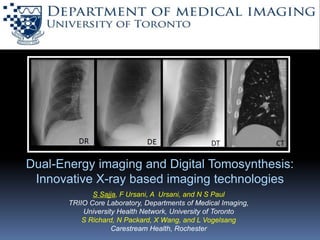
  • 1. Dual-Energy imaging and Digital Tomosynthesis: Innovative X-ray based imaging technologies S Sajja, F Ursani, A Ursani, and N S Paul TRIIO Core Laboratory, Departments of Medical Imaging, University Health Network, University of Toronto S Richard, N Packard, X Wang, and L Vogelsang Carestream Health, Rochester
  • 2. 2 Outline •Chest Radiography – Limitations of 2D imaging •Anatomical clutter •Dual-Energy (DE) X-ray •Anatomical clutter reduction via tissue discrimination •Factors that impact DE imaging performance •Clinical examples •Digital Tomosynthesis (DT) X-ray •Anatomical clutter reduction via spatial discrimination •Factors that impact DT imaging performance •Clinical examples
  • 3. 3 Chest Radiography (CXR) • Chest Radiography is the most common technique for 2D chest imaging •Limitation • A chest radiograph is a 2-dimensional projection of a complex 3- dimensional volume in which several tissues overlay each other. • Overlapping anatomy – ribs, lungs and vessels are background structures which may confound detection of a lung nodule on a single projection. Conspicuity (detectability) = Contrast of the object Complexity of the background
  • 4. 4 Limitation of Chest Radiography (CXR) 3-d object 2-d projection of the objectsCylinders made of material 1 Different objects made of material 2
  • 5. Overcoming anatomical noise via tissue discrimination •Removing overlapping ribs via tissue removal using dual-energy imaging. Material 1 only image2-d projection Tissue discrimination using x-ray imaging results in reduced background anatomical noise and improved feature conspicuity. DR - Digital Radiography DE - Dual Energy X-ray
  • 6. Tissue Discrimination – Dual-Energy (DE) imaging 6 High energy image Low energy image Bone image WbWs Soft-tissue image X-ray tube Anti-scatter bucky grid flat-panel digital detector
  • 7.  Energy selection: Choice of high and low energy values  Dose allocation:  Filtration:  Delay between two acquisitions: 7 Factors that affect DE imaging performance • DE soft tissue images of polyethylene nodule • High energy = 130 kVp (fixed) • Low energy = 60-90 kVp Patient (cm) equivalent phantom [acrylic] thicknesses: “slim patient” (18 cm) [7.5cm] “average patient” (24 cm) [10cm] “large patient” (30 cm) [12cm] Nodule contrast is highest at low kVp (60 kVp) and in a slim patient Nick Schumat, Masters Thesis, University of Toronto, 2008
  • 8.  Energy selection:  Dose allocation: Ratio of dose between high and low energies  Filtration:  Delay between two acquisitions: 8 Factors that affect DE imaging performance Aε = 0.06 Aε = 0.29 Aε = 0.72 Aε = 0.91 • DE bone-only images at different dose allocations (AƐ) • Noticeable increase in image noise at very low and very high allocations Nick Schumat, Masters Thesis, University of Toronto, 2008
  • 9. 9 Factors that affect DE imaging performance  Energy selection:  Dose allocation:  Filtration: helps in increasing the energy separation between the high and low energy images Fixed filtration: Same filtration for both high and low energy acquisitions.  Delay between two acquisitions: 4 0 50 100 150 0 2 4 6 8 10 12 x 10 Intensity 4 keV 0 50 100 150 0 2 4 6 8 10 12 x 10 Intensity 60 kVp 60 kVp 1 mm Cu keV No Filtration – high unused radiation Fixed Filtration – lower unused radiation
  • 10. 10 Factors that affect DE imaging performance Differential Filtration – very low unused radiation  Energy selection:  Dose allocation:  Filtration: helps in increasing the energy separation between the high and low energy images Differential filtration: Filtration is changed between high and low images to separate spectra.  Delay between two acquisitions: 4 keV 0 50 100 150 0 2 4 6 8 10 12 x 10 Intensity 4 keV 0 50 100 150 0 2 4 6 8 10 12 x 10 Intensity 60 kVp 1 mm Cu 120 kVp 1mm Cu 70 kVp 0.1 mm Cu 120 kVp 0.5 Ag Fixed Filtration – lower unused radiation
  • 11.  Energy selection:  Dose allocation:  Filtration:  Delay between two acquisitions: This has an impact on the motion artifacts observed in resultant DE image. 11 Factors that affect DE imaging performance ECG gating can be used to reduce motion artifacts. Timing diagram displaying the (ECG) trace, plethysmogram, and digital trigger. DE “soft-tissue” images acquired (a) with (b) without cardiac motion – custom built insert for motion simulation. Some motion blur is observed. Nick Schumat, Masters Thesis, University of Toronto, 2008
  • 12. 12 Modelling nodule conspicuity DR Objective function for system optimization - combines: MTF – System resolution GNPS- Noise and anatomical clutter Wtask- description of task i.e., nodule detection d‘ – Detectability index – surrogate for nodule conspicuity
  • 13. 13 Nodule detectability – fixed versus differential filtration DR DR DE fixed filtration DE differential filtration • Figure shows the detectability index values for DE fixed and differential filtration. • The results are normalized such that d’ = 1 for DR. • d’ = 1.2 and 1.3 for fixed and differential filtration respectively. • d’norm refers to detectability normalized by dose. • d’norm = 0.7 and 1.1 for fixed and differential filtration indicating fixed filtration less dose efficient than differential filtration. Richard S. et al., Diagnostic Imaging, 2015
  • 14. 14 DE Clinical patient imaging CXR of a patient with lung nodules: limited conspicuity due to overlapping structures DE-soft tissue of a patient with lung nodules: Improved conspicuity due to subtracted ribs.
  • 15. 15 DE – Cadaver Images Cadaveric chest x-rays were performed using conventional Digital Radiography (DR) (a) followed by DE projections decomposed into Bone only (b) and Soft tissue only (c) images to demonstrate improved conspicuity of bone and soft tissue details from DE projections compared to DR chest x-ray.
  • 16. How to overcome anatomical clutter via spatial discrimination •Separation of overlapping structures. Commonly used techniques – Computed tomography (CT) and Digital Tomosynthesis (DT) . Illustration of CT Illustration of DT Illustration of spatial discrimination in x-ray medical imaging resulting in reduced overlying clutter and improved feature conspicuity.
  • 17. 17 DT (Spatial discrimination) – Imaging Detector Detector X-ray tube Table top Wall Stand (a) Working of the tomosynthesis in wallstand position (b) Working of the tomosynthesis in table top position Phantom Phantom
  • 18. Factors that affect DT imaging performance  Angular range (Ɵ), number of projections (N) : A smaller range for image acquisition results in a lower tomo angle and reduced x-ray dose; but the depth resolution is also reduced.  Detector binning: The process of selecting regions of pixel and finding the mean value. Improves signal-to-noise ratio, speeds but results in loss of spatial resolution.  Scan time: The time taken to acquire the set of projections. This has an impact on motion artifacts due to cardiac and respiratory motion. (a) N=15, Ɵ=30o (b) N=60, Ɵ=30o (a) N=15, Ɵ=7.5o (b) N=60, Ɵ=30o Söderman C. et al., Medical Physics, 2015
  • 19. Factors that affect DT imaging performance  Angular range (Ɵ), number of projections: A smaller range for image acquisition results in a lower tomo angle and reduced x-ray dose; but the depth resolution is also reduced.  Detector binning: The process of selecting regions of pixel (NxN, N= number of pixels) and finding the mean value. Improves signal-to-noise ratio, speeds but results in loss of spatial resolution.  Scan time: The time taken to acquire the set of projections. This has an impact on motion artifacts due to cardiac and respiratory motion. Pacemaker generator: Greater detail is seen with 1X1 binning
  • 20. Factors that affect DT imaging performance  Angular range (Ɵ), number of projections: A smaller range for image acquisition results in a lower tomo angle and reduced x-ray dose; but the depth resolution is also reduced.  Detector binning: The process of selecting regions of pixel (NxN, N= number of pixels) and finding the mean value. Improves signal-to-noise ratio, speeds but results in loss of spatial resolution. .  Scan time: The time taken to acquire the set of projections. This has an impact on motion artifacts due to cardiac and respiratory motion. DT phantom images acquired (a) without motion (b) with respiratory (breathing) motion (c) with cardiac motion using a custom built insert for motion simulation.
  • 21. 21 Nodule detectability – Impact of dose in IQ DT 100% DT 50% DT 30% • Figure shows the detectability index values for DT at different doses (30%, 50% and 100%). • The results are normalized such that d’ = 1 for DR. • d’ = 11.8, 14.6 and 18.1 for 30%, 50% and 100% of the nominal doses respectively. • d’norm refers to detectability normalized by dose. • d’norm = 7.8, 7.7 and 5.9 for 30%, 50% and 100% respectively since DT becomes anatomical noise limited as opposed to quantum noise limited.Richard S. et al., Diagnostic Imaging, 2015
  • 22. 22 DT – Patient imaging Reconstructed DT patient image Corresponding coronal CT patient
  • 23. 23 DT – Cadaver Images Cadaveric DT through the pulmonary hila demonstrate high resolution images of the bifurcating left main bronchus (inset) using: Slice thickness = 5 mm Slice interval = 3 mm Dose = full Binning=1x1
  • 24. 24 DT – Cadaver Images Cadaveric DT through the pulmonary hila demonstrate high resolution images of the branch pulmonary arteries and veins (inset) using: Slice thickness = 5 mm Slice interval = 3 mm Dose = full Binning =1x1
  • 25. 25 DT – Limitations A nodule in a vessel branching point may be mistaken for an enlarged vessel in tomosynthesis – DT (left), CT (right) (Asplund S. , Acta Radiologica, 2011) •DT involves a limited angle of acquisition compared to CT. •This results in limited sampling of the signal in the frequency domain. • Depth resolution is poorer in DT than CT. • In some cases this may result in misinterpretation of structures as seen in the image here. •This can be alleviated by relating the location of the ribs as compared to the structure in question. •Also for this particular example, the potential use of contrast medium can be explored.
  • 26. 26 DT – Limitations Nodule close to pleura border – With X-ray beam tangential to ribs Nodule close to pleura border – With X-ray beam not tangential to ribs Nodule close to pleura border – With X-ray beam not tangential to ribs Nodule on vessel branching point – may be mistaken for an enlarged vessel on DT DT may provide a false anatomical location of a lung nodule when it is located close to the pleura; the effect depends on the angle of the incident X-ray beam relative to the nodule. Asplund S. , Acta Radiologica, 2011
  • 27. 27 DE-DT – Comparison Study – Low density objects • Low density objects (cotton spheres) were inserted into the anthropomorphic phantom: 1 dry cotton sphere (green arrow) and 2 cotton spheres dipped in distilled water (blue arrows) • The phantom was imaged using (a) DR (b) DE (c) and DT and low dose CT (1mSv). • CT images served as a reference standard (slide 28). • DR and soft tissue DE images demonstrate the wet cotton spheres with a faint projection of the dry cotton sphere. The DT images clearly demonstrate all of the spheres. • Lung pathologies (tumors) vary in their water content • DE-DT may improve lesion characterization Digital Radiograph Dual Energy Digital Tomosynthesis
  • 28. 28 DE-DT – Comparison Study – Low density object Coronal low dose CT images Digital Radiograph Dual Energy Digital Tomosynthesis
  • 29. 29 What is the future of DE and DT? Future innovation: Qualitative to quantitative Increased detector spatial resolution will facilitate extraction of quantitative image data to provide more accurate diagnosis. Application study: Volume estimation of thoracic water content • In critically ill patients, clinical assessment of change in thoracic fluid volume is necessary. • A method based on temporal subtraction of CXR is proposed to quantify the change in fluid volume • Proof of concept testing was done using a chest anthropomorphic phantom and solid water blocks. • The estimated volume based on the technique was compared with the actual volume. •Dual energy (DE)- soft tissue only images had the highest accuracy and correlated closely with the actual volume with a root mean square (RMS)=.4.74 ml Sajja et al., World Congress of Med. Phys. and Biomed. Eng. 2015
  • 30. 30 What is the future of DE and DT? DE- DT An integrated DE-DT system would be beneficial for increasing the scope of thoracic diseases which can be potentially diagnosed using chest radiography. Illustration of DE-DT – material 1 Illustration of DE-DT – material 2
  • 31. 31 Summary • For DR CXR, overlapping of structures confounds the detection of nodules. This results in anatomical noise. • Anatomical noise can be overcome either via tissue discrimination or spatial discrimination. • Tissue discrimination is achieved via Dual-Energy (DE) which involves acquisition of paired radiographs at 2 energies. • Spatial discrimination is achieved via Digital Tomosynthesis (DT) which involves acquisition of radiographs at different angles. • It would be beneficial to combine the spatial and tissue discrimination through an integrated DE-DT system.

Editor's Notes

  1. ECG gating -