Caris Centers of Excellence Virtual Molecular Tumor Board - October 28, 2015 (No Audio)
•
1 like•4,534 views

Slide deck from Caris Life Sciences' Virtual Molecular Tumor Board hosted by COE network member Barbara Ann Karmanos Cancer Center.

Recommended
Recommended
More Related Content
What's hot
What's hot (20)
Similar to Caris Centers of Excellence Virtual Molecular Tumor Board - October 28, 2015 (No Audio)
Similar to Caris Centers of Excellence Virtual Molecular Tumor Board - October 28, 2015 (No Audio) (20)
Recently uploaded
Recently uploaded (20)
Caris Centers of Excellence Virtual Molecular Tumor Board - October 28, 2015 (No Audio)
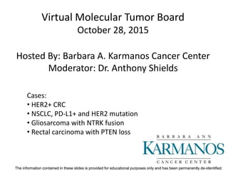
- 1. The information contained in these slides is provided for educational purposes only and has been permanently de-identified.The information contained in these slides is provided for educational purposes only and has been permanently de-identified. Virtual Molecular Tumor Board October 28, 2015 Hosted By: Barbara A. Karmanos Cancer Center Moderator: Dr. Anthony Shields Cases: • HER2+ CRC • NSCLC, PD-L1+ and HER2 mutation • Gliosarcoma with NTRK fusion • Rectal carcinoma with PTEN loss
- 2. The information contained in these slides is provided for educational purposes only and has been permanently de-identified.The information contained in these slides is provided for educational purposes only and has been permanently de-identified. Patient 1
- 3. The information contained in these slides is provided for educational purposes only and has been permanently de-identified.The information contained in these slides is provided for educational purposes only and has been permanently de-identified. Clinical History • Demographics: male approx. 60 years old • Rectal adenocarcinoma – Anterior-Poster resection – pT3 N1c (stage III) – Adjuvant radiation/capecitabine and CAPOX chemotherapy
- 4. The information contained in these slides is provided for educational purposes only and has been permanently de-identified.The information contained in these slides is provided for educational purposes only and has been permanently de-identified. Treatment History • 2 yr Follow-up: • CEA 27 • CT: normal • PET/CT: positive pre-sacral mass, nodes and 3 cm liver lesion - Liver Bx positive. • Started CAPOX/bevzcizumab • CT after 3 cycles (pending)
- 5. The information contained in these slides is provided for educational purposes only and has been permanently de-identified.The information contained in these slides is provided for educational purposes only and has been permanently de-identified. Pathology H&E 20x HER2 IHC 20x
- 6. The information contained in these slides is provided for educational purposes only and has been permanently de-identified.The information contained in these slides is provided for educational purposes only and has been permanently de-identified. HER2 Amplified IHC expression results: EGFR 1+60%, HER2 3+ 75%, cMET 2+60%, PTEN 1+40% (neg)
- 7. The information contained in these slides is provided for educational purposes only and has been permanently de-identified.The information contained in these slides is provided for educational purposes only and has been permanently de-identified. NGS Results • KRAS, NRAS, and BRAF wildtype • Mismatch Repair proficient (MSI 0/5 markers).
- 8. The information contained in these slides is provided for educational purposes only and has been permanently de-identified.The information contained in these slides is provided for educational purposes only and has been permanently de-identified. HER2 in rectal cancer • The Cancer Genome Atlas project identified HER2 somatic mutations and gene amplification in 7% of patients with colorectal cancer. • Preclinical xenograft models show these mutations can contribute to EGFR resistance, and targeting HER2 inhibitors (trastuzumab, neratinib, or lapatinib) can lead to tumor regression. Kavuri et al, Cancer Discovery, 2015
- 9. The information contained in these slides is provided for educational purposes only and has been permanently de-identified.The information contained in these slides is provided for educational purposes only and has been permanently de-identified. Discussion Points • Standard chemotherapy options at this point • Irinotecan and EGFR inhibitors still available • HER2 amplified by CISH and NGS CNV • Options: trastuzumab/lapatinib and trastuzumab emtansine (TDM1) on Match • EGFR and cMET expression may indicate resistance mechanisms
- 10. The information contained in these slides is provided for educational purposes only and has been permanently de-identified.The information contained in these slides is provided for educational purposes only and has been permanently de-identified. Patient 2
- 11. The information contained in these slides is provided for educational purposes only and has been permanently de-identified.The information contained in these slides is provided for educational purposes only and has been permanently de-identified. Clinical History • Demographics: male in late thirties • Relevant medical history: – Right sided chest pain – Underwent chest xray and then CT scan – Large 7 cm right lung mass – PET scan positive in multiple lymph nodes with a supraclavicular lymph node – EBUS showed adenocarcinoma – Brain MRI negative
- 12. The information contained in these slides is provided for educational purposes only and has been permanently de-identified.The information contained in these slides is provided for educational purposes only and has been permanently de-identified. Treatment History • Received chemotherapy+RT.
- 13. The information contained in these slides is provided for educational purposes only and has been permanently de-identified.The information contained in these slides is provided for educational purposes only and has been permanently de-identified. Pathology H&E 20x PD-L1 20x
- 14. The information contained in these slides is provided for educational purposes only and has been permanently de-identified.The information contained in these slides is provided for educational purposes only and has been permanently de-identified. Immune Markers • PD-L1 2+ in 100% of tumor cells • PD-1 Positive; 1 per HPF
- 15. The information contained in these slides is provided for educational purposes only and has been permanently de-identified.The information contained in these slides is provided for educational purposes only and has been permanently de-identified. PD-1 / PD-L1 Immune Checkpoint
- 16. The information contained in these slides is provided for educational purposes only and has been permanently de-identified.The information contained in these slides is provided for educational purposes only and has been permanently de-identified. PD-L1 as Biomarker in Lung • Nivolumab studies presented at ASCO 2015 • Checkpoint-017 – Squamous NSCLC – 41% OS improvement with nivolumab versus docetaxel in the squamous setting • Checkpoint-051 – Non-squamous NSCLC – OS benefit with nivolumab was 27% in patients with nonsquamous NSCLC – Stronger OS outcomes were observed in PD-L1–positives – Including 60% reduction in the risk of death for those with the highest PD-L1 levels.
- 17. The information contained in these slides is provided for educational purposes only and has been permanently de-identified.The information contained in these slides is provided for educational purposes only and has been permanently de-identified. PD-L1: Meta-analysis • Sensitivity analysis of 20 (12 nivolumab, 4 pembrolizumab, and 4 MPDL3280A) trials where a checkpoint inhibitor was utilized either as a single agent or in combination – ORR was found to be slightly higher in PD-L1 positive patients than in PD-L1 negative patients treated with nivolumab and pembrolizumab, – with an absolute difference of 16.4% and 19.5%, respectively. • A significant difference in activity of 22.8% and 8.7% according to PD-L1 was found for melanoma and NSCLC, respectively. Carbognin et al, 2015
- 18. The information contained in these slides is provided for educational purposes only and has been permanently de-identified.The information contained in these slides is provided for educational purposes only and has been permanently de-identified. Carbognin et al, 2015
- 19. The information contained in these slides is provided for educational purposes only and has been permanently de-identified.The information contained in these slides is provided for educational purposes only and has been permanently de-identified. Carbognin et al, 2015
- 20. The information contained in these slides is provided for educational purposes only and has been permanently de-identified.The information contained in these slides is provided for educational purposes only and has been permanently de-identified. HER2 mutated, non-ampified
- 21. The information contained in these slides is provided for educational purposes only and has been permanently de-identified.The information contained in these slides is provided for educational purposes only and has been permanently de-identified. Discussion Role for immunotherapy with PD-L1 expression Impact of sub-clone with HER2 kinase domain mutation
- 22. The information contained in these slides is provided for educational purposes only and has been permanently de-identified.The information contained in these slides is provided for educational purposes only and has been permanently de-identified. Patient 3
- 23. The information contained in these slides is provided for educational purposes only and has been permanently de-identified.The information contained in these slides is provided for educational purposes only and has been permanently de-identified. History •Female in early fifties with one month onset of behavioral changes, L visual field defects, L sided weakness, and gait disturbance. •Presented to the ER after a fall with blunt head trauma, L facial droop, and on-going L sided weakness, and visual complaints. •No Hx of LOC, seizures, bowel/bladder dysfunction, or neuro problems. •PE: Complete L homonymous hemianopsia, weak LUE, L facial drooping, L hemineglect •CT head and MRI brain: R temporo-parietal and R parieto-occipital mass lesion w/ extensive edema, and slight midline shift. •PET scan: Areas of increased uptake w/ necrotic centers in the lesion.
- 24. The information contained in these slides is provided for educational purposes only and has been permanently de-identified.The information contained in these slides is provided for educational purposes only and has been permanently de-identified. Treatment History • R temporo-parietal-occipital craniotomy, and tumor resection • PBRT: 6000 cGy over 30 fractions • Chemo: Temozolomide
- 25. The information contained in these slides is provided for educational purposes only and has been permanently de-identified.The information contained in these slides is provided for educational purposes only and has been permanently de-identified. Pathology Although the tumor appeared predominantly glioblastoma, and contained extensive areas of necrosis, solid tumor, and infiltrative tumor, the histologic patterns were heterogeneous and included areas of proliferating cellular connective tissue considered consistent with sarcoma. The tumor was thus classified as gliosarcoma. (H&E, next slide)
- 26. The information contained in these slides is provided for educational purposes only and has been permanently de-identified.The information contained in these slides is provided for educational purposes only and has been permanently de-identified. Pathology H&E 20x
- 27. The information contained in these slides is provided for educational purposes only and has been permanently de-identified.The information contained in these slides is provided for educational purposes only and has been permanently de-identified.
- 28. The information contained in these slides is provided for educational purposes only and has been permanently de-identified.The information contained in these slides is provided for educational purposes only and has been permanently de-identified.
- 29. The information contained in these slides is provided for educational purposes only and has been permanently de-identified.The information contained in these slides is provided for educational purposes only and has been permanently de-identified.
- 30. The information contained in these slides is provided for educational purposes only and has been permanently de-identified.The information contained in these slides is provided for educational purposes only and has been permanently de-identified.
- 31. The information contained in these slides is provided for educational purposes only and has been permanently de-identified.The information contained in these slides is provided for educational purposes only and has been permanently de-identified. Molecular Tumor Summary • NGS Mutations detected – PTEN – TP53 • CNV alterations detected – CMET amplification – EGFR amplification • EGFRvIII mutation detected • Absence of IDH1 expression and 1p19q
- 32. The information contained in these slides is provided for educational purposes only and has been permanently de-identified.The information contained in these slides is provided for educational purposes only and has been permanently de-identified. TRK Fusions in Cancer Investigational molecule: LOXO-101 “An oncogenic NTRK fusion in a soft tissue sarcoma patient with response to thetropomyosin-related kinase (TRK) inhibitor LOXO-101” (Doebele et al, 2015)
- 33. The information contained in these slides is provided for educational purposes only and has been permanently de-identified.The information contained in these slides is provided for educational purposes only and has been permanently de-identified. Discussion • Lack of MGMT promoter methylation may explain progression under temozolomide. • PTEN: Point mutation and intact protein expression may indicate presence of dysfunctional PTEN protein. • Cancer is EGFR driven (shown by EGFR overexpression, gene copy number increase and EGFRvIII presence), and is a typical fast-growing de novo grade IV glioma (also supported by lack of IDH1 mutation). • Potentially targetable genes: – EGFR (ABT-414, EGFR-targeted antibody-drug conjugate; EGFRvIII-targeted vaccine) – cMET-amplification may indicate resistance to EGFR-targeted therapy – NTRK re-arrangement (LOXO-1010, an NTRK inhibitor may be promising)
- 34. The information contained in these slides is provided for educational purposes only and has been permanently de-identified.The information contained in these slides is provided for educational purposes only and has been permanently de-identified. Patient 4
- 35. The information contained in these slides is provided for educational purposes only and has been permanently de-identified.The information contained in these slides is provided for educational purposes only and has been permanently de-identified. Clinical History • Demographics: male in early forties • Rectal adenocarcinoma –Diagnosed with limited liver mets –KRAS WT
- 36. The information contained in these slides is provided for educational purposes only and has been permanently de-identified.The information contained in these slides is provided for educational purposes only and has been permanently de-identified. Treatment History • Treated with FOLFOX/bevacizumab with stable disease • Treated with FOLFIRI/cetuximab with CEA decline – partial hepatectomy with extensive necrosis – low anterior resection – PET/CT new liver lesions • Treated with FOLFIRI/cetuximab with CEA decline – Radiofrequency ablation – Progressed in liver and FOLFIRI/cetuximab resume followed by partial hepatectomy • Further liver progression treated at times with FOLFIRI/cetuximab, FOLFIRI/panitumumab, and FOLFOXIRI/panitumumab • 3 yr post-DX: – Y-90 radiomebolization and regorafenib – Biliary drain placed for obstructive jaundice
- 37. The information contained in these slides is provided for educational purposes only and has been permanently de-identified.The information contained in these slides is provided for educational purposes only and has been permanently de-identified. Genomics History Previously testing: MYC, TP 53, and SMAD4 alterations Testing negative for Lynch syndrome. KRAS was wildtype. Biopsy of peritoneal lesion 4 years-post Dx sent for Match and Caris Molecular intelligence testing
- 38. The information contained in these slides is provided for educational purposes only and has been permanently de-identified.The information contained in these slides is provided for educational purposes only and has been permanently de-identified. Pathology H&E 20x PTEN IHC 20x
- 39. The information contained in these slides is provided for educational purposes only and has been permanently de-identified.The information contained in these slides is provided for educational purposes only and has been permanently de-identified.
- 40. The information contained in these slides is provided for educational purposes only and has been permanently de-identified.The information contained in these slides is provided for educational purposes only and has been permanently de-identified. PTEN Loss by IHC • Clinical Trials Connector • NCI Match study
- 41. The information contained in these slides is provided for educational purposes only and has been permanently de-identified.The information contained in these slides is provided for educational purposes only and has been permanently de-identified. NGS Findings • TP53 R273C pathogenic mutation • No other pathogenic mutations on 592 genes • KRAS, NRAS, HRAS wildtype
- 42. The information contained in these slides is provided for educational purposes only and has been permanently de-identified.The information contained in these slides is provided for educational purposes only and has been permanently de-identified. PTEN Pathway
- 43. The information contained in these slides is provided for educational purposes only and has been permanently de-identified.The information contained in these slides is provided for educational purposes only and has been permanently de-identified. Discussion Points • Now without standard therapy available except trifluridine/tipiracil (TAS-102, Lonsurf) • Tbili now 3.2 and stent being revised • PTEN loss on Match and Caris testing, possible new arm on Match trial • How and when to do both Match and Caris testing?
- 44. The information contained in these slides is provided for educational purposes only and has been permanently de-identified.The information contained in these slides is provided for educational purposes only and has been permanently de-identified. Next Molecular Tumor Board: Thursday, November 19, 2015 5pm ET (4pm CT) Host: MedStar Franklin Square Medical Center Leader: Dr. Albert J. Aboulafia