![point in embryology. The extraembryonic mesoderm is a fundamental tissue in embryonic development because it con-
tributes most of the bulk mass of the UC and the placental stroma. At day 13, there is evidence of the formation of the
connecting stalk between the embryo and the walls of the developing chorionic cavity. The UC originates from the con-
necting stalk [1,2], and then this anatomical formation connects fetus and placenta throughout pregnancy. The UC is formed
essentially by the closing in of the somatic stalk. The obliteration of the UC coelom is determined by a proliferation of the
fibrous tissue, which forms a ring at the embryonic attachment of the cord. Some faults of the junctional mesoderm may cause
congenital herniation, which may result from incorrect development of the cord [3]. Among the functions of UC, its closure
after birth is an important (and yet poorly understood) process that safeguards against blood loss of the newborn [2].
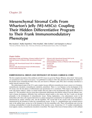
The UC is layered by cubic epithelial cells, forming the umbilical epithelium (Fig. 20.1A) that connects amniotic
epithelial cells and the tegumentary epithelium of the fetus [4,5]. As shown in Fig. 20.1, these cells are positive for keratin
expression.
The cord epithelial layer shows characteristic gestational age-related changes. Initially the epithelial cells have been
demonstrated to possess microvilli and cilia. Along the third month, between the 8th and 10th weeks, they constitute a
single-layered epithelium, which then becomes bilaminar by the end of the third month [6]. Between the sixth and seventh
months the epithelium is trilayered but keratinization is a rare occurrence, except in the cord region closer to fetus. At term,
this area is characteristically opaque, and the remaining part consists of a simple squamous epithelium. At the placental
end, there is a characteristic sudden transition with the cubical amniotic epithelium [2,6].
Human umbilical cord (HUC) epithelium covers the subamnion and a mature mucous connective tissue; the so-called
WJ that surrounds the adventitia and media of the fetal vessels is thought to act preventing vessel compression, torsion, and
bending (Fig. 20.1). WJ features cellular elements scattered into an abundant extracellular matrix, where the amorphous
(A) (B)
(C) (D)
FIGURE 20.1 Microscopic demonstration of the different zones of human umbilical cord. (A) The umbilical epithelium (UE) lines cord surface;
subamniotic zone (SA) is a narrow band immediately beneath epithelial layer. The Wharton’s jelly (WJ) occupies the rest of the stroma, both intervascular
(IV) and perivascular (PV). (B) Higher magnification of the PV area around umbilical vein. (C and D) Keratin immunohistochemistry shows positivity at
epithelial level and also in the scattered WJ stromal cells. Magnification: 50Â (A), 200Â (BeD).
272 SECTION j III Umbilical CordeDerived Cells](data:image/gif;base64,R0lGODlhAQABAIAAAAAAAP///yH5BAEAAAAALAAAAAABAAEAAAIBRAA7)
Recommandé
Recommandé
Contenu connexe
Tendances
Tendances (20)
Similaire à Ch 20 -_mesenchymal_stromal_cells_from_whartons_jelly_wj-ms_cs_coupling_their_hidden_differentiative_program_to_their_fra
Similaire à Ch 20 -_mesenchymal_stromal_cells_from_whartons_jelly_wj-ms_cs_coupling_their_hidden_differentiative_program_to_their_fra (20)
Dernier
Dernier (20)
Ch 20 -_mesenchymal_stromal_cells_from_whartons_jelly_wj-ms_cs_coupling_their_hidden_differentiative_program_to_their_fra
- 1. Chapter 20 Mesenchymal Stromal Cells From Wharton’s Jelly (WJ-MSCs): Coupling Their Hidden Differentiative Program to Their Frank Immunomodulatory Phenotype Rita Anzalone1 , Radka Opatrilova2 , Peter Kruzliak2 , Aldo Gerbino1 and Giampiero La Rocca1 1 University of Palermo, Palermo, Italy; 2 University of Veterinary and Pharmaceutical Sciences, Brno, Czech Republic Chapter Outline Embryological Origin and Histology of Human Umbilical Cord 271 Molecular Features of Wharton’s Jelly Mesenchymal Stromal Cells 273 Wharton’s Jelly Mesenchymal Stromal Cells Are Multipotent Stem Cells 273 Wharton’s Jelly Mesenchymal Stromal Cell Differentiation Toward Insulin-Producing Cells 274 Wharton’s Jelly Mesenchymal Stromal Cells Can Be Differentiated to Hepatocyte-Like Cells 275 Immunomodulatory Activity of Wharton’s Jelly Mesenchymal Stromal Cells 276 Conclusion 277 Acknowledgments 277 References 277 EMBRYOLOGICAL ORIGIN AND HISTOLOGY OF HUMAN UMBILICAL CORD The first complete description of the umbilical cord (UC) tissue was given by Thomas Wharton, whose book “Adenografia sive glandularum totius corporis descriptio,” was published in London in 1656. This allowed to first define the features of the peculiar tissue constituting the bulk of the cord, now known as Wharton’s jelly (WJ), that is nowadays classified as a mature mucoid connective tissue. The embryological origin of the UC is quite complex because different extraembryonic tissues concur to its formation: extraembryonic mesoderm, extraembryonic endoderm, amnioblasts. There is a vast literature on the development of the UC tissues since the first phases of differentiation and specification of its constituents. With respect to the difficulties to work with human embryos, studies on animal models shed new light on the developmental origin of the cells found in mature UC. The mesoblast is the key tissue that evolutionarily differentiates the triblastic embryos from the diblastic ones. And in human development, differently from well-known animal models as the mouse, the first 2 weeks are devoted principally to the development of extraembryonic tissues. During implantation, a key phenomenon occurs in the embryoblast cells: these cells organize themselves in a planar arrangement constituted by two epithelial layers, the epiblast, characterized by mainly columnar cells, and the hypoblast. The second half of the second week of development is characterized by the formation of other key extraembryonic tissues. At day 11, a deepithelization and cavitation process starts in the epiblast layer, giving rise to the formation of the first amnioblast cells. These will proliferate and cover the overlying cytotrophoblast to create the primordium of the amniotic cavity. Around days 12e13, another extraembryonic tissue appears, namely, extraembryonic mesoderm or mesoblast. The origin of this tissue in human embryo is still a debated Perinatal Stem Cells. https://doi.org/10.1016/B978-0-12-812015-6.00020-0 Copyright © 2018 Elsevier Inc. All rights reserved. 271
- 2. point in embryology. The extraembryonic mesoderm is a fundamental tissue in embryonic development because it con- tributes most of the bulk mass of the UC and the placental stroma. At day 13, there is evidence of the formation of the connecting stalk between the embryo and the walls of the developing chorionic cavity. The UC originates from the con- necting stalk [1,2], and then this anatomical formation connects fetus and placenta throughout pregnancy. The UC is formed essentially by the closing in of the somatic stalk. The obliteration of the UC coelom is determined by a proliferation of the fibrous tissue, which forms a ring at the embryonic attachment of the cord. Some faults of the junctional mesoderm may cause congenital herniation, which may result from incorrect development of the cord [3]. Among the functions of UC, its closure after birth is an important (and yet poorly understood) process that safeguards against blood loss of the newborn [2]. The UC is layered by cubic epithelial cells, forming the umbilical epithelium (Fig. 20.1A) that connects amniotic epithelial cells and the tegumentary epithelium of the fetus [4,5]. As shown in Fig. 20.1, these cells are positive for keratin expression. The cord epithelial layer shows characteristic gestational age-related changes. Initially the epithelial cells have been demonstrated to possess microvilli and cilia. Along the third month, between the 8th and 10th weeks, they constitute a single-layered epithelium, which then becomes bilaminar by the end of the third month [6]. Between the sixth and seventh months the epithelium is trilayered but keratinization is a rare occurrence, except in the cord region closer to fetus. At term, this area is characteristically opaque, and the remaining part consists of a simple squamous epithelium. At the placental end, there is a characteristic sudden transition with the cubical amniotic epithelium [2,6]. Human umbilical cord (HUC) epithelium covers the subamnion and a mature mucous connective tissue; the so-called WJ that surrounds the adventitia and media of the fetal vessels is thought to act preventing vessel compression, torsion, and bending (Fig. 20.1). WJ features cellular elements scattered into an abundant extracellular matrix, where the amorphous (A) (B) (C) (D) FIGURE 20.1 Microscopic demonstration of the different zones of human umbilical cord. (A) The umbilical epithelium (UE) lines cord surface; subamniotic zone (SA) is a narrow band immediately beneath epithelial layer. The Wharton’s jelly (WJ) occupies the rest of the stroma, both intervascular (IV) and perivascular (PV). (B) Higher magnification of the PV area around umbilical vein. (C and D) Keratin immunohistochemistry shows positivity at epithelial level and also in the scattered WJ stromal cells. Magnification: 50Â (A), 200Â (BeD). 272 SECTION j III Umbilical CordeDerived Cells
- 3. ground substance is predominant. Classical studies showed that the main components are sulfated glycosaminoglycans (GAGs), such as hyaluronic acid, and proteoglycans, such as decorin and biglycan [7,8]. Collagen types I, III, and VI and basement membrane molecules such as collagen type IV, laminin, and heparan sulfate have been demonstrated in the extracellular matrix throughout the cord stroma from the subamnion to the perivascular zone [9,10]. Collagen filaments are increased immediately beneath the epithelium and in the deeper part of the cord, especially near the large umbilical vessels, where they are oriented circularly to the vessels, an arrangement which is characteristically shown also for the cells (Fig. 20.1) [6,11]. The organized distribution of the various collagen types has been suggested to be responsible for the mechanical properties of the UC [12]. MOLECULAR FEATURES OF WHARTON’S JELLY MESENCHYMAL STROMAL CELLS The bone marrow (BM) is certainly the most common source of mesenchymal stromal cells (MSCs). Friedenstein and colleagues were the first to demonstrate that mesenchymal stem cells from bone marrow (BM-MSCs) are able to undergo ex vivo expansion, growing on a plastic surface, and differentiate in various cellular lineages such as adipocytes, osteocytes, chondrocytes, tenocytes, and nervous tissue cells [13]. According to the general consensus, BM-MSCs express typical “core” markers such as CD44, CD73, CD90, CD105, CD166, CD49e, CD51, CD54, CD59, CD71 [14,15]. Specific markers of the endothelial lineage, such as CD31 and vWF are absent in BM-MSCs, as well as hematopoietic markers (CD14, CD34, CD45, CD79, CD86, and CD235a) [16e18]. Various reports suggested that the use of MSCs in vivo should be safer with respect to formerly investigated embryonic stem cells (ESCs) because MSCs have higher chromosomal stability and do not induce neoplasm formation in the recipient host [19,20]. However, only a minor fraction of BM cells are useful for regenerative medicine applications (their frequency ranging between 0.0001% and 0.01% of nucleated cells) [21]. The abundant ECM of UC stroma contains dispersed stromal cells, now referred to as MSCs. Classical studies, such as that by Takechi and colleagues, identified the majority of stromal cells as myofibroblasts [12]. Even if the stroma can be divided into three different zones (subamnion, WJ, and perivascular zone) and data from ex vivo cells have shown dif- ferences between cells pertaining to the various zones, the term “Wharton’s jelly cells” (WJCs) is often extended to all umbilical stromal cells. As demonstrated by different groups, MSCs derived from HUC and other fetal/neonatal tissues share common features with MSCs derived from adult tissues (BM, adipose tissue, peripheral blood) such as self-renewal capability and differentiative potential toward different types of tissue cells. Considering the expression of surface mol- ecules, there is a significant overlap between the markers expressed by BM-MSCs and WJ-derived mesenchymal stromal cells (WJ-MSCs). Immunocytochemistry experiments have shown that WJ-MSCs (as well as BM-MSCs) lack expression of CD31, CD33, CD34, CD45. CD56 is expressed by BM-MSCs, but not by WJ-MSCs. Moreover, both BM-derived and WJ-MSCs lack expression of HLA-DR [13]. On the other hand, WJ-MSCs express at protein level: CD73, CD90, CD105, HLA class I, as well as CD10, CD13, CD29, CD44, CD49e, and CD166. All of them were also characterized as BM-MSC markers [13,22]. More recently, other markers have been reliably associated to the WJ-MSC populations ex vivo. Immune-related antigens such as CD68 and CD14 have been reported in WJ-MSCs and cord lining (CL)-MSCs, respectively, by us and other authors [23,24]. More recently, CD200 and its receptor, which are involved in immunomo- dulation processes, have been demonstrated to be expressed in WJ-MSCs [25]. In addition, CD271, an immunomodulatory molecule initially described in BM-MSCs, has also been shown to be expressed in fresh UC specimens [26]. Our group showed that WJ-MSCs express at mRNA and protein levels the key transcription factors GATA-4, GATA-5, GATA-6 [27]. All of these transcription factors are involved in different developmental pathways of mesoderm- and endoderm-derived organs [13,27]. We also demonstrated that WJ-MSCs express connexin-43 (Cx-43) [27], a molecule that is typically expressed in embryonic and myocardial cells. It is responsible for the formation of intercellular gap junctions. Recent reports indicated that Cx-43 expression along the myocardial differentiation pathway increases in a stage-related manner and is correlated to proliferation arrest and acquisition of a mature phenotype [13]. Moreover, we were the first to describe the expression of a subset of epithelial cytokeratins in WJ-MSCs isolated by nonenzymatic methods [27]. In particular, we demonstrated the expression of CK-8, CK-18, CK-19, whereas CK-7 was not detected. In addition, we confirmed the expression of neu- roectodermal markers such as glial fibrillar acidic protein and neuron-specific enolase also in undifferentiated cells, as described in earlier reports on WJ-MSCs and in BM-MSCs [13,27]. WHARTON’S JELLY MESENCHYMAL STROMAL CELLS ARE MULTIPOTENT STEM CELLS Several reports indicate that WJ-MSCs are multipotent cells, thus capable of giving rise to different mature cellular types. Most studies agree that WJ-MSCs can be successfully induced toward connective tissue phenotypes (osteoblasts, Mesenchymal Stromal Cells From Wharton’s Jelly (WJ-MSCs) Chapter | 20 273
- 4. adipocytes, and chondrocytes), thus opening new paths in regenerative medicine applications to the musculoskeletal system. This trilineage differentiation potential of WJ-MSCs fulfills the minimal criteria stated in 2006 to uniformly define MSC properties [28]. Phenotypical and morphological criteria can be used to define the effectiveness of the differentiation of MSCs toward the mature cytotypes of these connective tissues. The standardized protocols to obtain osteogenic differentiation of MSCs [29e31] result in the acquisition of a differentiated phenotype that may be confirmed by specific histological stains for extracellular calcium such as Alizarin Red S and Von Kossa [27]. Moreover, differentiated MSCs should express specific proteins such as osteonectin, osteo- calcin, periostin, runx2 [32]. After adipogenic differentiation protocols, differentiated adipocytes should be demonstrated by lipid-specific histo- logical stains such as Oil Red O [27]. In addition, newly differentiated adipocytes express specific proteins such as adiponectin, leptin, and PPAR-g. When chondrogenic differentiation of MSCs is performed by standardized methods, the differentiated cells can be specifically stained by Alcian blue or Safranin O-Fast Green [33]. In addition, the acquisition of the chondrocyte phenotype can be demonstrated by the expression of specific proteins such as collagen type II, cartilage oligomeric matrix protein, and aggrecan [13,34]. In addition, WJ-MSCs have been successfully differentiated toward cells of other mature organs such as neural cells [35], skin follicular cells [36], cardiomyocytes [37]. WHARTON’S JELLY MESENCHYMAL STROMAL CELL DIFFERENTIATION TOWARD INSULIN-PRODUCING CELLS WJ-MSCs recently gained much attention because of their easy sourcing, culture, and differentiability into several tissues. A few important pilot reports indicate that these cells can be successfully differentiated into IPCs (insulin producing cells). Chao and coworkers differentiated WJ-MSCs into IPCs through a stepwise culture protocol using neuron-conditioned medium. The authors transplanted the obtained differentiated cell clusters into livers of diabetic mice. Insulin was showed to be expressed in response to physiological glucose levels. The authors also assessed the secretion of C-peptide and the expression of specific genes such as Pdx-1, Nkx2.2, HLXB-9 and Glut-2 [38]. A comparative study was performed by Wu and colleagues to compare the differentiative ability of WJ-MSCs and BM-MSCs towards an IPC phenotype. Both cellular types were able to form islet-like clusters on the first day of culture in a medium containing nicotinamide, activin, hepatocyte growth factor (HGF), exendin-4, and pentagastrin. Pdx-1 was shown to be expressed at higher levels in differentiated WJ-MSCs than in differentiated BM-MSCs. Secretion of insulin and mRNA expression of insulin and C-peptide were comparably higher in the differentiated WJ-MSCs [39]. In a parallel report, Wang and coworkers further enriched these data with in vitro and in vivo experiments using differentiated human WJ cells to treat diabetes in NOD mice. After transplantation, IPCs were located in the liver and were able to normalize glycemia [40]. Taken collectively, these promising data suggest that WJ-MSCs possess the ability, both in vitro and in vivo, to differentiate into insulin-secreting cells [22]. In a later report, Tsai et al. performed differentiation experiments using MSCs from HUC, which were induced through a three step protocol to obtain IPCs. The features of differentiated cells were assessed by immunocytochemistry, real-time PCR, and ELISA. In vivo experiments were performed by transplanting differentiated cells into the liver of diabetic rats via portal infusion. In vitro data showed that pancreatic b-cell developmenterelated genes (such as PDX1, Pax4, and insulin) were expressed in the differentiated cells. In addition, C-peptide release was increased after glucose challenge in vitro. In vivo, human nuclei and C-peptide were detected in the rat livers by immunohistochemistry. Moreover, after trans- plantation of differentiated cells into the diabetic rats, blood sugar level decreased [41]. Prabakar and colleagues investigated the use of CB-MSCs for the treatment of diabetes mellitus through in vitro and in vivo experiments. Subsequently to a pancreatic differentiation protocol, the cells expressed key markers such as PDX1, NKX6.1, and NGN3 by immunofluorescence and RT-PCR, thus confirming that CB-MSCs may be successfully differ- entiated toward a pancreatic lineage [42]. Further reports provided evidence on the effects of PDX1 gene transfection in UC-MSCs to obtain insulin-producing cells in vitro. The pancreatic differentiation protocol comprised three steps. The authors showed that insulin and C-peptide were detected after the third step of differentiation. Dithizone stain provided a morphological formal evidence of the differentiation process at this stage. In addition, insulin, PDX1, and Nkx6.1 expressions were also confirmed by RT-PCR and western blot analyses in differentiated cells. Interestingly, the expression of such genes was restricted to transfected cells alone, whereas untransfected ones, or cells subjected only to the standard differentiation protocol, failed in expressing such genes [43]. 274 SECTION j III Umbilical CordeDerived Cells
- 5. Xiao and colleagues performed in vivo experiments aimed to investigate whether cotransplantation of UC-derived mesenchymal stromal cells (UC-MSCs) and cord blood mononuclear cells (CB-MNCs) could reverse hyperglycemia in type 1 diabetic mice. Authors also aimed to determine the appropriate ratio for cotransplantation [44]. UC-MSCs and CB-MNCs were transplanted into type 1 diabetic mice at different ratios, and blood glucose concentration was monitored in animals. Histology, immunohistochemistry, and human Alu PCR assays were performed to evaluate the presence of donor-derived cells and the repair extent of endogenous islets. In separate experiments, the authors also induced UC-MSC differentiation toward islet-like cells to determine their differentiation potential. Cotransplantation experiments showed that UC-MSCs and CB-MNCs at a ratio of 1:4 effectively reversed hyperglycemia in diabetic mice. Donor-derived cells were detected in pancreas and kidneys of transplanted animals. The in vitro data of the authors strongly suggested that the MSCs were able to be differentiated, in vitro, into insuling producing cells. However, human insulin was not detected in the regenerated pancreases. This may suggest that reactivation of local precursor cells, rather than cell replacement, should explain the mechanism of action of the infused cells [44]. Another work from Hu and coworkers showed that WJ-MSCs may be administered to type I diabetes patients, and the treatment is safe and prospectively effective [45]. The authors assessed the long-term effects of the implantation of WJ-MSCs in patients with newly onset T1DM. Patients were randomly divided into two groups; patients in group I were treated with WJ-MSCs and patients in group II were treated with normal saline and standard therapy. The long-term follow-up of patients was up to 21 months. No acute or chronic side effects were observed in group I patients compared with group II. Clinical parameters such as HbA1c and C peptide were significantly better in group I patients when compared either with pretherapy values or parallel values from group II during the follow-up period. These data suggested that the implantation of WJ-MSCs for the treatment of newly onset T1DM may safe and effective. WHARTON’S JELLY MESENCHYMAL STROMAL CELLS CAN BE DIFFERENTIATED TO HEPATOCYTE-LIKE CELLS MSCs are considered useful for liver regenerative medicine because of their key features such as self-renewal capacity, endodermal lineage differentiation potential, and immunomodulatory activity [46]. Different populations of MSCs have been used in in vitro experiments and preclinical studies to derive mature hepatocyte-like cells (HLCs). In a seminal report, Campard and coworkers showed that UC matrix stem cells (UC-MSCs) are able to differentiate toward hepatocyte-like cells. UC-MSCs were cultured in a medium supplemented with factors promoting hepatic dif- ferentiation [47]. The initial population of UC-MSCs expressed CK-8, CK-18, CK-19; was negative for CK-7; expressed G6Pase, PEPCK (phosphoenolpyruvate carboxykinase), a-1-antitrypsin (a-1AT), tryptophan 2,3-dioxygenase (TDO); and lacked HepPar1 positivity and HNF-4a or CYP3A4 expression. The UC-MSC-derived hepatocyte-like cells increasingly expressed markers such as tyrosine aminotransferase and TDO but remained negative for HNF-4. Nevertheless, functional assays showed that differentiated cells responded well to the differentiative stimulus, being able to store glycogen, pro- ducing urea and possessing active hepato-specific enzymes (CYP3A4, G6Pase). Moreover, in vivo experiments showed that after transplantation of undifferentiated UC-MSCs in liver of SCID (severe combined immunodeficiency) mice with partial hepatectomy, the engrafted cells expressed human hepatic markers such as albumin and AFP after 2, 4, and 6 weeks following transplantation [47]. Zhang and coworkers [48] performed an in vitro study on UC-MSC differentiation. The authors applied a single step protocol based on HGF and FGF-4 supplementation. The differentiated cells expressed liver-specific markers (albumin and AFP), stored glycogen, and showed uptake of LDL, thus reinforcing the concept of their usefulness as cellular therapy tools for liver diseases. Zhao and collaborators [49] demonstrated that WJ-MSCs maintain in vitro hypoimmunogenicity even after a hepatic differentiation protocol has been performed. In fact, differentiated hepatocyte-like cells, apart from expressing hepatocyte markers in vitro, and albumin in vivo, did not express HLA-DR following 2 or 4 weeks differentiation in vitro, thereby demonstrating that the differentiative process did not exert any change on the immunological features of these cells. This datum is of key importance because it provides a molecular confirmation of the low-immunogenic phenotype of WJ-MSCs in vivo, even after the application of a differentiation protocol. Other authors showed that WJ-MSC-derived hepatocyte-like cells may promote the resolution of acute liver injury [50]. WJ-MSCs were induced to differentiate toward HLCs by seeding them on different supports (plastic, matrigel, and human acellular matrix). After up to 4 weeks of differentiation, the expression of several hepatic markers was assessed. The authors analyzed the expression of albumin, AFP, microsomal triacylglycerol transfer protein, tryptophan 2,3 dioxygenase (TDO). In addition, some functional assays were performed. The authors showed that UC-derived cells can differentiate Mesenchymal Stromal Cells From Wharton’s Jelly (WJ-MSCs) Chapter | 20 275
- 6. into functional HLCs without any support. Moreover, the authors demonstrated that undifferentiated UC-MSCs, once transplanted in a murine model of acute liver injury (induced by CCl4), homed specifically at the injury site and attenuated the inflammatory process, resulting in a lower infiltrate, lower proinflammatory cytokines levels (TNF-a, TGF-b), and increased levels of IL-10. Interestingly, the authors suggested that UC-MSC transplantation ameliorated hepatic damage by stimulating the activity of catalase, one of the main liver protection systems against reactive oxygen species [50]. In a more recent report, Li and coworkers [51] pointed out the role of exosomes derived from human UC-MSCs, which were demonstrated to alleviate liver fibrosis in vivo. In particular, transplantation of the sole exosomes in mice with CCl4-induced liver fibrosis resulted in reduced inflammation and collagen deposition. In addition, treated mice showed a recovery in serum AST levels. Interestingly, the authors showed also an inhibition of the epithelial-to-mesenchymal transition, with a reduction of vimentin-positive cells and increase of E-cadherin positive ones, with a positive effect on hepatocyte protection. IMMUNOMODULATORY ACTIVITY OF WHARTON’S JELLY MESENCHYMAL STROMAL CELLS In the last years the interest for MSCs in regenerative medicine has been further justified by their striking features of hypoimmunogenicity and immune modulation [46]. The main proposed mechanisms of immunomodulation by MSCs involve secretion of soluble factors such as transforming growth factor-b (TGF-b), HGF, prostaglandin E2 (PGE-2), indolamine 2,3 dioxygenase (IDO). This may be due to the cross talk between MSCs and T lymphocytes [52]. Moreover, cellecell contacts may also have relevant roles for the immunomodulatory activities of MSCs [53]. As reported by numerous groups, main immune-related features of MSCs include the inhibition of T cell proliferation and dendritic cell (DC) maturation and migration [54]. In addition, some studies suggested that MSCs may modulate T cell proliferation because of their low expression of costimulatory molecules and the lack of class II HLA [27,53]. In addition, the immunosuppressive capacity of MSCs may also be mediated by the induction of T cell anergy and regulatory T cells (Tregs), with significant consequences for postinfusion therapies [55,56]. Several reports indicate that MSCs express nonclassical type I HLAs such as HLA-G (as well as its soluble form HLA-G5) [57,58], HLA-F, and HLA-E [27]. To date, HLA-E expression has been observed in BM-MSCs, WJ-MSCs, and heart-resident MSCs [59]. It has been also demonstrated that these class Ib MHC molecules are involved in the instauration of tolerance of the mother’s immune system toward the semiallogeneic embryo and in the induction of tolerance of NK cells toward self-cells [60,61], acting coordinately with other key molecules as early pregnancy factor [62]. In particular for WJ-MSCs, recent reports by us and others [63,64] demonstrated the expression of all three class Ib MHC molecules [65]. HLA-E expression has been also demonstrated in CL-MSCs and BM-MSCs, after TNF-a challenge [66]. Anergy is another mechanism underlying MSC-mediated T cell suppression. Further reports demonstrated that MSCs can induce immunosuppression, by stimulation of the production of CD8þ regulatory T cells, thereby inhibiting allogeneic lymphocyte proliferation [67]. MSCs may also affect DC differentiation, maturation, and activation [68]. A striking feature that emerged in WJ-MSCs is the possibility to maintain the promising expression of immuno- modulatory molecules also after the differentiation protocol, i.e., in mature differentiated progeny. We showed [65] that WJ-MSCs, subjected to osteogenic, adipogenic, and chondrogenic differentiations, in parallel to the acquisition of the morphofunctional features of the differentiated cells, presented a pattern of expression of immunomodulatory molecules which resembled that of undifferentiated cells. In particular, we demonstrated that WJ-MSCs do express B7eH3 (CD276) at both the protein and RNA levels. The expression of such marker seems to be unaffected by the differentiation protocol applied and recalls what we observed in human heartederived MSCs that are the first class of adult MSCs in which CD276 expression has been characterized [59,65]. These observations may provide a further point in the characterization of differentiated cells not only on the basis of the expression of desired markers of the mature cytotype but also for the maintenance of the immunomodulatory properties of naïve cells, which may further promote the reparative action of these cells if used in regenerative medicine applications. A number of diseases, which in their final stages require organ transplant or cellular therapy, derive from or are accompanied by an unbalance in the organ inflammatory or immune state. To this regard, the use of a cellular therapy vehicle, which may provide both organ recellularization and restoration of a physiological microenvironment, may be a further benefit for patients. In addition, as MSCs are globally recognized for their immune privilege, which allows to evade the host immune response also in allogeneic settings, the possibility that also differentiated cells may maintain this feature deserves further research and in vivo applications for the increasing potential beneficial outcomes it can reserve. Literature reports did suggest that the chondrogenic differentiation process often fails in maintaining the immunomodulatory features 276 SECTION j III Umbilical CordeDerived Cells
- 7. of undifferentiated cells, when compared with the adipogenic or osteogenic differentiations [69e71]. Our present data suggest that for some antigens (such as HLA-E), the alginate-embedding protocol may result in a phenotypical switch in the control cells. However, the positive correlation observed for the other MHC molecules, and the B7 costimulators monitored, strongly points to the maintenance of immune-related molecules as a global feature of differentiated WJ-MSCs [65]. CONCLUSION In the last 10 years the research on WJ MSCs witnessed an authentic explosion of data coming from laboratories throughout the world both on the basic biology of these cells and the clinical indications for the applications in cell therapy. The ways of interactions between WJ-MSCs and host microenvironment are many and complex. This is further shown by the studies that demonstrated that both cellecell contact and diffusible signals can be responsible for the observed cellular action. Cells isolated from human Wharton’s jelly constitute a particularly interesting population in that, albeit not exerting a frank stem cell function in the organ in which they reside, namely UC, they have shown an exceptional and unsurpassed plasticity in vitro and in vivo. This greatly expanded the clinical indications for these cells in regenerative medicine. More interestingly, WJ is a key part of the immunoprivileged placental tissues, and many immunomodulatory molecules that have been characterized in the tissue are continued to be expressed constitutively ex vivo. This suggests that, apart the repopulation-type cell therapy approach, based on the administration of fully competent mature cells, an alternative view can be imagined. In a support-type regenerative medicine approach, these cells may provide antiinflammatory and immunomodulatory activities that may promote the organ self-repair, even in diseases in which the physiological reparative processes are hampered by the underlying disease. More research is needed to fully understand the possibilities that these cells offer to the clinicians, and yet much has to be done to standardize isolation and differentiation protocols, to provide a safe and effective cell therapy agent to the patients. ACKNOWLEDGMENTS Authors’ results referred to in this paper were in part supported by University of Palermo grants (FFR 2012) to GLR. Conflict of Interest Dr. La Rocca is a member of the Scientific Board of Auxocell Laboratories, Inc. The funders had no role in article design, data collection, decision to publish, or preparation of the manuscript. REFERENCES [1] Karahuseyinoglu S, Cinar O, Kilic E, Kara F, Akay GG, Demiralp DO, et al. Biology of stem cells in human umbilical cord stroma: in situ and in vitro surveys. Stem Cell 2007;25:319e31. [2] Corrao S, La Rocca G, Lo Iacono M, Corsello T, Farina F, Anzalone R. Umbilical cord revisited: from Wharton’s jelly myofibroblasts to mesenchymal stem cells. Histol Histopathol 2013;28(10):1235e44. [3] Wyburn GM. The formation of the umbilical cord and the umbilical region of the anterior abdominal wall. J Anat 1939;73:289e310. [4] Copland IB, Adamson SL, Post M, Lye SJ, Caniggia I. TGF-ß3 expression during umbilical cord development and its alteration in pre-eclampsia. Placenta 2002;23:311e21. [5] Mizoguchi M, Suga Y, Sanmano B, Ikeda S, Ogawa H. Organotypic culture and surface plantation using umbilical cord epithelial cells: morphogenesis and expression of differentiation markers mimicking cutaneous epidermis. J Dermatol Sci 2004;35:199e206. [6] Hoyes AD. Ultrastructure of the epithelium of the human umbilical cord. J Anat 1969;150:149e62. [7] Yamada K, Shimizu S, Takahashi N. Histochemical demonstration of asparagine-linked oligosaccharides in glycoproteins of human placenta and umbilical cord tissues by means of almond glycopeptidase digestion. Histochem J 1983;15:1239e50. [8] Gogiel T, Bankowski E, Jaworski S. Proteoglycans in Wharton’s jelly. Int J Biochem Cell Biol 2003;35:1461e9. [9] Nanaev AK, Kohen G, Milovanov AP, Domogatsky SP, Kaufmann P. Stromal differentiation and architecture of the human umbilical cord. Placenta 1997;18:53e64. [10] Can A, Karahuseyinoglu S. Concise review: human umbilical cord stroma with regard to the source of fetus-derived stem cells. Stem Cell 2007;25:2886e95. [11] Bankowski E, Sobolewski K, Romanowicz L, Chyczewski L, Jaworski S. Collagen and glycosaminoglycans of Wharton’s jelly and their alterations in EPH-gestosis. Eur J Obstet Gynecol 1996;66:109e17. [12] Takechi K, Kuwabara Y, Mizuno M. Ultrastructural and immunohistochemical studies of Wharton’s jelly umbilical cord cells. Placenta 1993;14:235e45. [13] Anzalone R, Lo Iacono M, Corrao S, Magno F, Loria T, Cappello F, Zummo G, Farina F, La Rocca G. New emerging potentials for human Wharton’s jelly mesenchymal stem cells: immunological features and hepatocyte-like differentiative capacity. Stem Cells Dev 2010;19(4):423e38. [14] Hung SC, Chen NJ, Li H-S, Ma H-L, Lo W-H. Isolation and characterization of size-sieved stem cells from human bone marrow. Stem Cell 2002;20:249e58. Mesenchymal Stromal Cells From Wharton’s Jelly (WJ-MSCs) Chapter | 20 277
- 8. [15] Reger RL, Tucker AH, Wolfe MR. Differentiation and characterization of human MSCs. Methods Mol Biol 2008;449:93e107. [16] Kuang D, Zhao X, Xiao G, Ni J, Feng Y, Wu R, Wang G. Stem cell factor/c-kit signaling mediated cardiac stem cell migration via activation of p38 MAPK. Basic Res Cardiol 2008;103:265e73. [17] Liu CT, Yang YJ, Yin F, Wang X, Yu XH, Wang QH, Wang XL, Xie M. The immunobiological development of human bone marrow mesenchymal stem cells in the course of neuronal differentiation. Cell Immunol 2006;244:19e32. [18] Turnovcova K, Ruzickova K, Vanecek V, Sykova E, Jendelova P. Properties and growth of human bone marrow mesenchymal stromal cells cultivated in different media. Cytotherapy 2009;25:1e12. [19] Rao MS. Are there morally acceptable alternatives to blastocyst derived ESC? J Cell Biochem 2006;98:1054e61. [20] Vilalta M, Dégano IR, Bagò J, Gould D, Santos M, Garcia-Arranz M, Ayats R, Fuster C, Chernajovsky Y, Garcia-Olmo D, Rubio N, Blanco J. Biodistribution, long-term survival, and safety of human adipose tissue-derived mesenchymal stem cells transplanted in nude mice by high sensitivity non-invasive bioluminescence imaging. Stem Cells Dev 2008;17:993e1004. [21] Fukuchi Y, Nakajima H, Sugiyama D, Hirose I, Kitamura T, Tsuji K. Human placenta-derived cells have mesenchymal stem/progenitor cell potential. Stem Cell 2004;22:649e58. [22] Anzalone R, Lo Iacono M, Loria T, Di Stefano A, Giannuzzi P, Farina F, La Rocca G. Wharton’s jelly mesenchymal stem cells as candidates for beta cells regeneration: extending the differentiative and immunomodulatory benefits of adult mesenchymal stem cells for the treatment of type 1 diabetes. Stem Cell Rev 2011;7(2):342e63. [23] La Rocca G, Anzalone R, Farina F. The expression of CD68 in human umbilical cord mesenchymal stem cells: new evidences of presence in non- myeloid cell types. Scand J Immunol 2009;70:161e2. [24] Kita K, Gauglitz GG, Phan TT, Herndon DN, Jeschke MG. Isolation and characterization of mesenchymal stem cells from the sub-amniotic human umbilical cord lining membrane. Stem Cells Dev 2010;19:491e502. [25] Najar M, Raicevic G, Jebbawi F, et al. Characterization and functionality of the CD200-CD200R system during mesenchymal stromal cell interactions with T-lymphocytes. Immunol Lett 2012;146:50e6. [26] Margossian T, Reppel L, Makdissy N, Stoltz JF, Bensoussan D, Huselstein C. Mesenchymal stem cells derived from Wharton’s jelly: comparative phenotype analysis between tissue and in vitro expansion. Bio Med Mater Eng 2012;22(4):243e54. [27] La Rocca G, Anzalone R, Corrao S, et al. Isolation and characterization of Oct-4þ/HLA-Gþ mesenchymal stem cells from human umbilical cord matrix: differentiation potential and detection of new markers. Histochem Cell Biol 2009;131:267e82. [28] Dominici M, Le Blanc K, Mueller I, Slaper-Cortenbach I, Marini F, Krause D, Deans R, Keating A, Prockop DJ, Horwitz E. Minimal criteria for defining multipotent mesenchymal stromal cells. The International Society for Cellular Therapy position statement. Cytotherapy 2006;8(4):315e7. [29] Chen H, Zhang N, Li T, et al. Human umbilical cord Wharton’s jelly stem cells: immune property genes assay and effect of transplantation on the immune cells of heart failure patients. Cell Immunol 2012;276:83e90. [30] Schercroun N, Delloye C. Bone-like nodules formed by human bone marrow stromal cells: comparative study and characterization. Bone 2003;32:252e60. [31] Bellows CG, Aubin JE, Heersche JN, Antosz ME. Mineralized bone nodules formed in vitro from enzymatically released rat calvaria cell populations. Calcif Tissue Int 1986;38:143e54. [32] Ciavarella S, Dammacco F, De Matteo M, Loverro G, Silvestris F. Umbilical cord mesenchymal stem cells: role of regulatory genes in their differentiation to osteoblasts. Stem Cells Dev 2009;18:1211e20. [33] Mackay AM, Beck SC, Murphy JM, Barry FP, Chichester CP, Pittenger MF. Chondrogenic differentiation of cultured human mesenchymal stem cells from marrow. Tissue Eng 1998;4:415e28. [34] Steck E, Fischer J, Lorenz H, Gotterbam T, Jung M, Richter W. Mesenchymal stem cell differentiation in an experimental cartilage defect: restriction of hypertrophy to bone-close neocartilage. Stem Cells Dev 2009;18:969e78. [35] Troyer DL, Weiss ML. Concise review: Wharton’s jelly-derived cells are a primitive stromal cell population. Stem Cell 2007;26:591e9. [36] Buhring HS, Battola VL, Treml S, Schewa B, Kanz L, Vogel W. Novel markers for the prospective isolation for human MSC. Ann NY Acad Sci 2007;1106:262e71. [37] Deans RJ, Moseley AB. Mesenchymal stem cells: biology and potential clinical uses. Exp Hematol 2000;28:875e84. [38] Chao KC, Chao KF, Fu YS, Liu SH. Islet-like clusters derived from mesenchymal stem cells in Wharton’s Jelly of the human umbilical cord for transplantation to control type 1 diabetes. PLoS One January 16, 2008;3(1):e1451. [39] Wu LF, Wang NN, Liu YS, Wei X. Differentiation of Wharton’s Jelly primitive stromal cells into insulin-producing cells in comparison with bone marrow mesenchymal stem cells. Tissue Eng 2009;15:2865e73. [40] Wang HS, Shyu JF, Shen WS, et al. Transplantation of insulin producing cells derived from umbilical cord stromal mesenchymal stem cells to treat NOD mice. Cell Transplant 2011;20(3):455e66. [41] Tsai PJ, Wang HS, Shyr YM, Weng ZC, Tai LC, Shyu JF, Chen TH. Transplantation of insulin-producing cells from umbilical cord mesenchymal stem cells for the treatment of streptozotocin-induced diabetic rats. J Biomed Sci April 30, 2012;19:47. [42] Prabakar KR, Domínguez-Bendala J, Molano RD, Pileggi A, Villate S, Ricordi C, Inverardi L. Generation of glucose-responsive, insulin-producing cells from human umbilical cord blood-derived mesenchymal stem cells. Cell Transplant 2012;21(6):1321e39. [43] He D, Wang J, Gao Y, Zhang Y. Differentiation of PDX1 gene-modified human umbilical cord mesenchymal stem cells into insulin-producing cells in vitro. Int J Mol Med December 2011;28(6):1019e24. [44] Xiao N, Zhao X, Luo P, Guo J, Zhao Q, Lu G, Cheng L. Co-transplantation of mesenchymal stromal cells and cord blood cells in treatment of diabetes. Cytotherapy November 2013;15(11):1374e84. 278 SECTION j III Umbilical CordeDerived Cells
- 9. [45] Hu J, Yu X, Wang Z, Wang F, Wang L, Gao H, Chen Y, Zhao W, Jia Z, Yan S, Wang Y. Long term effects of the implantation of Wharton’s jelly-derived mesenchymal stem cells from the umbilical cord for newly-onset type 1 diabetes mellitus. Endocr J 2013;60(3):347e57. [46] La Rocca G, Anzalone R. Perinatal stem cells revisited: directions and indications at the crossroads between tissue regeneration and repair. Curr Stem Cell Res Ther 2013;8:2e5. [47] Campard D, Lysy PA, Najimi M, Sokal EM. Native umbilical cord matrix stem cells express hepatic markers and differentiate into hepatocyte-like cells. Gastroenterology 2008;134:833e48. [48] Zhang YN, Lie PC, Wei X. Differentiation of mesenchymal stromal cells derived from umbilical cord Wharton’s jelly into hepatocyte-like cells. Cytotherapy 2009;11:548e58. [49] Zhao Q, Ren H, Li X, Chen Z, Zhang X, Gong W, Liu Y, Pang T, Han ZC. Differentiation of human umbilical cord mesenchymal stromal cells into low immunogenic hepatocytelike cells. Cytotherapy 2009;11:414e26. [50] Jung KH, Shin HP, Lee S, Lim YJ, Hwang SH, Han H, Park HK, Chung J-H, Yim S-V. Effect of human umbilical cord blood-derived mesenchymal stem cells in a cirrhotic rat model. Liver Int 2009;29:898e909. [51] Li T, Yan Y, Wang B, et al. Exosomes derived from human umbilical cord mesenchymal stem cells alleviate liver fibrosis. Stem Cells Dev 2013;22:845e54. [52] Alma J, Nauta W, Fibbe E. Immunomodulatory properties of mesenchymal stromal cells. Blood 2007;110:3499e506. [53] Di Nicola M, Carlo-Stella C, Magni M, et al. Human bone marrow stromal cells suppress T-lymphocyte proliferation induced by cellular or nonspecific mitogenic stimuli. Blood 2002;99:3838e43. [54] Vija L, Fargec D, Gautier JF, et al. Mesenchymal stem cells: stem cell therapy perspectives for type 1 diabetes. Diabetes Metab 2009;35:85e93. [55] Li DS, Warnock GL, Tu HJ, et al. Do immunotherapy and b cell replacement play a synergistic role in the treatment of type 1 diabetes? Life Sci 2009;85:549e56. [56] Nichols J, Cooke A. Overcoming self-destruction in pancreas. Curr Opin Biotechnol 2009;20:511e5. [57] Weiss ML, Anderson C, Medicetty S, et al. Immune properties of human umbilical cord Wharton’s jelly-derived cells. Stem Cell 2008;26:2865e74. [58] Selmani Z, Naji A, Gaiffe E, et al. HLA-G is a crucial immunosuppressive molecule secreted by adult human mesenchymal stem cells. Transplantation 2009;87(9 Suppl.):S62e6. [59] Anzalone R, Corrao S, Lo Iacono M, et al. Isolation and characterization of CD276þ/HLA-Eþ human sub-endocardial mesenchymal stem cells from chronic heart failure patients: analysis of differentiative potential and immunomodulatory markers expression. Stem Cells Dev 2013;22:1e17. [60] Rouas-Freiss N, Goncalves RMB, Menier C, Dausset J, Carosella ED. Direct evidence to support the role of HLA-G in protecting the fetus from maternal uterine natural killer cytolysis. Proc Natl Acad Sci USA 1997;94:11520e5. [61] Fanchin R, Galiot V, Rouas-Freiss N, Frydman R, Carosella ED. Implication of HLA-G in human embryo implantation. Hum Immunol 2009;68:259e63. [62] Corrao S, Campanella C, Anzalone R, et al. Human Hsp10 and Early Pregnancy Factor (EPF) and their relationship and involvement in cancer and immunity: current knowledge and perspectives. Life Sci 2010;86:145e52. [63] Fong CY, Chak LL, Biswas A, et al. Human Wharton’s jelly stem cells have unique transcriptome profiles compared to human embryonic stem cells and other mesenchymal stem cells. Stem Cell Rev 2011;7:1e16. [64] Chen X, McClurg A, Zhou GQ, McCaigue M, Armstrong MA, Li G. Chondrogenic differentiation alters the immunosuppressive property of bone marrow-derived mesenchymal stem cells, and the effect is partially due to the upregulated expression of B7 molecules. Stem Cell 2007;25:364e70. [65] La Rocca G, Lo Iacono M, Corsello T, Corrao S, Farina F, Anzalone R. Human Wharton’s jelly mesenchymal stem cells maintain the expression of key immunomodulatory molecules when subjected to osteogenic, adipogenic and chondrogenic differentiation in vitro: new perspectives for cellular therapy. Curr Stem Cell Res Ther 2013;8:100e13. [66] Deuse T, Stubbendorff M, Tang-Quan K, et al. Immunogenicity and immunomodulatory properties of umbilical cord lining mesenchymal stem cells. Cell Transplant 2011;20:655e67. [67] Djouad F, Plence P, Bony C, et al. Immunosuppressive effect of mesenchymal stem cells favors tumour growth in allogeneic animals. Blood 2003;102:3837e44. [68] Aggarwal S, Pittenger MF. Human mesenchymal stem cells modulate allogeneic immune cell responses. Blood 2005;105:1815e8. [69] Liu H, Kemeny DM, Heng BC, Ouyang HW, Melendez AJ, Cao T. The immunogenicity and immunomodulatory function of osteogenic cells differentiated from mesenchymal stem cells. J Immunol 2006;176:2864e71. [70] Zheng ZH, Li XY, Ding J, Jia JF, Zhu P. Allogeneic mesenchymal stem cell and mesenchymal stem cell-differentiated chondrocyte suppress the responses of type II collagen-reactive T cells in rheumatoid arthritis. Rheumatology 2008;47:22e30. [71] Chen J, Hunt P, Mcelvain M, Black T, Kaufman ES, Choi H. Osteoblast precursor cell are found in CD34þ cells from human bone marrow. Stem Cell 1997;15:368e77. Mesenchymal Stromal Cells From Wharton’s Jelly (WJ-MSCs) Chapter | 20 279