
Recommandé
Contenu connexe
Tendances
Tendances (20)
Similaire à Excitable tissues nerve
Similaire à Excitable tissues nerve (20)
Dernier
Dernier (20)
Excitable tissues nerve
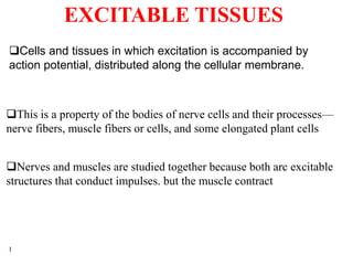
- 1. 1 EXCITABLE TISSUES Cells and tissues in which excitation is accompanied by action potential, distributed along the cellular membrane. This is a property of the bodies of nerve cells and their processes— nerve fibers, muscle fibers or cells, and some elongated plant cells Nerves and muscles are studied together because both arc excitable structures that conduct impulses. but the muscle contract
- 2. 2 NERVES Neurons in the mammalian central nervous system come in many different shapes and sizes.
- 3. 3 NERVES Neurons in the mammalian central nervous system come in many different shapes and sizes. Perikayon (soma) - nerve cell body, contains nucleus and typical cell organelles. It is the metabolic center of the neuron. a. Nucleus - large, central in most, large amount of euchromatin (intense synthetic activity), Barr body (Dormant X chromosome of females). b. Rough endoplasmic reticulum (RER) - lots for synthesis of structural and transport proteins, Nissl bodies are condensations of the RER and free ribosomes. c. Golgi apparatus - only found near nucleus in perikaryon. Expected, since intense synthetic activity of neurotransmitters and/or neurohormones.
- 4. 4 NERVES Dendrite - cell process, may be branched, forms receptive area for synaptic contacts from other neurons, Has tiny rough projections or spines called gemmules that may be points of synaptic contact, Dendrites from larger neurons may be lightly myelinated by oligodendroglia. Neurons may have more than one dendrite. Cytoplasm in these processes similar to that of perikaryon, but no golgi bodies.
- 5. 5 NERVES Axon – a single, long, cell process extending away from perikaryon, may be branched, Ends of branches form synapses with other neurons or muscle cells May be myelinated by either oligodendroglia in CNS or Schwann cells in PNS..
- 6. 6 NERVES Each neuron has only one axon.. Axon hillock (pyramid shaped region where axon originates from the perikaryon) Initial segment (unmyelinated intitial portion of axon) remainder of axon (may be myelinated or unmyelinated, may be branched)
- 7. 7 NERVES Axons carry electrical impulses (action potentials) to synapses at end of axon. Except for the axon hillock and the synaptic bouton, the axon cytoplasm (axoplasm) has few organelles, microtubules, or microfilaments. Not much synthetic activity in this part of neuron. The synaptic button granules or vesicles in which the synaptic transmitters secreted by the nerves are stored. The axon divides into presynaptic terminals, each ending in a number of synaptic knobs which are also called terminal buttons or boutons
- 8. 8 NERVES Based on the number of processes that emanate from their cell body, neurons can be classified as unipolar, bipolar, and multipolar Multipolar - more than two processes (one axon plus multiple dendrites), most of neurons in brain and spinal cord are of this type Bipolar - two major processes (axon and dendrite), but may be branched at ends, sensory neurons in retina, cochlea, and olfactory epithelium are of this type
- 9. 9 NERVES Pseudounipolar - two major processes that are fused along portions closet to perikaryon - found in spinal ganglia and some cranial ganglia. Unipolar have one process, with different segments serving as receptive surfaces and releasing terminals.
- 10. 10 NEURON CLASSIFICATION BASED ON FUNCTION Motor neurons - efferent, action potential moves from CNS to effector organ (e.g. muscle) Sensory neurons - afferent, action potential moves from sensory organ to CNS (e.g. neuron processes associated with pacinian corpuscles, touch, pressure) Interneurons - form connections between neurons
- 11. 11 GLIAL CELLS There are many more glial cells in the nervous system than there are neurons. These cells are situated among the neurons and are generally smaller. In sections stained with hematoxylin - eosin, only the glial cell nuclei show up. Special staining techniques are necessary if their cell bodies are to be easily differentiated from surrounding cells.
- 12. 12 GLIAL CELLS - Astrocytes Two types: 1. Protoplasmic astrocytes: a. Granular cytoplasm, many branches on short processes b. Some of processes are closely applied to neurons, while others form intimate contacts with blood vessels. c. Thought to form a conduit for nutrients from blood vessels to neurons. d. Found in gray matter. e. Protoplasmic astrocytes have a membrane potential that varies with the external K+ concentration but do not generate propagated potentials f. Produce substances that are tropic to neurons, and they help maintain the appropriate concentration of ions and neurotransmitters by taking up K+ and the neurotransmitters glutamate and γ-aminobutyrate (GABA)
- 13. 13 GLIAL CELLS - Astrocytes 2. Fibrous astrocytes: a. Contain many intermediate filaments, are found primarily in white matter b. Function not well understood Both types send processes to blood vessels, where they induce capillaries to form the tight junctions making up the blood–brain barrier.
- 14. 14 GLIAL CELLS - oligodendroglia Smaller than astrocytes, fewer processes In white matter, these cells form the myelin sheaths that are around many axons, in gray mater they may lightly myelinate some dendrites Also called oligodendrocytes Found in both gray and white matter Anaologous to Schwann cells of peripheral nervous system These cells must be cultured with neurons in order to get neurons to grow in tissue culture. Suggests intimate interactive association.
- 15. 15 GLIAL CELLS - microglia Elongate nucleus with mostly heterochromatin Small cell body that is elongated Can be differentiated from other glia by elongate nucleus. Other glia have a spherical nucleus
- 16. 16 GLIAL CELLS - ependymal cells Ciliary action acts to circulate cerebral spinal fluid. Ciliated cells forming single layer of cuboidal epithelium that lines the entire neurocoel Neurocoel is the cavity of the chordate cerebrospinal system, consisting of the ventricles of the brain and the central canal of the spinal cord, regarded as a unit.
- 17. 17 PROPERTIES OF NERVES This is the ability of living tissues to respond to various stimuli. It is an electric phenomenon, and the electric changes that accompany nerve excitation are called the action potential. Such changes are very small and very rapid. so their magnitudes are measured in millivolts (mV) while their durations are measured in milliseconds (msec). They are recorded by microelectrodes connected to either a galvanometer or a cathode ray oscilloscope (CRO ). 1. Excitability
- 18. 18 A stimulus is a change in the environment around the nerve (or muscle)which may be either chemical, thermal, mechanical or electrical. In laboratories, electrical stimuli are preferred because they can be accurately controlled (both in strength and duration) and, in addition, they leave the stimulated structures without damage. 2 types of electric currents can be used for stimulation of excitable tissues : a. The galvanic current: This is a constant (or direct) current (D.C.) which is obtained from a battery. b. The faradic current: This is an alternating current (A.C.) like the induction currents used in laboratories for nerve stimulation. The stimulus
- 19. 19 The physicochemical change produced by various stimuli in the nerve is called the nerve impulse. Such impulse is actively conducted along the nerve fibre and it can be conducted in both directions. In the body, each nerve conducts impulses in one direction only (motor nerves toward the effector organs and sensory nerves toward the nervous' system). 2. Conductivity Conduction in the normal direction is called orthodromic conduction, If it occurs in the opposite direction due to any cause, it will be called antidromic cmuluction Does antidromic conduction occur in the brain under normal conditions?
- 20. 20 States that "A threshold (minimal) stimulus produces a maximal response " i.e. a maximal action potential in nerve and muscle fibres and a maximal contraction in muscle fibres. Therefore as long as other factors that affect excitability remain constant: Increasing the intensity of the stimulus above the threshold value produces no further increase in the action potential or muscle contraction (3) All or none law (or rule)
- 21. 21 The all or none law is obeyed in the following structures: a. A single nerve fibre - A motor unit is made up of a motor neuron and the skeletal muscle fibers innervated by that motor neuron's axonal terminals. (3) All or none law (or rule) c. The cardiac muscle and some smooth muscles which act as one unit called syncytium b. A single skeletal muscle fibre and the motor unit - Groups of motor units often work together to coordinate the contractions of a single muscle; all of the motor units within a muscle are considered a motor pool.
- 22. 22 However, nerve trunks and whole skeletal muscles (which contain many fibres) do not obey the law. (3) All or none law (or rule) Why? The threshold intensity for stimulation varies in the different types of nerve and muscle fibres (i.e. it is not equal). Therefore if the intensity of stimulation is increased in these structures, the response will also increase till reaching a maximum.
- 23. 23 The nerve fibre adapts to stimulation by a constant current so no response occurs during passage of the current. (4) Accommodation (or adaptation) (5) Infatiguability Nerve fibres are not fatigued by continuous stimulation
- 24. 24 1. Intensity (strength) of the stimulus: Sub-threshold stimuli produce only local responses that don't initiate action potentials . Factors that determine the effectiveness of stimuli If the intensity is increased slowly the nerve will not respond because of the property of accommodation 2. Rate of increase in the intensity of stimuli; Sub-threshold stimuli that are gradually increased produce a response only with a rapid increase in the intensity of stimuli
- 25. 25 3. Duration of stimulus (duration of current): The relation between the intensity of a stimulating current and the duration (time) of its flow necessary to set up an impulse is shown in the strength-duration curve Factors that determine the effectiveness of stimuli
- 26. 26 Within limits, there is a reciprocal relationship between the current strength and duration of flow required to produce an impulse There is a minimal duration needed for excitation below which no excitation occurs whatever be the strength of the stimulus. RHEOBASE: This is the minimal strength (or threshold intensity ) of a galvanic current that can set up an impulse. CHRONAXIE: This is the duration of current flow required for excitation when using a strength equal to twice (or double) the rheobase. The time required for excitation when using the rheobase is called the utilization time
- 27. 27 What is the significance of the Chronaxie? Its measurement can be used to compare the excitability of different tissues, or that of the same tissue under different conditions. The chronaxie is a good index for the degree of excitability (the shorter the chronaxie the greater the excitability versa). In the strength-duration curve to the right, which nerve is more excitable?
- 28. 28 The curve for the slower fibres would be shifted to the right, indicating that for a given stimulus strength, a longer stimulus duration would be needed to bring the slower fibres to threshold. Q How would the strength-duration curve for a set of slow fibres (not very excitable) compare to the strength-duration curve for a set of quick fibres (very excitable)?
- 29. 29 RESTING MEMBRANE POTENTIAL REVIEW Outside the cell membranes there are mainly Na+, Cl- and HCO3 while inside the cell there are mainly K+ and organic protein anions In neurons the potential difference about -70 mV RM P is due to an unequal distribution of ions on both sides of the membrane with relatively excess cations outside and excess anions inside This is produced as a result or 2 main factors (a) Selective permeability of cell membranes (b) The N a+/K+ ATPase
- 30. 30 RESTING MEMBRANE POTENTIAL REVIEW (a) The electrical gradient is directed inwards K+ ions tend to diffuse outside the cells. However, this is limited because: (b) The +ve charge on the outside of the membranes repels K+ ions inwards. (c) The sodium-potassium pump actively drives K+ ions inwards The concentration gradient for Cl- and HCO3 is directed inwards, so these anions tend to diffuse into the cells. This is limited because the interior of the cells has a great -ve charge, and accordingly, they are expelled out of the cell along this electrical gradient
- 31. 31 CHANGES THAT ACCOMPANY PROPAGATION OF A NERVE IMPULSE The Action Potential (AP) (1) ELECTRIC CHANGES This refers to the changes in potential that occur in excitable tissues when stimulated It is transmitted as a self-propagated disturbance called impulse Stimulating the nerve (by an electric stimulator) is marked by a stimulus artifact, which is due to current leakage from the stimulating electrode to the recording (external) electrode. Latent period (which is an isopotcntial interval representing the time taken by the impulse to reach the recording electrode) after which the AP is recorded.
- 32. 32 Various stages of the action potential (AP).
- 33. 33 The AP consists of 2 main stages (called depolarization and repolarization) This is followed by 2 other stages known as after- depolarization and after-hyperpolarization.
- 34. 34 Depolarization (DP) This is loss of the normal resting polarized state of the membrane It is recorded as a rise of the membrane potential in the positive direction from -70 mV towards the isopotential line (zero potential) It produces the ascending limb of the A.P DP develops slowly. but after an initial 15 mV of DP (i.e. MP becomes about -55 mV). the rate of DP suddenly increases (so this point is called the firing level)
- 35. 35 Depolarization (DP) DP then proceeds rapidly till the resting membrane potential is lost The potential difference between both sides of the membrane becomes zero this change is called overshoot or reversal of polarity, and it results in an A.P. having a magnitude of 105 mV (from -70 to +35 mV) The membrane potential the reaches +35m V (indicating that the inner surface of the membrane becomes positive relative to the outer surface.
- 36. 36 Repolarization (RP) This is restoration or the normal resting polarized state of the membrane It is recorded as a fall of the membrane potential in the negative direction from +35 mV to -70 mV. RP proceeds immediately and rapidly after the overshoot is reached It produces the descending limb of the A.P. When RP is 70-80% completed, its rate decreases for about 4 msec. This stage is called after-depolarization (or negative after-potential)
- 37. 37 Repolarization (RP) After RP is completed the membrane potential overshoots to the negative side (by about 1-2 mV) leading to hyperpolarization of the membrane This stagc is called after-hyperpolarization (or positive after-potential). It lasts about 40 msec but its magnitude gradually declines till the normal resting membrane potential is restored
- 38. 38 Ionic basis (or mechanisms) of DP and RP The initial slow DP is produced by the stimulating current itself . How? Stimulating current arc cathodic in nature, which adds negative charges outside the nerve membrane Thus the potential difference between both sides of the membrane is decreased The membrane potential becomes less -vc than at the resting state. This is called electrotonic DP. Depolarization
- 39. 39 The rapid phase of DP and the overshoot are produced by an increase in Na+ influx (= entrance) into the nerve fiber as a result of increased Na+ conductance of the nerve membrane Increased permeability occurs secondary to marked increase in the Na+ permeability of the membrane through opening of specific Na+ channels in the membrane. Each Na+ channel has an activation gate at the outer surface of the membrane and an inactivation gate at its inner surface. The membrane Na+ conductance (and consequently the Na influx ) is increased only when both gates are opened.
- 40. 40 In the resting state, only the inactivation gates are open so the membrane permeability to Na+ is low When the nerve is stimulated the Na+ activation gates also open thus the membrane permeability and conductance to Na+ as well as Na+ influx are markedly increased Opening of the Na+ activation gates is voltage-dependent. They start to open when the initial electrotonic DP becomes 7 mV This allows Na influx. which further decreases the membrane polarity leading to opening of more gates.
- 41. 41 This results in more Na+ influx and more decrease of the membrane polarity, which leads to opening of more and more gates and more Na+ influx. Such process continues in the form of a vicious circle (i.e. by a positive feedback mechanism ) till all gates open The rate of opening of the Na+ activation gates is slow between 7 and I5 mV of DP (i.e. till a membrane potential of about -55 mV) leading to slow DP. After15 mV of DP, the rate of opening of these gates suddenly increases leading to much acceleration of DP (the firing level)
- 42. 42 (a) Closure of the Na+ inactivation gates (b) Reversal ofthe direction of eleclrical gradient for Na+ (2) K+ efflux (exit) from the nerve fibre: Repolarization (RP) and after-potentials RP of the membrane takes place rapidly after DP as a result of: (1) Stoppage of Na+ influx due to: This occurs through specific K+ channels that contain a single gate located toward the inside of the membrane. The decrease in membrane polarity during DP leads to opening of the K+ gates, thus the K+ conductance is markedly increased and K+ ef1ux occurs.
- 43. 43 The negative after-potential stage is clue to slowing of the rate of K+ efflux. The positive after-potential stage is due to slow return of the K+ channels to the closed state (which allows prolonged K+ efflux) The process is however slower than opening of the Na+ channels so the increase in K+ conductance is slightly delayed The slow opening and delayed closure of the K+ channels may explain the phenomenon of accommodation that occurs in nerves Following the AP, the resting Na+ and K+ ionic gradients is restored by the action of the Na+/K+ pump.
- 44. 44 Normally, both the concentration and electrical gradients for Ca2+are directed inwards The Ca2+ conductance increases during nerve excitation leading to Ca2+ influx. Role of Ca2+ in Nerve excitation Ca2+ contributes to DP (and in some invertebrates. it is primarily responsible for the AP) Ca2+ influx occurs via voltage-gated Ca2+ channels which are also slightly permeable to Na+ (so they arc called Ca2+- Na+ channels)
- 45. 45 They are however very slow to become activated, so they are also called slow channels (in contrast to the voltage-gated Na+ channels which arc called fast channels) Role of Ca2+ in Nerve excitation The extracellular Ca2+ concentration also affects nerve excitability Its decrease increases the excitability while its increase decreases the excitability and stabilizes the nerve membrane
- 46. 46 ETPs are localized potential changes that occur in nerves when stimulated by sub-threshold constant currents Electrotonic potentials (ETPs) and the local response There are 2 types of the ETPs both of which are passive changes in the membrane polarization This is produced by addition of subtraction of charges through the stimulating current) that decay (i.e. disappear) gradually
- 47. 47 This is the potential change that occurs when using anodal (+ve) currents for stimulation (A) Anelectrotonic potential (or anelectrotonus) It is a state of hyperpolarization caused by addition of +ve charges at the outer surface of the nerve membrane. The magnitude of the potential change is proportionate to the strength of the stimulus It is associated with a decrease of excitability of the nerve and with strong anodal currents, the nerve excitability may be completely lost ( anodal block) It takes the membrane potential away from firing level (which inhibits discharge of impulses).
- 48. 48 This is the potential change that occurs when using cathodal (-ve) currents for stimulation (A) Catelectrotonic potential (or catelectrotonus) It is a state of partial DP caused by addition of –ve charges at the outer surface of the nerve membrane. The magnitude of the potential change varies with the strength of the stimulating current It is associated with an increase of excitability of the nerve,
- 49. 49 MECHANISM OF NERVE IMPULSE CONDUCTION
- 50. 50 (A) In unmyelinated nerve fibres Nerve impulses are propagated along unmyelinated nerve fibres in the form of a wave of APs Local circular currents flow between the activated point and the neighbouring inactive areas of the nerve membrane The initial stimulus causes reversal of polarity and an AP at the point of stimulation +ve charges from the inactive areas flow into the initial area of negativity produced by the AP (= area of current sink )
- 51. 51 This decreases the polarity at the inactive areas ( electrotonic depolarization) which produces an AP initiating on reaching the firing level. The latter areas, in turn, electrotonically depolarize the membrane in front of it through local circular currents and this sequence of events moves regularly along the nerve fibre to its end. the nerve impulse is self-propagated and once it leaves a point this point will soon repolarize thus a repolarization wave starts after the depolarization wave and is propagated in the same direction
- 52. 52 (B) In myelinated nerve fibres Nerve impulses are propagated along myelinated nerve fibres by a mechanism called the saltatory conduction Myelin surrounds the nerve axon and is interrupted at regular intervals at the nodes of Ranvier It is an insulator to current flow (in contrast to the nodes of Ranvier which easily permit current flow because of their high permeability to Na) Circular currents also flow in myelinated nerve fibres but the +ve charges jump from the inactive nodes to the area of current sink at the active node
- 53. 53 This leads to electrotonic depolarization and production of an AP at the inactive nodes, which in turn activates the neighbouring nodes. This jumping of DP from node to node is called saltatory conduction and it results in: (a) Increasing velocity of conduction (b) Conservation of energy (because excitation occurs only in the nodes and not allover the nerve membrane)
- 54. 54 (2) EXCITABILITY CHANGES During propagation of a nerve impulse (i.e. during an AP). The excitability of nerve fibres passes in the following phases (a) Absolute refractory period (ARP): During this period, the nerve is completely inexcitable. No stimulus can excite it whatever its strength It corresponds to the ascending limb of the AP from the time the firing level is reached That is during DP and overshoot and upper part of the descending limb (until RP is about 1/3 complete)
- 55. 55 (b) Relative refractory period (RRP): During this period, nerve excitability is partiality recovered Stronger stimuli than normal are required for excitation It corresponds to the remaining part of the descending limb of the AP till the start of after-depolarization That is during the later 2/3 of RP
- 56. 56 (c) Supernormal phase (or period): During this period nerve excitability is increased. Weaker stimuli than normal can excite the nerve It corresponds to the period of after-depolarization
- 57. 57 (d) Subrnormal phase (or period): During this period nerve excitability is decreased. Stronger stimuli than normal are required for excitation It corresponds to the period of afler-hyperpolarization.
- 58. 58
- 59. 59 Factors that affect nerve excitability (1) Temperature: Cooling decreases nerve excitability while warming increases it (2) Pressure: Mechanical pressure on a nerve reduces its excitability. (3) Blood supply: Nerve excitability is decreased in cases of ischemia. (4) Oxygen supply: O2 lack decreases nerve excitability. (5) H+ concentration: Alkalinity increases while acidity decreases excitability of nerves.
- 60. 60 (6) Chemicals: Nerve excitability is decreased by excess CO2 and alcohol as well as by anesthetic drugs e.g. ether, chloroform and cocaine. (8) Electrolytes: The concentrations of Na+ K+ and Ca2+ in the extracellular fluid affect nerve excitability as follows
- 61. 61 Ionic changes that increase nerve excitability 1- Decreased Ca2+ concentration: This increases the membrane permeability to Na+ and decreases the amount of DP necessary to initiate the changes in Na+ and K+ conductances that produce the AP. 2- Increased Na concentration: This facilitates the process of DP 3- Increased K+ concentration: This favours K influx which leads to DP thus the nerve excitability will increase
- 62. 62 Ionic changes that decrease nerve excitability 1- Increased Ca2+ concentration : This decreases the membrane permeability to Na+ and increases the amount of DP necessary to initiate the changes in Na+ and K+ conductances that produce the AP 2-Decreased Na concentration : This decreases nerve excitability by delaying the process of DP 3- Decreased K+ concentration : This favours K+ efflux which leads to hyperpolarization, thus the nerve excitability will decrease
- 63. 63 Methods of producing nerve block Propagation of impulses by nerves can be blocked (i.e. prevented) by one of the following methods : (1) Physical methods e.g. application of cold or a strong anelectrotonus (2) Mechanical methods (e.g. application of pressure on the nerve). (3) Chemical methods: These include: (a) The ionic changes that decrease nerve excitability i.e. increased Ca2+ and decreased Na+ or K+ concentration in the extracellular fluid
- 64. 64 (b) Use of certain chemical substances known as the membrane stabilizers which include mainly the local anesthetic drugs (e.g. cocaine and novocaine) These drugs markedly decrease the membrane permeability to Na+ (by preventing opening of the Na+ channel activation gales) So the depolarization process is inhibited and nerve impulses fail to be produced
- 65. 65 (3) METABOLIC CHANGES The metabolic processes that occur in nerves are generally similar to those occurring in muscles (O2 and ATP) In resting nerves, they occur at a low rate (mainly to maintain the polarized state) but they are much increased during transmission of nerve impulses The breakdown of ATP supplies the energy required for the sodium potassium pump and nerve impulse propagation It is resynthesized by energy derived from breakdown of glycogen. Nerve fibres are also rich in vitamin B1 which is necessary for their metabolic activities
- 66. 66 (4) THERMAL CHANGES Resting nerves liberate little heat as a result of their low metabolic rate. During nerve impulse transmission the heat production by nerves is markedly increased, and is liberated in the following 2 stages : (1) Initial heat: This coincides with propagation of spike (i.e. during ionic migration), and it is due to anaerobic breakdown of ATP (2) Recovery (delayed) heat: This follows propagation of the spike, and it is due to aerobic reactions that liberate the energy required for ATP resynthesis.
- 67. 67 Types of mammalian nerve fibres The mammalian nerve fibres are classified according to their diameters into the following types: ( 1) Group A nerve fibres : These have the largest diameters (3-20 microns) The have highest speeds of conduction ( 15-120 m/sec) They are further subdivided into alpha, beta, gamma & delta nerve fibres E.g. the somatic nerve fibres that transmit motor impulses & deep sensations They are most sensitive to pressure (i.e. the conduction of impulses in these nerves can be readily blocked by pressure)
- 68. 68 ( 2) Group B nerve fibres : These have the smaller diameters (1.3 - 3 microns) They have moderate speeds of conduction (3-15 m/sec) E.g. the myelinated preganglionic autonomic nerves They arc most sensitive to O2 lack
- 69. 69 ( 3) Group C nerve fibres : These have the smallest diameters (0.3-1.3 microns) The have slowest speeds of conduction (0.5-3 m/sec) E.g. the unmyelinated postganglionic autonomic nerves They arc most sensitive to local anaesthetics
- 71. 71 The area of contact between a nerve fibre and a muscle fibre is called the motor end plate (M.E.P. ) or neuromuscular junction As the nerve approaches the muscle, it loses its myelin sheath and its axon is branched. The tip of each branch (sole foot ) is covered by the neurolemma which continues with the sarcolemma ((the outer membrane of the n1uscle fibre) Each sole foot lies in a depression in the plasma membrane (the inner membrane of the muscle fibre) called the synaptic gutter
- 72. 72 Where the membrane is thrown into many folds called the palisades
- 73. 73 Where the membrane is thrown into many folds called the palisades There is no protoplasmic continuity between the nerve and muscle fibres. They are separated by the synaptic cleft in which the chemical transmitter acetylcholine is released from the vesicles present in the sole feet. The sarcoplasm at the motor end plate is granular and contains the cholinesterase enzyme which hydrolyzes acetylcholine.
- 74. 74 Miniature end plate potential ( M. E.P.P.) This is a state of persistent localized subthreshold DP (0.5 mV in amplitude) of the muscle fibres at the M.E.P. during rest. It does not reach the firing level (so it does not lead to an AP) It is due to continuous release of small amounts of acetylcholine Arising from the continuous rupture of a few vesicles that contain this chemical transmitter at the nerve terminals
- 75. 75 Mechanism of neuromuscular transmission When a motor nerve is stimulated, the generated impulse is propagated toward the M.E.P it causes movement of'Ca2+ from the extracellular fluid into the nerve terminals Ca2+ cause rupture of acetylcholine vesicles leading to release (exocytosis) of this transmitter into the synaptic cleft Acetylcholine then combines with nicotinic receptors in the muscle membrane
- 76. 76 This increases its permeability to Na+ through opening of ligand-gated Na+ channel. Na+ influx increases resulting in DP of the muscle at the M.E.P.. which is called the end plate potential This normally reaches the firing level which starts an A P along the surface of the muscle The released acetylcholine is rapidly hydrolyzed by the cholinesterase enzyme so that the re-excitation of the muscle wouldn’t occur.
- 77. 77
- 78. 78 Properties of neuromuscular transmission 1. At the M.E.P. impulses are conducted in one direction only 2. Transmission of impulses at the M. E.P. is delayed 0.5- 0. 7 millisecond due to the time required for: (a) Release of acetylcholine (b) Passage of acetylcholine across the synaptic cleft and its combination with the nicotinic receptors in the muscle (c) Increase in the permeability of the muscle membrane to Na (d) Increase in Na+ influx till the firing level 3. Transmission of impulses at the M.E.P. readily fatigues after repeated stimulations (mostly due to exhaustion of Ach) 4. Transmission of impulses at the M.E.P. is readily affected by drugs
- 79. 79 Drugs that affect neuromuscular transmission (A) Drugs that block neuromuscular transmission 1. Curare (tubocurarine) competitively inhibits Ach interaction at the Nm receptors. α- neurotoxins (α- bungarotoxin, α- cobrotoxins) irreversibly blocks. 2. Botulinum toxin: From Clostridium botulinum. The toxin inhibits the release of ACh
- 80. 80 (B) Drugs that stimulate neuromuscular transmission 1. Acetylcholine-like drugs that are not rapidly hydrolyzed by the cholinesterase enzyme and act mainly at the M. E.P. e.g. carbacol. 2. Drugs that preserve the liberated acetylcholine at the M.E.P. by antagonizing the cholinesterase enzyme e.g. prostigmine and eserine
Notes de l'éditeur
- +256786014374
- +256786014374
- Electrotonic potential (or graded potential) — a non-propagated local potential, resulting from a local change in ionic conductance (e.g. synaptic or sensory that engenders a local current). When it spreads along a stretch of membrane, it becomes exponentially smaller (decrement).