Recommended

Recommended
More Related Content
Recently uploaded
Recently uploaded (20)
Featured
Featured (20)
Dependence of Contrast-Enhanced Lesion Detection in Contrast-Enhanced Digital Breast Tomosynthesis on Imaging Chain Design
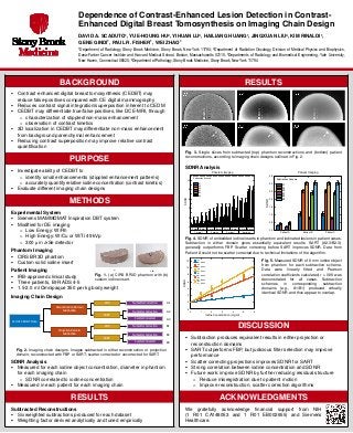
- 1. DAVID A. SCADUTO1, YUE-HOUNG HU2, YIHUAN LU3, HAILIANG HUANG1, JINGXUAN LIU4, KIM RINALDI1, GENE GINDI1, PAUL R. FISHER1, WEI ZHAO1 1Department of Radiology, Stony Brook Medicine, Stony Brook, New York 11794; 2Department of Radiation Oncology, Division of Medical Physics and Biophysics, Dana-Farber Cancer Institute and Harvard Medical School, Boston, Massachusetts 02115; 3Departments of Radiology and Biomedical Engineering, Yale University, New Haven, Connecticut 06520; 4Department of Pathology, Stony Brook Medicine, Stony Brook, New York 11794 Dependence of Contrast-Enhanced Lesion Detection in Contrast- Enhanced Digital Breast Tomosynthesis on Imaging Chain Design BACKGROUND Contrast-enhanced digital breast tomosynthesis (CEDBT) may reduce false-positives compared with CE digital mammography Reduces contrast signal integration/superposition inherent to CEDM CEDBT may differentiate true/false positives, like DCE-MRI, through o characterization of stippled/non-mass enhancement o observation of contrast kinetics 3D localization in CEDBT may differentiate non-mass enhancement from background parenchymal enhancement Reducing contrast superposition may improve relative contrast quantification PURPOSE Investigate ability of CEDBT to o identify small enhancements (stippled enhancement patterns) o accurately quantify relative iodine concentration (contrast kinetics) Evaluate different imaging chain designs METHODS Experimental System Siemens MAMMOMAT Inspiration DBT system Modified for DE imaging o Low Energy: W/Rh o High Energy: W/Cu or W/Ti 49 kVp o 300 μm a-Se detector Phantom Imaging CIRS BR3D phantom Custom solid iodine insert Patient Imaging IRB-approved clinical study Three patients, BI-RADS 4-5 1.5-2.0 ml Omnipaque 350 per kg body weight Imaging Chain Design SDNR Analysis Measured for each iodine object concentration, diameter in phantom for each imaging chain o SDNR correlated to iodine concentration Measured in each patient for each imaging chain RESULTS Subtracted Reconstructions Six weighted subtractions produced for each dataset Weighting factor derived analytically and tuned empirically RESULTS SDNR Analysis DISCUSSION Subtraction produces equivalent results in either projection or reconstruction domains SART outperforms FBP, but judicious filter selection may improve performance Scatter correcting projections improves SDNR for SART Strong correlation between iodine concentration and SDNR Future work: improve SDNR by further reducing residual structure o Reduce misregistration due to patient motion o Improve reconstruction, scatter correction algorithms ACKNOWLEDGMENTS We gratefully acknowledge financial support from NIH (1 R01 CA148053 and 1 R01 EB002655) and Siemens Healthcare. 2 mm 8 mm 2 mm 8 mm 2 mm 8 mm 2 mm 8 mm 1 mg/ml 2 mg/ml 3 mg/ml 5 mg/ml 0 2 4 6 8 10 Phantom Imaging Subtraction Scheme A1 A2 A3 B1 B2 B3 SDNR Patient A Patient B Patient C 0.0 0.5 1.0 1.5 2.0 2.5 3.0 3.5 Patient Imaging SDNR Subtraction Scheme A1 A2 A3 B1 B2 B3 Fig. 4. SDNR of embedded iodine inserts in phantom and iodinated lesions in patient cases. Subtraction in either domain gives essentially equivalent results. SART (A2-3/B2-3) generally outperforms FBP. Scatter correcting before SART improves SDNR. Data from Patient A could not be scatter corrected due to technical limitations of the algorithm. Fig. 5. Measured SDNR of 8 mm iodine object from phantom for each subtraction scheme. Data were linearly fitted and Pearson correlation coefficients calculated; r > 0.98 was demonstrated for all cases. Subtraction schemes in corresponding subtraction domains (e.g., A1/B1) produced virtually identical SDNR and thus appear to overlap. Fig. 3. Single slices from subtracted (top) phantom reconstructions and (bottom) patient reconstructions, according to imaging chain designs outlined in Fig. 2. Fig. 1. (a) CIRS BR3D phantom with (b) custom iodine insert. Fig. 2. Imaging chain designs. Images subtracted in either reconstruction or projection domain; reconstructed with FBP or SART; scatter corrected or uncorrected for SART. 0 1 2 3 4 5 6 -2 0 2 4 6 8 10 12 A1 r = 0.994 A2 r = 0.986 A3 r = 0.992 B1 r = 0.985 B2 r = 0.985 B3 r = 0.992 SDNR Iodine Concentration (mg/ml) LE/HE CEDBT Data A1 A2 A3 B1 B2 B3 Reconstruction Domain Subtraction Projection Domain Subtraction SART No Scatter Correction Scatter Corrected FBP No Scatter Correction SART No Scatter Correction Scatter Corrected FBP No Scatter Correction