
Recommandé
Contenu connexe
Tendances
Tendances (20)
Similaire à connective tissue.pptx
Similaire à connective tissue.pptx (20)
Dernier
Dernier (20)
connective tissue.pptx
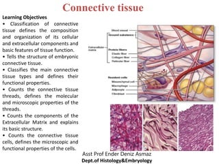
- 1. Connective tissue Asst Prof Ender Deniz Asmaz Dept.of Histology&Embryology Learning Objectives • Classification of connective tissue defines the composition and organization of its cellular and extracellular components and basic features of tissue function. • Tells the structure of embryonic connective tissue. • Classifies the main connective tissue types and defines their functional properties. • Counts the connective tissue threads, defines the molecular and microscopic properties of the threads. • Counts the components of the Extracellular Matrix and explains its basic structure. • Counts the connective tissue cells, defines the microscopic and functional properties of the cells.
- 2. Connective tissue is one of the four tissues found in the human body. Connective tissue provides a matrix that supports and physically connects other tissues and cells together to form the organs of the body. Connective tissue is found in between other tissues everywhere in the body, including the nervous system. 2
- 3. Organ Components: 3 Most organs can be divided into: The parenchyma, which is composed of the cells responsible for the organ’s specialized functions. The stroma, the cells of which have a supporting role in the organ. Except in the brain and spinal cord, the stroma is always connective tissue.
- 4. Most Organs Parenchyma Stroma 4
- 5. 5 Connective tissue originates from the mesenchyme. Atissue developing mainly from the middle layer of the embryo, the mesoderm. . Embryonic mesenchyme producing all types of connective tissue proper and the specialized connective tissues bone and cartilage, they also include stem cells for other tissues such as blood, the vascular endothelium, and muscle.
- 6. Origin of connective tissue cells 6
- 7. Origin of connective tissue cells 7
- 9. 9 Connective tissue is composed of cells and an extracellular matrix that binds the cells and organs, integrating all parts of the body. Connective tissue is the most widespread tissue of the body and can be found in every organ. Avariety of cell types are found in connective tissues.
- 10. 10
- 11. 11
- 12. Connective tissue vs Epithelium: Unlike the other tissue types (epithelium, muscle, and nerve), which consist mainly of cells, the main component of connective tissue is the extracellular matrix (ECM). 12
- 13. 13
- 14. Connective tissue that connects, supports, binds, or separates other tissues or organs, typically having relatively few cells embedded in an amorphous matrix, often with or other fibres, and including cartilaginous, fatty, and elastic tissues. The variety of connective tissue types in the body reflects differences in composition and amount of the cells, fibers, and ground substance which together are responsible for the remarkable structural, functional, and pathologic diversity of connective tissue. 14
- 15. Classes of Connective Tissue 15 Connective tissue proper Dense connective tissue Loose connective tissue Cartilage Bone Blood
- 16. 16
- 17. Protect Structural support Bind Trsansport Immunity Connective tissue binds skin to muscles 17
- 18. 18
- 19. Fibroblasts Adipocytes Macrophages & the Mononuclear Phagocyte System Mast Cells Plasma Cells Leukocytes 19 Fixed (permanent residents) 1. Fibroblasts 2. Adipose (fat) cells 3. Tissue Macrophages** 4. Mast cells** 5. Lymphocytes & Plasma Cells (differentiated B-cells) ** 6. “Leukocytes”** Free (transient residents) ** ( derived from hematopoietic stem cells and involved in immune function and inflammation. Specifically, neutrophils, eosinophils, & basophils)
- 20. 20 Fibroblasts are the key cells in connective tissue proper. Fibroblasts originate locally from mesenchymal cells and are permanent residents of connective tissue. Other cells found here, such as macrophages, plasma cells, and mast cells, originate from hematopoietic stem cells in bone marrow, circulate in the blood, and then move into connective tissue where they function.
- 21. The most common cells in connective tissue proper. Produce and maintain most of the tissue’s extracellular components. Fibroblasts synthesize and secrete collagen (the most abundant protein of the body) and elastin, which both form large fibers, as well as the GAGs, proteoglycans, and multiadhesive glycoproteins that comprise the ground substance. 24
- 22. C: Connective tissue arrows: Fibroblasts 22
- 23. 23
- 24. 24
- 25. 28
- 26. Active vs quiescent cells 26
- 27. Active vs quiescent cells 27 The inactive form of fibroblasts is called fibrocyte.
- 28. Adipocytes, or fat cells, are found in the connective tissue of many organs. These large, mesenchymally derived cells are specialized for cytoplasmic storage of lipid as neutral fats, or less commonly for the production of heat. Tissue with a large population of adipocytes, called adipose connective tissue, serves to cushion and insulate the skin and other organs. 28
- 31. 31 Macrophages & the Mononuclear Phagocyte System Macrophages a large phagocytic cell found in stationary form in the tissues or as a mobile white blood cell, especially at sites of infection. Size and shape vary considerably, corresponding to their state of functional activity.
- 32. Macrophage ultrastructure N: nucleus L: lysosomes Nu: nucleolus 36
- 33. 37 Macrophage
- 34. Macrophages arise from monocytes. Monocytes originate in the bone marrow from where they are released into the blood stream. They are actively mobile and leave the blood stream to enter connective tissues, where they differentiate into macrophages. 34
- 35. 35
- 36. Distribution and main functions of the cells of the mononuclear phagocyte system 36
- 37. 37 Mast Cells Mast cells are oval or irregularly shaped cells of connective tissue, between 7 and 20 μm in diameter. Mast cells filled with basophilic secretory granules that often obscure the central nucleus. Mast cells are components of loose connective tissues, often located near small blood vessels.
- 38. 42 BV: blood vessels Mast cells
- 39. M: mitochondria G: cytoplasmic granule N: nucleus C: collagen fibers E: elastic fibers 43 Ultrastructurally mast cells
- 40. Mast cell 44
- 41. Function of Mast cells Localized release of many bioactive substances important in the local inflammatory response, innate immunity, and tissue repair. A partial list of molecules released from these cells’secretory granules includes the following: Heparin Histamine Cytokines Phospholipid 45
- 42. Like macrophages, mast cells originate from progenitor cells in the bone marrow, which circulate in the blood, cross the wall of small vessels called venules, and enter connective tissues, where they differentiate. 42
- 43. 43 Plasma Cells Plasma cells are lymphocyte-derived, antibody- producing cells. These relatively large, ovoid cells have basophilic cytoplasm rich in RER and a large Golgi apparatus near the nucleus that may appear pale in routine histologic preparations. The nucleus of the plasma cell is generally spherical but eccentrically placed. Their average life span is only 10-20 days.
- 44. 44
- 46. 46 Leukocytes leukocyte is a colourless cell which circulates in the blood and body fluids and is involved in counteracting foreign substances and disease; a white (blood) cell.
- 48. The fibrous components of connective tissue are elongated structures formed from proteins that polymerize after secretion from fibroblasts. The three main types of fibers are: Collagen Reticular fibers Elastic fibers 48
- 49. • is a type of fiber found in connective tissue that provides strength Collagen fiber 49 • is a type of fiber found in connective tissue that has a netlike formation and provides support • is a type of fiber found in connective tissue that can stretch and recoil Reticular fiber Elastic fiber
- 50. Collagen and reticular fibers are both formed by proteins of the collagen family, and elastic fibers are composed mainly of the protein elastin. 50
- 51. 51
- 52. 52
- 53. 60
- 54. 54 Collagen The collagens can form various extracellular fibers, sheets, and networks, all of which extremely strong and resistant to normal shearing and tearing forces. Collagen is a key element of all connective tissues, as well as epithelial basement membranes and the external laminae of muscle and nerve cells.
- 55. 55 Collagen is the most abundant protein in the human body, representing 30% of its dry weight. A family of 28 collagens exists in vertebrates, numbered in the order they were identified. They can be categorized according to the structures formed by their interacting α-chains subunits:
- 56. 63
- 57. Type I is the most common - 90% of all collagens are type I. This type is found in bone, skin, tendon, ligaments, cornea etc. Types I, II, III, V and IX are fibrillar, which means that the protein chains assemble into fibrils, 10-300nm in diameter. Types V & VI form networks in the basal lamina. Type VI helps to anchor basal lamina of skin to underlying connective tissue. 64
- 58. C: type I collagen fibrils arrows: fibroblast Type I collagen fibrils 58
- 59. Collagen synthesis: 59 Collagen synthesis occurs in many cell types but is a specialty of fibroblasts. The Collagen subunit
- 60. This is a scanning EM of collagen fibres 60
- 61. 61
- 62. 62
- 63. 63
- 64. 64 Reticular fibers Reticular fibers found in delicate connective tissue of many organs, notably in the immune system. Reticular fibers consist mainly of collagen type III, which forms an extensive network (reticulum) of thin fibers (diameter 0.5-2 μm) for the support of many different cells.
- 65. Reticular fibers are rarely visible in hematoxylin and eosin (H&E) preparations but are characteristically stained black after impregnation with silver salts and are thus termed argyrophilic. 72
- 66. 66 Reticular fibers produced by fibroblasts. Reticular fibers occur in the reticular lamina of basement membranes and typically also surround adipocytes, smooth muscle and nerve fibers, and small blood vessels.
- 67. 67
- 68. Reticular vs Collagen Fibers 68 Reticular fibers Collagen fibers
- 69. Reticular vs Collagen Fibers 69 Reticular fibers Collagen fibers
- 70. Elastic fibers Elastin An elastic fiber Elastic fibers (or yellow fibers) are bundles of proteins (elastin) found in extracellular matrix of connective tissue. Elastic fibers produced by fibroblasts and smooth muscle cells in arteries. 70
- 71. Elastic fibers are thinner than the type I collagen fibers and form rare networks interspersed with collagen bundles in many organs, particularly those subject to regular stretching or bending. Elastic fibers can stretch up to 1.5 times their length, and snap back to their original length when relaxed (e.g. the stroma of the lungs) 78
- 72. 72
- 74. Molecular basis of elastic fiber elasticity 74
- 75. 75
- 76. Reticular vs Elastic vs Collagen Fibers 76 Reticular fibers Collagen fibers Elastic fibers
- 77. Reticular vs Elastic vs Collagen Fibers 77 Reticular fibers Elastic fibers Collagen fibers
- 78. 85
- 79. Ground substance is an amorphous gel-like substance in the extracellular space. The ground substance of the ECM is a highly hydrated (with much bound water), transparent, complex mixture of three major kinds of macromolecules: Glycosaminoglycans (GAGs) Proteoglycans Multiadhesive glycoproteins 86
- 80. 80 Ground substance functions: Ground substance filling the space between cells and fibers in connective tissue. Ground substance allows diffusion of small molecules and, because it is viscous, acts as both a lubricant and a barrier to the penetration of invaders.
- 81. 81
- 82. Glycosaminoglycans (GAGs) GAGs (also called mucopolysaccharides) are long polymers of repeating disaccharide units, 89 • binds a considerable amount of water • it has an important role in allowing molecular diffusion through connective tissue and in lubricating various organs and joints.
- 83. Proteoglycans Proteoglycans are GAG's that are covalently attached to a 'core' protein. 83
- 84. 84
- 85. Classifications used for the various types of connective tissue typically denote either a structural characteristic or a major component. 85
- 86. 94 Classification of connective or supporting tissues
- 87. 95 CT proper Binds Dense CT Regular Lig.+Tend. Irregular Epidermis Elastic Aorta Loose CT Adipose Fat Areolar Protects Reticular lymph organs Supporting CT Protection/ Structure Cartilage Elastic Ear Hyaline Ribs Fibrocartilage Vertibral discs Bone Fluid CT Transport Blood Connective tissue (CT)
- 88. Connective tissue proper Loose connective tissue Dense connective tissue Supporting connective tissue Cartilage Bone Fluid connective tissue Blood 88
- 89. 89 Connective Tissue Proper Connective tissue proper is broadly classified as: Loose connective tissue Dense connective tissue Regular dense connective tissue Irregular dense connective tissue
- 90. 90 Loose connective tissue Also called areolar tissue, is common, a layer beneath the epithelial lining of many organs and filling the spaces between fibers of muscle and nerve. The loose connective tissue typically contains cells, fibers, and ground substance in roughly equal parts.
- 91. 91 Charastaristics of Loose connective tissue: Delicate, flexible, not very resistant to stress. Well vascularized. All types of connective tissue cells present. (Majority are fibroblasts and macropahges) Collagen, elastic, and reticular fibers present. In certain organs (intestine) and in certain disease conditions, numerous lymphocytes may be present.
- 92. 100 Loose connective tissue (areolar tissue)
- 93. 93
- 95. 103 Loose connective tissue (areolar tissue)
- 96. Reticular tissue: a specialized loose connective tissue with reticular cells that form a fine matrix of reticular fibers. Provides a structural framework for hematopoietic organs such as bone marrow and spleen. . 104
- 97. Reticular tissue is characterized by abundant fibers of type III collagen This collagen is also known as reticulin and is produced by modified fibroblasts called reticular cells. Reticular tissues have a high cellular content.
- 99. Collagen bundles and Reticular cell processes of the Lymph Node, 200x 99
- 100. 100 Dense connective tissue Dense connective tissue has similar components as loose connective tissue, but with fewer cells, mostly fibroblasts, and a clear predominance of type I collagen fibers over ground substance. Less flexible and more resistant to stress When collagen bundles are present without apparent orientation, called dense irregular connective tissue. When oriented in parallel arrays, called dense regular connective tissue.
- 101. 101
- 102. Dense regular connective tissue 102 Dense irregular connective tissue
- 103. 103
- 104. Dense regular connective tissue 104 Loose connective tissue
- 105. 105 The best examples of dense regular connective tissue are the very strong and flexible tendons, cords connecting muscles to bones and ligaments, bands or sheets that hold together components of the skeletal system. .
- 106. Elastic connective tissue: is a modified dense connective tissue that contains numerous elastic fibers in addition to collagen fibers, which allows the tissue to return to its original length after stretching. 106
- 107. D: Dense irregular connective tissue L: Loose connective tissue 107
- 108. Loose Connective Tissue: The major function of the loose connective tissue is to serve as a supporting matrix for the blood vessels, lymphatic vessels, nerves, muscle fibers, organs, and the skin. Dense Connective Tissue: Dense connective tissue produces tendon and ligaments by forming strong, rope-like structures. 108