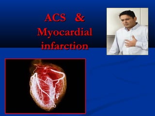
2. American heart association’s definition:American heart association’s definition:
This is group of clinical symptoms compatibleThis is group of clinical symptoms compatible
with acute myocardial ischemia. with acute myocardial ischemia.
Acute myocardial ischemia is chest painAcute myocardial ischemia is chest pain
due to insufficient blood supply to the heartdue to insufficient blood supply to the heart
musclemuscle
that results from coronary artery diseasethat results from coronary artery disease
(also called(also called coronary heartcoronary heart
diseasedisease).).
What is Acute
Coronary Syndrome?
3. Inside a ACS
The plaque deposited in
arteries is hard on the outside
and soft and mushy on the
inside. Sometimes the hard
outer shell cracks.
When this happens, a blood
clot forms around the plaque.
If the clot completely blocks
the artery, it cuts off the blood
supply to a portion of the
heart.
Without immediate treatment,
that part of the heart muscle
could be damaged or
destroyed.
6. Risk Factors ACS
The risk factors of acute coronary syndromeThe risk factors of acute coronary syndrome
involve the followinginvolve the following
AgingAging
Elevated blood pressureElevated blood pressure
Elevated blood cholesterolElevated blood cholesterol
Cigarette smokingCigarette smoking
Sedentary lifestyleSedentary lifestyle
Type 2 diabetesType 2 diabetes
Family history of chest pain, heart disease orFamily history of chest pain, heart disease or
strokestroke
7. PathophysiologyPathophysiology
The plaques inside a narrowed blood vesselThe plaques inside a narrowed blood vessel comes apart,comes apart,
splits, or ulcerate and cause development of thrombus.splits, or ulcerate and cause development of thrombus.
This will lead in unexpectedThis will lead in unexpected full or partial blockage of thefull or partial blockage of the
arteries.arteries.
Systemic issues and swelling can also add toSystemic issues and swelling can also add to changes inchanges in
hemostatic and coagulation pathshemostatic and coagulation paths. Inflammatory acute. Inflammatory acute
stage proteins, cytokines, long-time infections andstage proteins, cytokines, long-time infections and
catecholamine rushes maycatecholamine rushes may improveimprove pro-coagulantpro-coagulant
movement ormovement or thrombocyte hyperaggregability.thrombocyte hyperaggregability.
In any cases it may be brought about by coronary arteryIn any cases it may be brought about by coronary artery
occlusion by emboli, congenital defects, spasm of theocclusion by emboli, congenital defects, spasm of the
heartheart
At first, the infarcted muscle is tone down that willAt first, the infarcted muscle is tone down that will resultresult
in an elevation in the compliance of the ventriclesin an elevation in the compliance of the ventricles,,
however, as scarring happens, compliance reduces.however, as scarring happens, compliance reduces.
8. Therefore,
what is Acute Coronary Syndrome?
Is the acute medical situation that requires an
immediate response
causes rapid and diminished blood circulation towardcauses rapid and diminished blood circulation toward
the heartthe heart
is characterized by a chest pain more than 20 minutes,is characterized by a chest pain more than 20 minutes,
which is not removed two doses nitroglycerinewhich is not removed two doses nitroglycerine
patient requires immediate hospitalizationpatient requires immediate hospitalization
is manageable if confirmed immediately, differ, basedis manageable if confirmed immediately, differ, based
on the patient manifestations and general health status.on the patient manifestations and general health status.
9. What is a possible consequence ofWhat is a possible consequence of
ACS?ACS?
deathdeath
complicationcomplication
recoveryrecovery
What does it
depend?
From the patient'sFrom the patient's
awareness 40%awareness 40% andand competence doctor 60%competence doctor 60%
10. Introduction/Introduction/ classificationclassification
Stable anginaStable angina arise when lumen stenosis < 70%stenosis < 70%→→
impaired blood supply to heart only during on exertion or
increased metabolic demand
Acute coronary syndrome (ACSAcute coronary syndrome (ACS)) arise whenarise when
vessel becomes occluded by thrombusvessel becomes occluded by thrombus
Unstable anginaUnstable angina –– when atherosclerotic plaque shoot ofwhen atherosclerotic plaque shoot of
embolus downstream to cause microinfarctembolus downstream to cause microinfarct
NSTEMINSTEMI –– when necrosis confined to endocardialwhen necrosis confined to endocardial
layers (most susceptible to ischaemialayers (most susceptible to ischaemia))
STEMISTEMI –– when full thickness necrosis of thewhen full thickness necrosis of the
ventricular wall occursventricular wall occurs
11. depends on whether artery shut completely or partially we
have manifestations ACS
12. ST ElevationST Elevation
or new LBBBor new LBBB
STEMISTEMI
Non-specific ECGNon-specific ECG
Unstable AnginaUnstable Angina
ST Depression or dynamicST Depression or dynamic
T wave inversionsT wave inversions
NSTEMINSTEMI
13. ST Depression orST Depression or
dynamicdynamic
T wave inversionsT wave inversions
NSTEMINSTEMI
ST ElevationST Elevation
STEMISTEMI
Q
Acute coronary syndrome (ECG)
14. What we differentiate the manifestations ACS?What we differentiate the manifestations ACS?
Because are different treatment guidelinesBecause are different treatment guidelines
different managing
approaches
Similar pathophysiology,Similar pathophysiology,
presentation and earlypresentation and early
management rulesmanagement rules
STEMI requires evaluation forSTEMI requires evaluation for
acute reperfusion interventionacute reperfusion intervention
Unstable AnginaUnstable Angina
Non-ST-SegmentNon-ST-Segment
Elevation MIElevation MI
(NSTEMI)(NSTEMI)
ST-SegmentST-Segment
Elevation MIElevation MI
(STEMI)(STEMI)
15. Typical angina—All three of the followingTypical angina—All three of the following
Substernal chest discomfort
Onset with exertion or emotional stress
Relief with rest or nitroglycerin
Stable anginaStable angina FCI, FCII,
FCIII, FC I V
Atypical angina
2 of the above criteria
Noncardiac chest pain
1 of the above
16. I. Patients with typical angina
Increased in severity or duration
(FCII→FCIII, FCIV)
Has onset at rest or at a low level of exertion
Unrelieved by the 2 tab of nitroglycerin or rest
comorbidity worsened condition
II. Patients not known to have typical angina
First episode with usual activity or at rest within the
previous two weeks
Prolonged pain at rest
17. WHO criteria's MIWHO criteria's MI
At least 2 of the followingAt least 2 of the following
Ischemic symptomsIschemic symptoms
Diagnostic ECG changesDiagnostic ECG changes
Serum cardiac markerSerum cardiac marker
elevationselevations
18. An ECG can be usedAn ECG can be used
to detect patterns ofto detect patterns of
ischemia
injury
infarction
The ElectrocardiogramThe Electrocardiogram
of ACSof ACS
19. Myocardial ischemiaMyocardial ischemia
ST segment depressions of 2 mm or more for a
duration of 0.08 second may indicate
myocardial ischemia.
Ischemia also should be suspected when a flat
or depressed ST segment makes a sharp angle when
joining an upright T wave rather than merging smoothly
and imperceptibly with the T wave
On the ECG, myocardial ischemia results in T-wave inversion or
ST segment depression in the leads facing the ischemic area.
ST segment
depression
20. Myocardial Injury.
the injury process begins in the subendocardial layer and
moves throughout the thickness of the heart wall and if is
not interrupted, results in a transmural MI.
the hallmark of acute myocardial injury is thethe hallmark of acute myocardial injury is the
presence of ST segment elevations.presence of ST segment elevations.
With an acute injury, the ST segments in the leads facing the
injured area are elevated. The elevated ST segments also
have a downward concave or coved shape and merge
unnoticed with the T wave
In the normal ECG, the ST segment should not beIn the normal ECG, the ST segment should not be
elevated more televated more than 1 mm in the standard leads orhan 1 mm in the standard leads or
more than 2 mm in the precordial leads.more than 2 mm in the precordial leads.
21. Myocardial Injury.
A. ST segment elevation
without T-wave inversion.
B. ST segment elevation with
T-wave inversion.
A B
22. Infarction.Infarction.
When myocardial injury persists, MI is the result.
the earliest stage is thethe earliest stage is the hyperacute phase of MIhyperacute phase of MI
the T waves become tall and narrowthe T waves become tall and narrow ( 5-min-1h)( 5-min-1h)
acute phase of MIacute phase of MI
the ST segments elevatethe ST segments elevate (lasts from several hours to(lasts from several hours to
several days )several days )
In addition to the ST segment elevations in the leads of the ECGIn addition to the ST segment elevations in the leads of the ECG
facing the injured heart, the leads facing away from the injuredfacing the injured heart, the leads facing away from the injured
area may show ST segment depressionarea may show ST segment depression.. This finding is
known as
reciprocal ST segment changes
to be seen at the onset of infarction, may be a mirror image of
the ST segment elevations.
23. Infarction.Infarction.
The last stage in evolutionThe last stage in evolution acute phase of MIacute phase of MI of an MI isof an MI is
development of Q waves, the initial
downward deflection of the QRS complex. (develop
within several hours of the onset of MI)
Q waves compatible with an MI are
0.04 second or more in width or
one-fourth to one-third the height
of the R wave, deeper, than a 4 mm
Q waves represent the flow of electrical forces toward the septum.
Small, narrow Q waves may be seen in the normal ECG in leads I, II,
III, aVR, aVL, V5, and V6.
ST segments elevateST segments elevate
25. Within a few days after the MI is thethe subacute phase of MIsubacute phase of MI
the elevated ST segments return to baseline.
Persistent elevation of the ST segment more than 10 days may
indicate the presence of a ventricular aneurysm.
the T waves may remain inverted for several weeks,
indicating areas of ischemia near the infarct region. Eventually, the T
waves should return to their upright configuration.
After the MI isis thethe scarscar phase of MIphase of MI
The Q waves do not disappear
and therefore always provide ECG
evidence of a previous MI.
MI ECG dyamic
26. A normalA normal
BB hyperacutehyperacute
C –acuteC –acute
D, E – subacuteD, E – subacute
F -F - scarscar
T waves become tall
and narrow
the ST segments elevate
Q waves
evidence of a previous
MI.
downward
deflection
of the QRS
Q waves
normal
27. MI of RV, posterior wall
To attain an accurate view of the right ventricle, right-
sided chest leads are recorded by placing the six chest
electrodes on the right side of the chest using
landmarks analogous to those used on the left
side( Rv1-Rv6)
To detect posterior wall MI, three of the precordial
electrodes are placed posteriorly over the heart, a view
known as V7, V8, V9.
V7 is positioned at the posterior axillary line;
V8 at the posterior scapular line;
V9 at the left border of the spine
28. ST Elevation or
new LBBB
ST Depression or dynamic T
wave inversions
diagnostic algorithm ACSdiagnostic algorithm ACS
ECG
29. RResumeesume
Stable anginaStable angina –– normal ECG, normal troponinnormal ECG, normal troponin
Unstable anginaUnstable angina ––ST depression or dynamic
T wave inversionsee && normal troponin (<1,0)normal troponin (<1,0)
NSTEMINSTEMI –– ST depression or dynamic
T wave inversionse, troponin ↑(>1,0)e, troponin ↑(>1,0)
STEMISTEMI –– elevated ST segment, Q, troponin ↑elevated ST segment, Q, troponin ↑
31. Myocardial infarction (MI)Myocardial infarction (MI)
commonly known as a heart attackcommonly known as a heart attack
results from the interruption of blood supply to a partresults from the interruption of blood supply to a part
of the heart, causing heart cells to die. This is mostof the heart, causing heart cells to die. This is most
commonly due to occlusion (blockage) of a coronarycommonly due to occlusion (blockage) of a coronary
artery following the rupture of a atheroscleroticartery following the rupture of a atherosclerotic
plaqueplaque
The resulting ischemia (restriction in blood supply)The resulting ischemia (restriction in blood supply)
and ensuing oxygen shortage, if left untreated for aand ensuing oxygen shortage, if left untreated for a
sufficient period of time, can cause damage or deathsufficient period of time, can cause damage or death
(infarction) of heart muscle tissue (myocardium).(infarction) of heart muscle tissue (myocardium).
32.
33. ClassificationClassification
based on pathology:
Transmural: associated with atherosclerosis involving a major coronary
artery, extend through the whole thickness of the heart muscle.
On ECG ST elevation and Q waves are seen(QS).
Subendocardial: involving a small area in the subendocardial wall of the left
ventricle, ventricular septum, or papillary muscles,
ST depression is seen on ECG.
based on ECG changes
ST elevation MI (STEMI)
a non-ST elevation MI (non-STEMI) based on ECG
Q wave MIQ wave MI
on Q wave MIon Q wave MI
The phrase heart attack is sometimes used incorrectly to describe
sudden cardiac death, which may or may not be the result of acute
myocardial infarction.
34. The differential diagnosisThe differential diagnosis
The differential diagnosis includes other catastrophicThe differential diagnosis includes other catastrophic
causes of chest pain, such ascauses of chest pain, such as
pulmonary embolism,pulmonary embolism,
aortic ruptureaortic rupture
pericardial effusion causing cardiac tamponadepericardial effusion causing cardiac tamponade
tension pneumothoraxtension pneumothorax
esophageal rupture.esophageal rupture.
gastroesophageal refluxgastroesophageal reflux
Tietze's syndrome.Tietze's syndrome.
35. HISTORYHISTORY
Patients describe a heaviness, squeezing, choking, orPatients describe a heaviness, squeezing, choking, or
smothering sensation. Patients often describe thesmothering sensation. Patients often describe the
sensation as “someone sitting on my chest.”sensation as “someone sitting on my chest.”
The substernal pain canThe substernal pain can radiate to the neck, left arm,radiate to the neck, left arm,
back, or jaw.back, or jaw.
Unlike the pain of angina, theUnlike the pain of angina, the pain of an MI is oftenpain of an MI is often
more prolonged and unrelieved by rest or sublingualmore prolonged and unrelieved by rest or sublingual
nitroglycerin.nitroglycerin.
Associated findings includeAssociated findings include nausea and vomiting,nausea and vomiting, forfor
the patient with anthe patient with an inferior wall MIinferior wall MI..
These gastrointestinal complaints to be related to theThese gastrointestinal complaints to be related to the
severity of the pain and the resulting vagal stimulation.severity of the pain and the resulting vagal stimulation.
36.
37. PHYSICAL EXAMINATIONPHYSICAL EXAMINATION
patients usually appearpatients usually appear restless and in distress.restless and in distress.
TheThe skin is warm and moistskin is warm and moist..
Breathing may be difficulty and rapid.Breathing may be difficulty and rapid. CoarseCoarse
crackles, or rhonchi may be heard when auscultatingcrackles, or rhonchi may be heard when auscultating
the lungs.the lungs.
AnAn increased blood pressure related to anxietyrelated to anxiety oror
a decreased blood pressurea decreased blood pressure caused by heartcaused by heart
failurefailure..
The HR may vary fromThe HR may vary from bradycardia to tachycardia.bradycardia to tachycardia.
When the patient is placed in the left lateral decubitusWhen the patient is placed in the left lateral decubitus
position,position, precordial pulsations can be feltprecordial pulsations can be felt..
38. PHYSICAL EXAMINATIONPHYSICAL EXAMINATION
first heart sound may be diminishedfirst heart sound may be diminished as a result ofas a result of
decreased contractility.decreased contractility.
AA fourth heart sound is heard in almost all patientsis heard in almost all patients
with MI, whereas awith MI, whereas a third heart sound is detectedthird heart sound is detected inin
only about 10% to 20% of patients.only about 10% to 20% of patients.
Transient systolic murmursTransient systolic murmurs may be heardmay be heard
After about 48 to 72 hours, many patients acquire aAfter about 48 to 72 hours, many patients acquire a
pericardial friction rub
Patients with right ventricular infarctsPatients with right ventricular infarcts may presentmay present
with jugular vein distension,with jugular vein distension, peripheral edema, andperipheral edema, and
an elevated central venous pressure.an elevated central venous pressure.
39. InvestigationsInvestigations
Resting ECGResting ECG (on arrival)(on arrival)
Stable angina – normalStable angina – normal
Unstable angina or NSTEMI – ST depression or TUnstable angina or NSTEMI – ST depression or T
wave inversionwave inversion
STEMI – ST elevation → Q wave (permanent) → TSTEMI – ST elevation → Q wave (permanent) → T
wave inversion (in this order)wave inversion (in this order)
Cardiac enzymesCardiac enzymes – Troponin– Troponin,, CK MB/CKCK MB/CK
ratio, AST, LDHratio, AST, LDH
Stable angina and unstable angina – normalStable angina and unstable angina – normal
NSTEMI, STEMI – raisedNSTEMI, STEMI – raised
40. InvestigationsInvestigations
CBC – eukocytosis may beeukocytosis may be
observed within several hoursobserved within several hours
afterafter
urinalysis, coagulation
study – ability to take contrast
and undergo PCI
lipid profile (within 24h)
AST↑/ALT
C- reactive protein is a marker
of acute inflammation
Note: Troponin / CKMB
CKMBCKMB – rise in 4hr, levated– rise in 4hr, levated
for 72hr/ CKMB can befor 72hr/ CKMB can be
used to detect secondused to detect second
infarctsinfarcts
Troponin – rise in 8hr,– rise in 8hr,
elevated for 5 days (elevated for 5 days (trop Itrop I))
and 10 days (and 10 days (trop Ttrop T))
If trop –ve → repeat in 8hr →
last serial trop done 8hr after
sx resolves
41. Cardiac enzymesCardiac enzymes
Creatine Kinase CK-MBCreatine Kinase CK-MB appears in the serumappears in the serum in 6 to 12 hours,in 6 to 12 hours,
peaks between 12 and 28 hours, and returns topeaks between 12 and 28 hours, and returns to normal levels in aboutnormal levels in about
72 to 96 hours72 to 96 hours..
Creatine Kinase IsoformsCreatine Kinase Isoforms:: CK-MB1 is the isoform found in theCK-MB1 is the isoform found in the
plasma, and CK-MB2 is found in the tissuesplasma, and CK-MB2 is found in the tissues
MyoglobinMyoglobin is found in skeletal and cardiac muscle. Myoglobin’s release from ischemicis found in skeletal and cardiac muscle. Myoglobin’s release from ischemic
muscle occursmuscle occurs earlier than the release of CK. The myoglobin level elevate. The myoglobin level elevate within 1within 1
to 2 hours of acute MI and peaks within 3 to 15 hours.to 2 hours of acute MI and peaks within 3 to 15 hours. It ‘It ‘s not specific for the
diagnosis of MI.
Troponin <1,0 normal level
Troponin ITroponin I levelslevels rise in about 3 hours, peak at 14 to 18 hours, andrise in about 3 hours, peak at 14 to 18 hours, and
remain elevated for 5 to 7 days.remain elevated for 5 to 7 days.
Troponin TTroponin T levels rise in 3 to 5 hours and remain elevated for 10 tolevels rise in 3 to 5 hours and remain elevated for 10 to
14 days14 days
43. Testing: EchocardiogramTesting: Echocardiogram
An echocardiogramAn echocardiogram
uses 2-dimentional and Muses 2-dimentional and M
mode echocardiography whenmode echocardiography when
evaluating overallevaluating overall
ventricular functionventricular function
( OVF<52%)( OVF<52%)
wall hypokinesiawall hypokinesia
complications of MIcomplications of MI
(Valvular or pericardial(Valvular or pericardial
effusion, VSD)effusion, VSD)
44. Testing: Chest X-ray, Holter MonitorTesting: Chest X-ray, Holter Monitor
CXR eliminates
aortic dissection, pneumonia,
pneumothorax,
interstitial lung disease
A Holter monitorA Holter monitor
It captures the circadian andIt captures the circadian and
reveals silent ischemia.reveals silent ischemia.
45. Testing:Testing:
Cardiac CTCardiac CT
Cardiac computerizedCardiac computerized
tomographytomography
A cardiac CT can beA cardiac CT can be
used to look for plaqueused to look for plaque
or calcium buildup in theor calcium buildup in the
coronary arteries, heartcoronary arteries, heart
valve problems, andvalve problems, and
other types of heartother types of heart
diseasedisease
46. Testing: Cardiac CatheterizationTesting: Cardiac Catheterization
Cardiac catheterizationCardiac catheterization
helps diagnose and treat somehelps diagnose and treat some
heart conditions.heart conditions.
The doctor guides a narrowThe doctor guides a narrow
tube, called a catheter, through atube, called a catheter, through a
blood vessel in arm or leg untilblood vessel in arm or leg until
it reaches the coronary arteries.it reaches the coronary arteries.
Dye is injected into eachDye is injected into each
coronary artery, making themcoronary artery, making them
easy to see with an X-ray.easy to see with an X-ray.
Treatments such as
angioplasty or stenting can be
done during this procedure.
48. MIs can be located inMIs can be located in
AnteriorAnterior I, V1-V2I, V1-V2
Septal – ApexSeptal – Apex V3-V4V3-V4
LateralLateral I, avL, V5-V6I, avL, V5-V6
PosteriorPosterior V7-V9V7-V9
InferiorInferior ( II, III, avF)( II, III, avF) walls of thewalls of the
left ventricleleft ventricle
Right ventricular wallRight ventricular wall RV1-RV6RV1-RV6
49.
50. Localizing Infarcts on the 12 Lead ECG
Lateral -Anttrior Lateral - Lateral -
Lateral/
Posterior
Anttrior
Inferior - RCA Inferior - RCA
Anttrior
Anttrior
Anttrior septal
Anterior -apex
Anttrior
52. A 2007 consensus document classifiesA 2007 consensus document classifies
myocardial infarction into five mainmyocardial infarction into five main
types:types:
Type 1Type 1 –– Spontaneous myocardial infarction related to ischemiaSpontaneous myocardial infarction related to ischemia
due to adue to a primary coronary eventprimary coronary event
Type 2 –Type 2 – Myocardial infarctionMyocardial infarction secondary to ischemiasecondary to ischemia due todue to
either increased oxygen demand or decreased supplyeither increased oxygen demand or decreased supply
( coronary artery spasm, coronary embolism, anaemia,( coronary artery spasm, coronary embolism, anaemia,
arrhythmias, hypertension, or hypotension)arrhythmias, hypertension, or hypotension)
Type 3 –Type 3 – Sudden unexpected cardiac death,Sudden unexpected cardiac death, includingincluding
cardiac arrest, often with symptoms suggestive ofcardiac arrest, often with symptoms suggestive of
myocardial ischaemia accompanied by new STmyocardial ischaemia accompanied by new ST
elevation, or new LBBB,elevation, or new LBBB,
or evidence of fresh thrombus in a coronary artery byor evidence of fresh thrombus in a coronary artery by
angiography and/or at autopsyangiography and/or at autopsy
53. A 2007 AHA consensus document classifiesA 2007 AHA consensus document classifies
myocardial infarction into five main types:myocardial infarction into five main types:
Type 4Type 4 –– Associated with coronaryAssociated with coronary angioplasty or stents:angioplasty or stents:
Type 4a –Type 4a – Myocardial infarction associated with PCIMyocardial infarction associated with PCI
Type 4b –Type 4b – Myocardial infarction associated with stentMyocardial infarction associated with stent
thrombosisthrombosis as documented by angiography oras documented by angiography or
at autopsyat autopsy
Type 5Type 5 – Myocardial infarction– Myocardial infarction associated with CABGassociated with CABG
54. Heart Attack Symptoms in Women
Women don't always feel chest pain with a
heart attack.
Women are more likely than men to have
heartburn,
loss of appetite,
tiredness or weakness,
coughing,
heart flutters.
These symptoms should not be ignored.
55. Electrocardiographic evolution
of myocardial infarction.
After the first few minutes the
T waves become tall, pointed and
upright and ST segment elevation
develops.
After the first few hours the
T waves invert, the R wave voltage is
decreased and Q waves develop.
After a few days the
ST segment returns to normal.
After weeks or months the
T wave may return to upright but the
Q wave remains.
56. Differential MI ECG patternDifferential MI ECG pattern
WPWWPW negative Δ wave may mimic pathologic Q waveswave may mimic pathologic Q waves
IHSSIHSS septal hypertrophy may make Q(V3) "fatter" therebyseptal hypertrophy may make Q(V3) "fatter" thereby
mimicking pathologic Qmimicking pathologic Q
LVHLVH may have QS pattern or low R in V1-V3may have QS pattern or low R in V1-V3
RVHRVH tall in V1 or V2 may mimic true posterior MItall in V1 or V2 may mimic true posterior MI
Complete / incomplete LBBBComplete / incomplete LBBB QS waves or low R wave in V1-V3QS waves or low R wave in V1-V3
PneumothoraxPneumothorax loss of right precordial R wavesloss of right precordial R waves
Pulmonary emphysema and cor pulmonalePulmonary emphysema and cor pulmonale loss of R waves V1-3 andloss of R waves V1-3 and
or inferior Q waves with right axis deviationor inferior Q waves with right axis deviation
Left anterior fascicular blockLeft anterior fascicular block may see small q-waves in V1-V4 leadsmay see small q-waves in V1-V4 leads
Acute pericarditisAcute pericarditis the ST segment elevation may mimic acute STEMIthe ST segment elevation may mimic acute STEMI
Central nervous system diseaseCentral nervous system disease may mimic non-Q wave MI bymay mimic non-Q wave MI by
causing diffuse ST-T wave changescausing diffuse ST-T wave changes
Pancreatitis, bleedingPancreatitis, bleeding ischemic changes inferior lateral zoneischemic changes inferior lateral zone
60. Initial evaluation &Initial evaluation &
stabilizationstabilization
Assessment risk stratificationAssessment risk stratification
Immediate cardiac careImmediate cardiac care
Criteria for thrombolysis or PCI (i.e. STEMI)Criteria for thrombolysis or PCI (i.e. STEMI)
> 2mm elevation ST in 2 contiguous precordial leads
New onset LBBB
61. Risk Stratification
Risk stratification ofRisk stratification of NSTE ACSNSTE ACS ––
““HEART DOC”HEART DOC”
HHaemodynaic compromiseaemodynaic compromise
EECG changes ArrhythmiaCG changes Arrhythmia
RRenal failureenal failure
TTroponin riseroponin rise
DDiabetes mellitusiabetes mellitus
OOngoing chest painngoing chest pain
CCardiac bypass anytime or PCI in last 6monthsardiac bypass anytime or PCI in last 6months
Having 1 of these → high risk group
62. Risk StratificationRisk Stratification
TIMI, ScoreTIMI, Score
HistoricalHistorical “Para Sea”“Para Sea”
PPHx – known CAD (stenosis ≥ 50%)Hx – known CAD (stenosis ≥ 50%)
AAge>65ge>65
≥≥33 RRFs for CADFs for CAD
AAspirin use in past 7dspirin use in past 7d
PresentationPresentation
SST segment deviation ≥0.5mmT segment deviation ≥0.5mm
↑↑cardiaccardiac eenzymesnzymes
Recent (≤24hr) severeRecent (≤24hr) severe AnginaAngina
63. UA and NSTEMIUA and NSTEMI
Stabilize acute coronary lesionStabilize acute coronary lesion
Anti-plateletAnti-platelet (aspirin and clopidogrel ± GPIIb/IIIa(aspirin and clopidogrel ± GPIIb/IIIa
inhibitor)inhibitor)
Anti-thrombinAnti-thrombin (UFH or LMWH)(UFH or LMWH)
Anti-ischaemiaAnti-ischaemia (β-blocker if CI then CCB, consider(β-blocker if CI then CCB, consider
nitrates, morphine, ACE)nitrates, morphine, ACE)
High riskHigh risk – urgent angiography ± PCI– urgent angiography ± PCI
Low riskLow risk – arrange stress tests– arrange stress tests
Subsequent managementSubsequent management (start during this hospital admission)(start during this hospital admission)
Statins, aspirin and clopidogrel, ACEIStatins, aspirin and clopidogrel, ACEI (or ARB)(or ARB), β-, β-
blockerblocker (if CI then CCB)(if CI then CCB)
Nitrates PRNNitrates PRN
Cardiac rehabilitationCardiac rehabilitation
64.
65. EARLY MANAGEMENTEARLY MANAGEMENT STEMISTEMI
STEP 1STEP 1
After recording the initial 12-lead ECG, place the patientAfter recording the initial 12-lead ECG, place the patient
on a cardiac monitor and obtain serial ECGs.on a cardiac monitor and obtain serial ECGs.
Oxygen therapyOxygen therapy ggived by nasal cannulaived by nasal cannula
GTNGTN (½ sublingual tab)(½ sublingual tab)
AspirinAspirin 325mg325mg
IV MorphineIV Morphine 2.5~5mg + IV2.5~5mg + IV MetoclopramideMetoclopramide
10mg10mg
Monitor oximetry, BP, continuous ECGMonitor oximetry, BP, continuous ECG
66. STEP 2STEP 2
Treated with reperfusion therapy(RTTreated with reperfusion therapy(RT))
such as percutaneous coronary intervention (PCI) or thrombolysissuch as percutaneous coronary intervention (PCI) or thrombolysis
Immediate PCI or fibrinolytic therapy!!!Immediate PCI or fibrinolytic therapy!!!
PCI has higher reperfusion rate and is better if pt
present > 1hr , but
Thrombolysis is gold standard if pt arrive within an
12hrs of onset of MI12hrs of onset of MI
Antiplatelet therapyAntiplatelet therapy AAspirin and Clopidogrelspirin and Clopidogrel
GPIIb/IIIa inhibitor (GPIIb/IIIa inhibitor (AbciximabAbciximab,, EptifibatideEptifibatide TirofibanTirofiban
0,25mgkg iv 12- 24hs0,25mgkg iv 12- 24hs
Anticoagulation agentAnticoagulation agent unfractionated heparin or LMWHunfractionated heparin or LMWH
67. STEP 3STEP 3
Subsequent managementSubsequent management
Statins, aspirin and clopidogrel,Statins, aspirin and clopidogrel,
ACEIACEI (or ARB)(or ARB),,
β-blockerβ-blocker (if CI then CCB)(if CI then CCB)
Anticoagulation therapyAnticoagulation therapy to prevent thromboembolism (to prevent thromboembolism (warfarinwarfarin
for 6mos if large anterior MIfor 6mos if large anterior MI, esp if, esp if echoecho show largeshow large
akinetic/dyskinetic area, aneurysm or mural thrombus)akinetic/dyskinetic area, aneurysm or mural thrombus)
Nitrates PRNNitrates PRN
Cardiac rehabilitationCardiac rehabilitation
Antiplatelet post stentAntiplatelet post stent
AspirinAspirin for lifefor life
ClopidogreClopidogrel for at least 6wks for metal stentl for at least 6wks for metal stent
ClopidogrelClopidogrel for at least 12mos for drug eluting stentfor at least 12mos for drug eluting stent
Drug eluting stent have lower early re-stenosis rate,
but metal stent however have a problem of late thrombosis
68. Don't Wait to Be SureDon't Wait to Be Sure
The best time to treat a heartThe best time to treat a heart
attack is as soon as symptomsattack is as soon as symptoms
begin. Waiting to be sure canbegin. Waiting to be sure can
result in permanent heartresult in permanent heart
damage or even death.damage or even death.
Don't try driving yourselfDon't try driving yourself
to the hospitalto the hospital..
When you call 911, the EMSWhen you call 911, the EMS
staff can start emergency care asstaff can start emergency care as
soon as they reach you.soon as they reach you.
69. Options for Transport of Patients WithOptions for Transport of Patients With
STEMI and Initial Reperfusion TreatmentSTEMI and Initial Reperfusion Treatment
EMS Transport
Onset of
symptoms of
STEMI
9-1-1
EMS
Dispatch
EMS on-scene
• Encourage 12-lead ECGs.
• Consider prehospital fibrinolytic if
capable and EMS-to-needle within
30 min.
GOALS
PCI
capable
Not PCI
capable
Hospital fibrinolysis:
Door-to-Needle
within 30 min.
EMS
Triage
Plan
Inter-
Hospital
Transfer
Golden Hour = first 60 min. Total ischemic time: within 120 min.
Patient EMS Prehospital fibrinolysis
EMS-to-needle
within 30 min.
EMS transport
EMS-to-balloon within 90 min.
Patient self-transport
Hospital door-to-balloon
within 90 min.
Dispatch
1 min.
5
min.
8
min.
70. It should be managed in a manner similarIt should be managed in a manner similar
to other patients with acute coronaryto other patients with acute coronary
syndromesyndrome except beta blockersexcept beta blockers
shouldshould not be used benzodiazepinesnot be used benzodiazepines
should be administered early.should be administered early.
71. MorphineMorphine MORPHINI HYDROCHLORIDUM 1% - 1,0MORPHINI HYDROCHLORIDUM 1% - 1,0
Analgesia. Reduce pain/anxiety—decrease sympatheticAnalgesia. Reduce pain/anxiety—decrease sympathetic
tone, systemic vascular resistance and oxygen demandtone, systemic vascular resistance and oxygen demand
OxygenOxygen
Up to 70% of ACS patient demonstrate hypoxemia. May limitUp to 70% of ACS patient demonstrate hypoxemia. May limit
ischemic myocardial damage by increasing oxygen delivery/reduceischemic myocardial damage by increasing oxygen delivery/reduce
ST elevationST elevation
A 2009 and 2010 review of high flow oxygen in myocardial infarction found increasedA 2009 and 2010 review of high flow oxygen in myocardial infarction found increased
mortality and infarct size, calling into question the recommendation about its routinemortality and infarct size, calling into question the recommendation about its routine
use.use.
72. NitroglycerinNitroglycerin 0,5mg every 5 min up to 4 SL0,5mg every 5 min up to 4 SL
Sublingual, oral or intravenousSublingual, oral or intravenous
Dilates coronary vessels—increase blood flowDilates coronary vessels—increase blood flow
Reduces systemic vascular resistance and preloadReduces systemic vascular resistance and preload
Pharmacokinetics:Pharmacokinetics:
well awell absorbed from buccal mucosa, intestine, skin and alveoli.bsorbed from buccal mucosa, intestine, skin and alveoli.
Rapid onset of action (few minutes)Rapid onset of action (few minutes)
Short duration (15-30 min)Short duration (15-30 min)
Mechanism of action:Mechanism of action:
When metabolizedWhen metabolized donates three Nomoleculesdonates three Nomolecules NONO
stimulates guanylyl cyclasestimulates guanylyl cyclase increases cGMPincreases cGMP
Dephosphorylate MLCDephosphorylate MLC SmoothSmooth musclemuscle RelaxationRelaxation
& Vasodialation.& Vasodialation.
73. NitroglycerinNitroglycerin
The first line treatment for symptomatic relief of angina is
sub-lingual nitroglycerin.
Nitroglycerin should not be given if any phosphodiesteraseNitroglycerin should not be given if any phosphodiesterase
type 5 inhibitors such as Viagra (type 5 inhibitors such as Viagra ( Cialis, Stondra, andCialis, Stondra, and
Levitra) have been taken by the casualty within theLevitra) have been taken by the casualty within the
previous 24-48 hours, as the combination of the two couldprevious 24-48 hours, as the combination of the two could
cause a serious drop in blood pressure.cause a serious drop in blood pressure.
It should not be given to patients with systolic bloodIt should not be given to patients with systolic blood
pressure (SBP) less than 90mmHgpressure (SBP) less than 90mmHg
74. Arterial V.D decreasing the after-load (blood pressure) and the
ejection time.
Vento-dilatation decreasing the pre-load and the end diastolic
volume.
& the myocardial oxygen consumption decreases but there is
reflex increase of HR and contractility which could be avoided
by adding beta-blockers.
Nitrats: Pharmacological Effects
Decrease oxygen demand by
Decreasing HR, contractility, cardiac work.
Increase oxygen supply by:
Increasing diastolic coronary perfusion time.
Shifting of subepicardial blood flow to subendocardial flow. Inhibition of
platelet aggregation.
Shifting of oxygen hemoglobin dissociation curve to right i.e more oxygen
delivery to tissue.
75. NitroglycerineNitroglycerine 1,0- 0, 1% in 200,0 5% GS iv 2-17 drops in a minute1,0- 0, 1% in 200,0 5% GS iv 2-17 drops in a minute
ISOSOIRBIT DININRAT (Isoket)ISOSOIRBIT DININRAT (Isoket) 0,1% - 10,0 - in0,1% - 10,0 - in
200,0- 5% GS /Nch-0,9% 2-17 drops in a minute200,0- 5% GS /Nch-0,9% 2-17 drops in a minute
Aerosol form of nitroglycerinAerosol form of nitroglycerin (nitrosprey-ICN, nitromint,(nitrosprey-ICN, nitromint,
Nitroglycerin patchesNitroglycerin patches:: deponit, nitroderm, nitrodisk, nitrodurdeponit, nitroderm, nitrodisk, nitrodur
Isosorbst monitratIsosorbst monitrat monosan , mononitrosid 5mgmonosan , mononitrosid 5mg
Isosorbst dinitratIsosorbst dinitrat nitrorsorbid, Isoket, Isonit, Isorbid izodinit 10 mg,nitrorsorbid, Isoket, Isonit, Isorbid izodinit 10 mg,
isomak, isoket kardiket 20 mg.isomak, isoket kardiket 20 mg.
Preparations with prolonged action:Preparations with prolonged action: 6-24 h6-24 h
izoket retard or cardiket retard tablets 20, 40, 60, 80 120 mgizoket retard or cardiket retard tablets 20, 40, 60, 80 120 mg
izomak retard capsules of 20, 40, 60 and 120 mg,izomak retard capsules of 20, 40, 60 and 120 mg,
MolsidominMolsidomin 00,002-0,004,002-0,004 (korvaton, sidnofarm) differs from nitrate has(korvaton, sidnofarm) differs from nitrate has
vasodilating given to patients who can not tolerate nitratesvasodilating given to patients who can not tolerate nitrates
76. • Meningeal arteries V.D produces
temporal artery pulsation and
throbbing headache
• The venodilatation may produce
orthostatic hypotension and
syncope.
• Increased cGMP inhibits platelet
aggregation.
• Other organs: insignificant
relaxation of bronchi, GIT, &
genitourinary tract smooth muscles.
SIDE Efects of Nitroglycerins:
Adverse Effects:
Postural hypotension,
dizziness and weakness.
Tachycardia, headache
and flushing.
Methemoglobinemia
and cyanosis (common
with nitrites).
Allergic reactions like
skin rashes.
Tolerance fromfrom
prolonged useprolonged use
Sudden stop of nitratesSudden stop of nitrates
may produce MImay produce MI
77. Reperfusion therapyReperfusion therapy
The reperfusion has become soThe reperfusion has become so
central to the modern treatmentcentral to the modern treatment
of MIof MI
Patients who present withPatients who present with
suspectedsuspected acute MIacute MI andand STEMISTEMI
oror new bundle branch blocknew bundle branch block..
They are therefore candidatesThey are therefore candidates
forfor immediate reperfusionimmediate reperfusion,,
either witheither with
thrombolytic therapythrombolytic therapy
percutaneous coronarypercutaneous coronary
intervention (PCI)intervention (PCI)
or when these therapies areor when these therapies are
unsuccessful,unsuccessful, bypass surgerybypass surgery..
Some commonly usedSome commonly used
thrombolytics are:thrombolytics are:
streptokinasestreptokinase
urokinaseurokinase
RecombinantRecombinant
tissuetissue
plasminogenplasminogen
activatorsactivators
alteplase (rtPA)alteplase (rtPA)
reteplasereteplase
tenecteplasetenecteplase
78. Mechanism:
theythey activate the conversion ofactivate the conversion of
plasminogen to plasmin,plasminogen to plasmin, which in turnwhich in turn
converts fibrin into soluble products.converts fibrin into soluble products.
PlasminogenPlasminogen plasminplasmin
FibrinFibrin soluble productsoluble product
79. NB:NB:
Streptokinase, anistreplaseStreptokinase, anistreplase andand urokinaseurokinase areare
non fibrin-selectivenon fibrin-selective i.e. they activate plasminogeni.e. they activate plasminogen
in thrombi & circulation. So, they increase the riskin thrombi & circulation. So, they increase the risk
of bleeding.of bleeding.
Pro-urokinasePro-urokinase andand altiplasealtiplase areare fibrin-selectivefibrin-selective
i.e. their action is more localized on thei.e. their action is more localized on the
plasminogen of the thrombi and less likely toplasminogen of the thrombi and less likely to
produce bleeding.produce bleeding.
80. DrugDrug SourceSource
(min.)(min.)
tt ½½
Min.Min.
Adverse EffectAdverse Effect
StreptokinaseStreptokinase
1,5 mln u sn 200,01,5 mln u sn 200,0
GS/Ncl 30-60minGS/Ncl 30-60min
AnistreplaseAnistreplase
(APSAC)(APSAC)
Beta-Beta-
hemolytichemolytic
streptococcistreptococci
Pro-drugPro-drug
releasesreleases
streptokinastreptokina
se slowlyse slowly
2020
7070
Allergy,Allergy,
Anaphylactic reactionAnaphylactic reaction
Rapid injection causesRapid injection causes
abrupt hypotension.abrupt hypotension.
As streptokinaseAs streptokinase
81. DrugDrug SourceSource t ½t ½
Min.Min.
AdverseAdverse
EffectsEffects
Tenekteplasa (TNKr t-PA )
singl bolus10mg-/10kg 5 min
RecombinaRecombina
nt DNAnt DNA
technologytechnology
55
No allergy,No allergy,
but morebut more
expensiveexpensive
Reteplasa (r t-PA) 10u iv + 10u
given 30min
RecombinaRecombina
nt DNAnt DNA
technologytechnology
77
Alteplase (r t-PA)
15mg iv+75mg for30min
RecombebiRecombebi
nant DNAnant DNA
technologytechnology
2-102-10
83. AbsoluteAbsolute
Previous intracranial bleeding at any time,Previous intracranial bleeding at any time,
stroke in less than 6 months,stroke in less than 6 months,
suspected aortic dissection,suspected aortic dissection,
ischemic stroke within 3 months (except in ischemic strokeischemic stroke within 3 months (except in ischemic stroke
within 3 hours time)within 3 hours time)
active bleeding diathesis,active bleeding diathesis,
uncontrolled high blood pressure (>180 systolic or >100uncontrolled high blood pressure (>180 systolic or >100
diastolic),diastolic),
known structural cerebral vascular lesion, aneurysm, brainknown structural cerebral vascular lesion, aneurysm, brain
tumors,tumors,
Current use of anticoagulants in therapeutic doses
(international normalized ratio [INR] ≥2:3);
Recent trauma (within 2–4 weeks), including head trauma or
traumatic or prolonged (>10 minutes
arterio-venous malformations,arterio-venous malformations,
thrombocytopenia,thrombocytopenia,
known cogulation disorders,known cogulation disorders,
pericardial effusion.pericardial effusion.
84. RelativeRelative
Current anticoagulant use,Current anticoagulant use,
Invasive or surgical procedure in the last 2 weeks,Invasive or surgical procedure in the last 2 weeks,
Noncompressible vascular puncturesNoncompressible vascular punctures
Prolonged cardiopulmonary resuscitation (CPR) defined as more thanProlonged cardiopulmonary resuscitation (CPR) defined as more than
10 minutes,10 minutes, or major surgery (<3 weeks)
Arrhythmias.
Known bleeding diathesisKnown bleeding diathesis
PregnancyPregnancy
Hypotension.
For streptokinase : prior exposure (especially within 5 days to 2 years)
or prior allergic reaction
Hemorrhagic or diabetic retinopathies,Hemorrhagic or diabetic retinopathies,
Active peptic ulcer,Active peptic ulcer,
Controlled severe hypertensionControlled severe hypertension
85. Aspirin 75 - 100 –325 mgAspirin 75 - 100 –325 mg
Irreversible inhibition of platelet aggregationIrreversible inhibition of platelet aggregation
Stabilize plaque and arrest thrombusStabilize plaque and arrest thrombus
Reduce mortalityReduce mortality in patients with STEMIin patients with STEMI
Beta-BlockersBeta-Blockers
Reduce mortalityReduce mortality and of progression to MI in patientsand of progression to MI in patients
with threatening or evolving MI symptomwith threatening or evolving MI symptom
Atenolol 25mg, Metoprolol 50mg, Bisaprolol 5mg
are probably the drugs of choiceare probably the drugs of choice
Contraindications (Asthma & Hypotension)Contraindications (Asthma & Hypotension)
Used for prophylaxis of anginaUsed for prophylaxis of angina Except variant anginaExcept variant angina
86. ACE-Inhibitors / ARBACE-Inhibitors / ARB
Used in patients with anterior MI, pulmonary congestion.It’s decreaseUsed in patients with anterior MI, pulmonary congestion.It’s decrease
ventricular remodelling post-MIventricular remodelling post-MI
Start in first 24 hoursARB as substitute for patients unable to use ACE-Start in first 24 hoursARB as substitute for patients unable to use ACE-
I.I.
HeparinHeparin 1000/1h – 24000/ 24hs iv in infusomath1000/1h – 24000/ 24hs iv in infusomath
or 12 500sc/12hs, or 5000 sc /5hor 12 500sc/12hs, or 5000 sc /5h
control is a test of blood on PATT, increases twice
LMWHLMWH ((EnoxaparinEnoxaparin)) 2,5-5,0u s.c or i.v once/day2,5-5,0u s.c or i.v once/day..
It inhibits activated factor X.It inhibits activated factor X.
It is given 24-48 hours of treatment. Used in combo with aspirin and/orIt is given 24-48 hours of treatment. Used in combo with aspirin and/or
other platelet inhibitorsother platelet inhibitors
87. ClopidodrelClopidodrel 75 mg75 mg
Irreversible inhibition of platelet aggregation by ADF receptorsIrreversible inhibition of platelet aggregation by ADF receptors
Used in support of cath / PCI intervention or if unable to takeUsed in support of cath / PCI intervention or if unable to take
aspirin 3 to 12 month duration depending on scenarioaspirin 3 to 12 month duration depending on scenario
Glycoprotein IIb/IIIa inhibitorsGlycoprotein IIb/IIIa inhibitors ((AbciximabAbciximab))
0.25 mg/kg intravenous bolus administered 10-60 minutes before the start of PCI, followed by a
continuous intravenous infusion of 0.125 µg/kg/min (to a maximum of 10 µg/min) for 12 hours
inhibit theinhibit the fibrinogenfibrinogen-mediated cross linkage of platelets, which-mediated cross linkage of platelets, which
is the final common pathway of platelet aggregation.is the final common pathway of platelet aggregation.
Calcium antagonistCalcium antagonist (diltiazem 90-120mg, verapamil 60-(diltiazem 90-120mg, verapamil 60-
120mg)120mg) Are used for patients who cannot tolerate a beta-Are used for patients who cannot tolerate a beta-
blocker or in addition to a beta-blocker.blocker or in addition to a beta-blocker.
Verapamil should not be combined with a beta-blockerVerapamil should not be combined with a beta-blocker..
88. Therapy aspirin and/or clopidogrel should beTherapy aspirin and/or clopidogrel should be
continued to reduce the risk of plaque rupture.continued to reduce the risk of plaque rupture.
AspirinAspirin is first-line, owing to its low cost andis first-line, owing to its low cost and
comparable efficacy,comparable efficacy,
ClopidogrelClopidogrel reserved for patients intolerant ofreserved for patients intolerant of
aspirin.aspirin.
The combination of clopidogrel and aspirin mayThe combination of clopidogrel and aspirin may
further reduce risk of cardiovascular events, however thefurther reduce risk of cardiovascular events, however the
risk of hemorrhage is increasedrisk of hemorrhage is increased
Antiplatelet drugAntiplatelet drug
89.
90. ASPIRINASPIRIN
U.S. guidelines recommendU.S. guidelines recommend
a dose of 162–325 mg.a dose of 162–325 mg.
Australian guidelines recommend aAustralian guidelines recommend a
dose of 150–300 mgdose of 150–300 mg
Aspirin:Aspirin:
AspirinAspirin irreversibly inhibit COXirreversibly inhibit COX (up to the life-time of the(up to the life-time of the
platelets 8-10 days).platelets 8-10 days).
Both PGI2 and TXA2 synthesis are inhibited.Both PGI2 and TXA2 synthesis are inhibited.
Aspirin inAspirin in small dose 75-100 mg/daysmall dose 75-100 mg/day inhibits TXA2inhibits TXA2
synthesis without significant effect on the endothelialsynthesis without significant effect on the endothelial
PGI2.PGI2.
ToxicityToxicity: peptic ulcer: peptic ulcer bleeding in patients > 60 years.bleeding in patients > 60 years.Prof. M. Adel
91. ClopidodrelClopidodrel
7575 -150 -300 mg
It is a thienopyridine derivative.It is a thienopyridine derivative.
It irreversiblyIt irreversibly inhibits the P2Y12 purinergic receptorinhibits the P2Y12 purinergic receptor onon
platelets, there by blockingplatelets, there by blocking ADP-mediated plateletADP-mediated platelet
activation.activation.
Usually given in combination with aspirin because ofUsually given in combination with aspirin because of
their complementary mechanism of action.their complementary mechanism of action. It howeverIt however
may worsen outcomes in those who need urgent coronary arterymay worsen outcomes in those who need urgent coronary artery
bypass surgerybypass surgery
ToxicityToxicity : bleeding, dyspepsia, and rashes.: bleeding, dyspepsia, and rashes.
92. Myocardial Energy MetabolismMyocardial Energy Metabolism
RegulatorRegulator
Mildronate 10%-Mildronate 10%- 5,5,0 acts as a myocardial energy metabolism0 acts as a myocardial energy metabolism
regulator by inhibiting fatty acid oxidationregulator by inhibiting fatty acid oxidation
Covitin (blocker leukotrienes) 0,5mg ivCovitin (blocker leukotrienes) 0,5mg iv is antioxidant, anti-is antioxidant, anti-
inflammatory vasodilationinflammatory vasodilation
Riboxin 20,0-2% ( inosin) ivRiboxin 20,0-2% ( inosin) iv increases energy reserves heartincreases energy reserves heart
Thiotriazolinum 2,5% 2,0Thiotriazolinum 2,5% 2,0 -- Protective cellular metabolicProtective cellular metabolic
antioxidant effectantioxidant effect
TrimetazidineTrimetazidine (PREDUCTAL MR(PREDUCTAL MR) 35mg) 35mg improves energyimproves energy
metabolism in the heart due to the partial suppression of fattymetabolism in the heart due to the partial suppression of fatty
acid oxidation This leads to increased glucose oxidationacid oxidation This leads to increased glucose oxidation
Mexycor 50 mg / ml. 2 mlMexycor 50 mg / ml. 2 ml -- Protective cellular metabolicProtective cellular metabolic
antioxidant effectantioxidant effect
93. SSurgical turgical treatment: Angioplastyreatment: Angioplasty
Angioplasty is used to open aAngioplasty is used to open a
blocked heart artery andblocked heart artery and
improve blood flow to theimprove blood flow to the
heart. The doctor inserts a thinheart. The doctor inserts a thin
catheter with a balloon on thecatheter with a balloon on the
end into the artery. When theend into the artery. When the
balloon reaches the blockage, itballoon reaches the blockage, it
is expanded, opening up theis expanded, opening up the
artery and improving bloodartery and improving blood
flow. The doctor may alsoflow. The doctor may also
insert a small mesh tube, calledinsert a small mesh tube, called
a stent, to help keep the arterya stent, to help keep the artery
open after angioplasty.open after angioplasty.
94. Stenosises of LCA and circumflex branch of the left coronal artery. 2
еluting stents of TAXUS(CYPER) is implante
Before a stenting
Then a stenting
95. Bypass SurgeryBypass Surgery
Bypass surgery is anotherBypass surgery is another
way to improve the heart'sway to improve the heart's
blood flow.blood flow.
It gives blood a newIt gives blood a new
pathway when the coronarypathway when the coronary
arteriesarteries have become toohave become too
narrow or blockednarrow or blocked. During. During
the surgery, athe surgery, a blood vessel is
first moved from one area
of the body -- such as the
chest, legs, or arms -- and
attached to the blocked
artery, allowing it to
bypass the blocked part.
96. RehabilitationRehabilitation
Physical exercise is an important part of rehabilitation after a ACS
the advice is a gradual increase in physical exercise during about
6–8 weeks following an MI and exercise is at least 20–30 minutes
of moderate exercise on most days to the extent of getting slightly
short of breath..
Most people can resume sexual activities after 3 to 4 weeks. The
amount of activity needs to be dosed to the patient's possibilities.
100. Sudden Cardiac DeathSudden Cardiac Death
SCDSCD occurs when the heart'soccurs when the heart's
electrical system goeselectrical system goes
haywire, causing it to beathaywire, causing it to beat
irregularly and dangerouslyirregularly and dangerously
fast. The heart's pumpingfast. The heart's pumping
chambers may quiver insteadchambers may quiver instead
of pumping blood out to theof pumping blood out to the
body. Without CPR andbody. Without CPR and
restoration of a regular heartrestoration of a regular heart
rhythm, death can occur inrhythm, death can occur in
minutes.minutes.
ventricles fibrilation
101. Primary prevention ofPrimary prevention of atherosclerosisatherosclerosis by:by:
Healthy eating.Healthy eating.
Exercise.Exercise.
Treatment forTreatment for hypertensionhypertension andand diabetesdiabetes..
AvoidingAvoiding smokingsmoking..
ControllingControlling cholesterolcholesterol levels); in patients withlevels); in patients with
significant risk factors,significant risk factors,
AAspirinspirin has been shown to reduce the risk ofhas been shown to reduce the risk of
cardiovascular events.cardiovascular events.
102. Life After a Heart AttackLife After a Heart Attack
lastslasts
thank you for attentionthank you for attention