Foramen Magnum Meningioma
•Télécharger en tant que PPTX, PDF•
19 j'aime•3,247 vues

Foramen Magnum Meningioma: Presentation, Diagnosis & Treatment

Recommandé
Contenu connexe
Tendances
Tendances (20)
Similaire à Foramen Magnum Meningioma
Similaire à Foramen Magnum Meningioma (20)
Plus de Farrukh Javeed
Plus de Farrukh Javeed (19)
Dernier
Dernier (20)
Foramen Magnum Meningioma
- 1. F A R R U K H J A V E D FORAMEN MAGNUM MENINGIOMA
- 2. ANATOMY The occipital bone surrounds the foramen magnum and is composed of two parts: the posterior squamosal and the narrower anterior part (basal extension of the clivus).
- 3. ANATOMY The foramen magnum (FM) comprises a bony channel formed: anteriorly by the lower third of the clivus, the anterior arch of the atlas, and the odontoid process. the lateral limits are the jugular tubercle (JT), the occipital condyle (OC), and the lateral mass of the atlas. Lastly, the FM is limited posteriorly by the lower part of the occipital bone, the posterior arch of the atlas, and the two first intervertebral spaces.
- 4. ANATOMY Oval shaped, wider posteriorly than anteriorly The contents of the foramen magnum consist of: the vertebral arteries (VAs) and their meningeal branches the anterior and posterior spinal arteries the lower part of the medulla the lower cranial nerves (IX, X, and XI) the roots of the C1 and C2 vertebrae. the cervicomedullary junction the cerebellar tonsils, the inferior vermis and the fourth ventricle. surrounded by veins, venous sinuses, and the jugular bulb.
- 7. EPIDEMIOLOGY account for only 0.3–3.2% of all meningiomas and between 4% and 15% of all posterior fossa meningiomas altogether. they are slow growing tumors. the mean length of symptoms before diagnosis is 30.8 months.
- 8. CLINICAL FEATURES The clinical presentation is insidious. Earlier complains are Occipital headache and cervical pain. This pain is described as deep and is aggravated by neck motion, coughing, and straining. As the tumor grows, sensory and motor deficits develop
- 9. There is an asymmetrical deficit defined by weakness, paresthesis, and spasticity, first in the ipsilateral arm and progressing to the ipsilateral leg, then to the contralateral leg, and finally to the contralateral arm. Long tract signs of upper-motor lesions are the presence of atrophy in the intrinsic muscles of the hands. Later findings include spastic quadriparesis, respiratory dysfunction and lower cranial nerve deficits.
- 10. CLASSIFICATION Among the many classifications of meningiomas of the FM,6-8 the one most frequently used by neurosurgeons is the classification from Bruneau and George. The main objective of this system is to define the surgical strategy preoperatively
- 11. Bruneau and George Classification Based on this classification, meningiomas of the FM are classified as intradural, extradural intra- and extradural.
- 12. According to their insertion on the dura, meningiomas are: anterior if insertion happens on both sides of the anterior midline, anterolateral if insertion occurs between the midline and the dentate ligament or posterior if insertion is posterior to the dentate ligament
- 13. The other landmark used for classification is the relation to the Vertebral Arteries. Meningiomas of the Foramen Magnum can develop: above the vertebral arteries below the vertebral arteries on both sides of the vertebral arteries
- 15. Intradural meningiomas are the most common type most of them arise anterolaterally, these are followed in frequency by posterolateral tumors. Tumors that arise purely posteriorly and anteriorly are rare.
- 16. DIAGNOSIS
- 17. All patients should undergo a detailed study of their neurological function, independent of their clinical neurological examination. Preoperative imaging studies allow for planning of the surgery
- 18. the following information must be retrieved from the radiology: the nature of the tumor (intra- and/or extradural) its location and attachment its relationship with the cervicomedullary junction its caudal and rostral extension the position and possible involvement of the VAs and their branches the shape of the FM the dominance of the VAs the venous drainage patterns and dominance bony involvement
- 19. MRI T1-weighted MRI with contrast enhancement clearly defines the tumor and the dural attachment site and discriminates between the tumor and the brain stem. T2-weighted MRI provides information on the arachnoid plane between the tumor and the cervicomedullary junction
- 20. CT SCAN CT using sagittal, coronal, and axial viewing and bone window remains the tool of choice for the study of: bone involvement the shape of the FM the surgical corridor
- 21. Angiography Should be considered in all patients with suspected foramen magnum meningioma to demonstrate the vascular supply of the neoplasm define the position of major vessels with respect to the tumor determine the venous drainage of the posterior fossa eliminate the possibility of an aneurysm of the posterior circulation
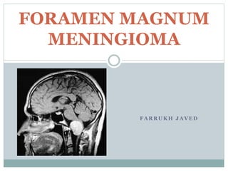
- 22. MRI sagital section MRI T1:a large anterior foramen magnum meningioma isointense to surrounding brain severely compresses the neuraxis MRI T2: pocess hyperintense to surrounding brain
- 23. homogeneously enhancing tumor arises predominantly in an anterior location
- 24. MRI. Sagittal T2
- 25. Contrast-enhanced T1-weighted MRI scans
- 26. INTRAOPERATIVE MONITORING Somatosensory evoked potentials auditory evoked responses facial nerve monitoring monitoring of the X, XI, and XII cranial nerves
- 27. Surgical Corridor the space between the cervicomedullary junction and the lateral wall of the FM.
- 28. Surgical approach the location of the tumor the extent of the tumor (above the foramen magnum) the relation of the tumor with the vertebral artery and with the origin of posterior inferior cerebellar artery
- 29. SURGICAL APPROACHES Three general approaches to the foramen magnum are employed: o Posterior o Anterior o Lateral
- 31. 31 Posterior approach – • Tumors located posteriorly or posterolaterally to the cervicomedullary junction. Ant. approach – • Extradural lesions situated anterior to foramen magnum. Lat. Approach – • Anterior or anterolateral lesions especially when involve or are located adjacent to temporal bone and clivus.
- 33. POSITION Pt is kept in the three quarter–prone position. The side of the approach is ipsilateral to the lesion. If the lesion is placed midline, the side of the approach is usually the side of the nondominant vertebral artery and the nondominant jugular bulb.
- 34. POSITION The results of this positioning are the cerebellum falling away from the operating field and the contents of the lateral aspect of the FM and posterior fossa being placed right under the surgeon’s view.
- 35. INCISION An inverted hockey stick–shaped incision is made as it provides good exposure of the muscular layers.
- 36. MUSCULAR STAGE (1) elevation of the superficial muscles to expose the suboccipital triangle (2) dissection of the suboccipital triangle to expose the VAs.
- 37. The first muscular layer: the sternocleidomastoid and trapezius muscles. The second or middle muscular layer: the splenius capitis, longissimus capitis, and semispinalis capitis muscles. the third layer forms the suboccipital triangle: the rectus capitis posterior major muscle, the inferior oblique muscle and the superior oblique muscle.
- 38. VENOUS NETWORK The venous system of the posterior neck is divided into two connected plexuses: (1) the suboccipital venous plexus and (2) the plexus around the VAs. This is the main source of bleeding and air embolism in this region.
- 39. EXPOSURE OF THE EXTRADURAL VAs The VAs is divided into 4 segments: V1 is the pre-foraminal segment. V2 is foraminal segment. V3 is from C2 to dura. V4 is the intradural segment of the VAs and joins the opposite side vessel to form the basilar artery
- 40. MOBILIZATION OF THE VA Multiple periosteal attachments of the vertebral artery into the foramen superiorly and inferiorly may be present; these should be sharply divided. The vertebral artery is mobilized away from the occipital condyle with a vessel loop and protected.
- 41. The V3 segment of the VAs has some branches that need to be coagulated during the approach. the anterior vertebral artery the posterior meningeal artery Care should be taken not to coagulate a posteroinferior cerebellar artery (PICA) or a posterior spinal artery that arises extradurally from the V3.
- 42. Osseous Stage: Suboccipital Craniectomy The landmarks for orientation of the craniotomy are (1) the asterion (2) the midline (3) the posterior border of the mastoid (4) the inion and (5) the superior nuchal line.
- 43. The inferior margin of the transverse sinus is the upper limit of the lateral suboccipital approach. The mastoid air cells are the lateral limit of the suboccipital approach.
- 44. Osseous Stage: Hemilaminectomy A C1 hemilaminectomy is necessary to lengthen the dural incision to achieve the desired exposure in this approach. The hemilaminectomy is performed either piecemeal or using a side cutting bur with a footplate
- 45. Retrosigmoid Mastoidectomy The goal is to expose the transverse and sigmoid sinuses defining the superior and lateral extent of the dural incision.
- 46. Drilling of Occipital Condyle Removal of the occipital condyle and associated lip of foramen magnum allows the additional anterior visualization and reduces brain stem retraction.
- 47. A high-speed drill is used to remove the posterior portion of the condyle after displacement of the VAs to avoid injury of the vessel. Additional condylar removal provides increased visualization at the cost of decreased stability of the atlantooccipital joint. Roughly 8 mm of condyle can be safely removed posteriorly before occipitocervical fusion should be considered
- 48. Dural Incision The dura is opened in a lazy J-shaped fashion from the transverse sigmoid junction curving medial and inferiorly so that it crosses the foramen magnum just posteriorly to the intradural entry point of the vertebral artery
- 49. The cervical dura should be opened in a linear and paramedian fashion down to at least the upper edge of the C2 lamina.
- 50. Intradural Stage The first step of the intradural stage, before beginning tumor resection, is to identify several important structures. The VA is identified by following the course of the V3 segment where it pierces the dura matter.
- 51. TUMOR EXCISION In general, tumors that encase the VAs can be removed via an arachnoid plane. In tumors located below the VAs, the lower cranial nerves may be identified in the superior part of the tumor. In contrast, the position of these nerves cannot be anticipated in tumors with superior extension.
- 52. The tumor is approached first via the side of the main vasculature at the dural attachment. The tumor is devascularized and removed piecemeal with protection of the neurovascular structures involved. The bone and the dura involved by the meningioma attachment are also removed, if possible, to avoid recurrence.
- 54. CLOSURE A meticulous homeostasis is performed The dura is closed in a watertight manner with the aid of patches from the pericranium or of dural substitutes. The mastoid bone, if open, is filled with bone wax, pieces of muscle, and fibrin glue. To avoid dead space, the posterior part of the aponeurotic-muscle flap is made and is sutured onto the dura.
- 55. Preoperative contrast-enhanced T1-weighted MRI showed the presence of a hyperintense lesion located at the anterolateral surface of the FM and that compressed the brain stem
- 56. Postoperative contrast-enhanced T1-weighted MRI confirmed the gross total resection of the tumor. G, Coronal and sagittal CT showing complete preservation of the OC
- 57. Three-dimensional CT reconstruction showing the suboccipital approach.
- 58. COMPLICATIONS Haemorrhage Psuedomeningocoele CSF leakage Lower cranial nerve injury especially IX, X and XI. Craniocervical instability.
- 59. THANK YOU