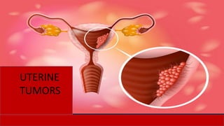
Dr. IMRANA TANVIR's Guide to Uterine Tumors
•Télécharger en tant que PPTX, PDF•
1 j'aime•1,021 vues

1) Uterine tumors include endometrial polyps, hyperplasia, and carcinomas. Endometrial hyperplasia is classified based on complexity and presence of atypia, with complex hyperplasia with atypia carrying the highest risk of developing into carcinoma. 2) Endometrial carcinomas are classified into Type I (endometrioid) and Type II (non-endometrioid). Type I carcinomas are associated with estrogen excess and hyperplasia, while Type II carcinomas arise in an atrophic endometrium and have a poorer prognosis. 3) Leiomyomas are the most common benign uterine tumor, while leiomyosarcom

Recommandé
Contenu connexe
Tendances
Tendances (20)
Similaire à Dr. IMRANA TANVIR's Guide to Uterine Tumors
Similaire à Dr. IMRANA TANVIR's Guide to Uterine Tumors (20)
Plus de imrana tanvir
Plus de imrana tanvir (20)
Dernier
Dernier (20)
Dr. IMRANA TANVIR's Guide to Uterine Tumors
- 1. UTERINE TUMORS Dr. IMRANA TANVIR Associate Professor of Pathology Faculty of Medicine Rabigh KAU 2020 UTERINE TUMORS
- 2. Objectives Identify morphology of endometrial polyp Correlate morphological types of endometrial hyperplasia with associated and risk of malignancy Describe pathogenesis, morphology and grading of endometrial carcinoma Identify morphology of leiomyoma and leiomyosarcoma and criteria of malignancy Definition of mixed Mullerian tumor 2
- 3. Endometrial Hyperplasia Endometrial proliferation; glandular architectural abnormalities resulting in glandular crowding, may give rise to either; Simple Hyperplasia; gland to stroma ratio slightly increased greater than 1:1 with prominent variability in size of the gland, glandular budding and cystic glandular dilatation Complex Hyperplasia; crowded, architecturally complex glands with little intervening stroma, the gland to stroma is elevated at least 3:1 Based on presence or absence of cytologic atypia it is further classified as; - Simple hyperplasia with/without atypia - Complex hyperplasia with/without atypia 3
- 4. Simple Endometrial Hyperplasia Tubular/Cystic without atypia With atypia Thick soft folds of endometrium
- 5. Complex Endometrial Hyperplasia With out atypia With atypia
- 6. Causes of Endometrial Hyperplasia An excess of estrogen relative to progestin, if sufficiently prolonged or marked, can induce exaggerated endometrial proliferation (hyperplasia), an important precursor of endometrial carcinoma Potential causes of estrogen excess: Failure of ovulation (such as is seen in perimenopause) Prolonged administration of estrogenic steroids without counterbalancing progestin Estrogen producing ovarian lesions (such as polycystic ovary disease and granulosa-theca cell tumors) Obesity, a common cause, as adipose tissue converts steroid precursors into estrogens
- 7. Risk of Carcinoma in Endometrial Hyperplasia Complex hyperplasia without cellular atypia; carries a low risk (less than 5%) Complex hyperplasia with cellular atypia; associated with higher risk (20% to 50%) In a significant number of cases, the hyperplasia is associated with inactivating mutations in the PTEN tumor suppressor gene which is believed to be one of several key steps in the transformation of hyperplasia to endometrial carcinoma
- 8. Endometrial Polyp Sessile, range from 0.5 to 3 cm in diameter, may project into the uterine cavity Histologically, composed of normal to cystically endometrial glands resembling the basalis, frequently with small muscular arteries Clinically present with abnormal uterine bleeding rarely risk of giving rise to a cancer
- 9. Endometrial carcinomas Peak age incidence 55 to 65 year, they classified into; Type I (80%): Majority well differentiated (Endometrioid carcinoma); Grade1 Associated with estrogen excess, endometrial hyperplasia, inactivation of DNA mismatch repair genes and PTEN tumor suppressor gene in 30-80% Associated risk factors; Obesity, DM, HTN and Infertility Type II (15%): Poorly Differentiated; Grade 3 Usually arise in the setting of endometrial atrophy, mutation in p53, and PIK3CA, KRAS seen Histological subtypes; Clear cell and Papillary serous carcinomas, aggressive behavior 9
- 10. Endometrioid carcinomas Morphology Gross; exophytic or infiltrative growth Histology; Well defined glandular pattern, graded I to III Sub types, mucinous, tubal (ciliated), squamous differentiation May infiltrate myometrium and enter vascular spaces Metastasize to regional lymph nodes Serous carcinomas, form small tufts and papillae and exhibit much greater cytologic atypia and behave aggressively
- 11. Leiomyomas Most common benign tumor of uterine myometrium of smooth muscle origin Affects 30% to 50% of women of reproductive age Tumors monoclonal, associated with several different recurrent chromosomal abnormalities, including rearrangements of chromosomes 6 and 12 that are also found in endometrial polyps and lipomas Estrogens and possibly oral contraceptives stimulate their growth and these tumors shrink in post menopausal
- 12. Leiomyomas Gross Circumscribed, firm gray-white single often multiple masses with a characteristic whorled cut surface Ranging in size from small nodules to large tumors Intramural, submucosal or subserosal in location Microscopy Interlacing bundles of uniform smooth muscle cells Foci of fibrosis, calcification, and degenerative softening may be present
- 13. Leiomyosarcoma Occur in postmenopausal women Gross; Single, soft, hemorrhagic, necrotic masses Microscopy; interlacing bundles of atypical smooth muscle fibers Diagnostic criteria of malignancy includes; coagulative necrosis, cytologic atypia, and increased mitotic activity (increased mitotic activity alone and sometimes cellular atypia can be seen in degenerated leiomyoma) Borderline category called; smooth muscle tumors of uncertain malignant potential 13
- 14. Other Malignant Tumors of Myometrium Malignant mixed mullarian tumour (MMMT) i- Homologus like; carcinosarcoma ii- Heterologus like; chondrosarcoma, rhabdomyosarcoma, or ostesarcoma Adenosarcoma Stromal sarcomas 14
- 15. Self Assessment Q1. What is the histologic diagnostic criteria of malignancy in uterine smooth muscle tumors? Q2. What is the difference between adenomyosis and endometriosis? Q3. Enlist organic causes of abnormal uterine bleeding in reproductive age group.
- 16. References Basic Pathology, 10th Edition 2017; Kumar, Abbas, Aster www.webpathology.com