SlideShare a Scribd company logo
1 of 4
Download to read offline
ORIGINAL ARTICLE ISRA MEDICAL JOURNAL | Volume 11 - Issue 3 | May – Jun 2019
163
Influence of dioptric power on retinal nerve fiber layer thickness in myopic subjects
Iqra Nehal1
, Umer Kazi2
, Asiya Saleem1
ABSTRACT
Objective: To evaluate myopic impact on thickness of nerve fiber layer of the retina in healthy myopic subjects.
Study Design: Prospective Observational study.
Place and Duration: Investigative Department of Ophthalmology of Al-Ibrahim Eye Hospital, Karachi from 1st
May 2018 to 30th
October
2018.
Methodology: In this study 80 eyes of myopic subjects (SE -0.5 to -11.0 DS) were enrolled. Each eye underwent through comprehensive
ocular examination beginning with visual acuity, refraction, fundoscopy by slit lamp and ending up to optical coherence tomography
of Nidek. Mean average peripapillary thickness of nerve fiber layer and thickness in superior, inferior, nasal and temporal quadrants
was taken into consideration, calculated by Spectral Domain Optical Coherence Tomography (version 1.5.5.0).
Results: Forty subjects volunteered for study protocol among which 21 were male and 19 were female with a degree of refractive
breakdown of 30% mild myopic, 50% moderately myopic and 20% highly myopic. The calculated average age was 25.0 ± 5.0 years
(range 16-40 years). The average total nerve fiber layer thickness in myopic respondents was 90.85µm; superiorly 112.37µm; inferiorly
117.52µm; temporally 71.85µm and in nasal quadrant was 61.55µm. Retinal nerve fiber layer thickness was statistically significant in
superior and temporal quadrant. In high myopes thickness was clinically significant in inferior quadrant in terms of quantity as
compared to mild and moderate myopia
Conclusion: Average retinal nerve fiber layer thickness was significantly decreased in high myopia as compared to mild myopia while
moderate group had slightly thicker thickness than high myopic group. Hence impact of dioptric power on nerve fiber layer thickness
in myopic patients is significant.
Keywords: Myopia, RNFL thickness, Optical coherrence tomography, Superior quadrant, Temporal quadrant.
How to Cite This:
NehalI, Kazi U, SaleemA. Influence of dioptric power on retinal nerve fiber layer thickness in myopic subjects. Isra Med J. 2019; 11(3):
163-166.
This is an Open Access article distributed under the terms of the Creative Commons Attribution-Noncommercial 4.0 International License
(http://creativecommons.org/licenses/by-nc/4.0/), which permits unrestricted use, distribution, and reproduction in any medium, provided the
original work is properly cited.
INTRODUCTION
From the last past decades, myopic prevalence had been
increasing day by day and leading to decreased vision
worldwide. Myopia has a higher prevalence in East Asia1
.
Degeneration of macula is a serious issue in short-sighted
patients particularly in East Asia and it is also the leading cause
of visual morbidity. Higher prevalence of 80 to 90% was reported
in East Asian countries like South Korea, Taiwan, Singapore,
China and Japan but an astounding prevalence of 42%was also
found even in USA2
. In 2006, the National Blindness and Visual
Impairment Survey, reported myopia in 36.5% of adult Pakistani
population3
. In 2015, worldwide survey of refractive error shows
myopic prevalence of 21.3% in which Pakistan had contributed
3.7%. With the increasing progression of myopia, entire length
of the eye ball also increases, causing thinning of Retinal Nerve
Fiber Layer (RNFL)4
. Myopic individuals often have enlarged
optic discs with more oval configuration and large areas of
peripapillary atrophy, hence similar to glaucomatous damage5
.
Retinal changes were extensively studied which were related to
both high myopia and concomitant glaucoma. A retinal change
in isolated high myopia is still unclear and this emphasizes for
measurements of RNFL thickness even in early stage6
. Retinal
nerve fiber layer is a sensitive indicator for early glaucomatous
damage, but it is uncertain whether RNFL thickness would vary
with the refractive error7
.
Optical coherence tomography (OCT) is a visual signal
acquirement and processing approach that acquires micro-
meter resolved images from within the visual scattering media5
.
The modern production spectral domain (SD) OCT with rapid
image acquisition, superior axial-resolution (1-5 µm) and feature
of stereoscopic images provides comprehensive measurements
1. Optometrist,
Isra School of Optometry, Isra University, Karachi.
2. Associate Professor of Ophthalmology,
Al-Tibri Medical College, Isra University, Karachi.
Correspondence to:
Umer Kazi
Associate Professor of Ophthalmology,
Al-Tibri Medical College, Isra University, Karachi.
Email: docuk1982@yahoo.com
Received for Publication: 09-11-18
Accepted for Publication: 20-05-19
Iqra Nehal et al ISRA MEDICAL JOURNAL | Volume 11 - Issue 3 | May – Jun 2019
164
of retinal nerve fiber layer and the macula.
Rationale of this study was that myopia is a highly significant
problem in Pakistani population3
, not only because of its high
prevalence, but also because it can contribute to visual
morbidity and increase the risk for vision-threatening
conditions. It is important to investigate RNFL changes in myopic
patients considering the risk of developing glaucoma with the
severity of myopia. This study aids in diagnosing and managing
early glaucomatous changes. Hence this study was conducted
for the fulfillment of the degree program so glaucomatic
parameters like IOP measurement and visual field
measurements were the limitations of the study. On the other
hand optic neuropathies were excluded and healthy myopic
subjects were included due to these limitations. Further the
objective of this study was to evaluate the myopic impact on
retinal nerve fiber layer thickness in healthy myopic subjects.
METHODOLOGY
In this prospective observational study, 80 myopic eyes of 40
subjects i.e. both male and female were assessed. This study was
conducted at Al-Ibrahim Eye Hospital, Karachi, Pakistan, in the
time period of 1st
May 2018 to 30th
October 2018.
The myopic patients were randomly selected attending the
general outdoor patient department of Al-Ibrahim Eye Hospital.
Mean age was considered as between 20-40 years in both
genders. Three groups on the basis of refractive error were
made among the respondents were; mild myopia (range= <-3.0
D), moderate myopia (range= -3.0 D to -6.0 D), and high myopia
(range= >-6.0 D). Patients were included without any cylindrical
component in first two groups, and ≤ 2.0 DC in high myopes.
Subjects were excluded having any other ocular pathology such
as any ocular media opacity, retinopathy, maculopathy, optic
neuropathy, amblyopia, diabetes and hypertension. Subjects
having visual acuity after best possible refraction less than 20/30
(6/9) were also excluded. Further, those patients who were not
giving consent and those who have age more than 40 were
excluded because of increased risk of systemic diseases like
hypertension, diabetes mellitus, sarcopenia (age-related
degeneration of muscular mass), muscular fibrosis etc. which
can affect ocular health.
Ethical confirmation of the study has been given by the ethical
committee of the institution. An informed approval has been
signed by every patient to participate in this study. Non-
probability sampling technique was used for sampling. In this
study new generation spectral domain Optical Coherrence
Tomography used was of Nidek Company (software version was
1.5.5.0). The data was collected by the researcher through the
general OPD and investigative department of the hospital. All
patients were undergone full ophthalmic examination, starting
with visual acuity on Snellen chart, objective refraction,
subjective refraction and fundoscopy. After that, scans of SD-
OCT were taken on each eye of each patient comprising a single
map, i.e. Disc map was taken into consideration to evaluate
RNFL thickness, CD ratio, disc area, rim area, RNFL symmetry.
Hence conclusively disc map is very important in the
interpretation and administration of glaucoma.
In objective refraction refractive error was ruled out by Nidek
autorefractometer. The mid points of the horizontal and vertical
axis were aligned subjectively by the operator to focus
centration around the optic nerve head. RNFL thickness was
calculated according to 4 types of divisions; average thickness,
superior and inferior thickness, four quadrant thickness and
clock hour thickness. A 6.0x6.0 mm cube was selected with fine
quality to measure the ONH parameters. Scans including
artifacts whether central or peripheral, quality index less than
3/5 and signal strength index less than 7/10 were excluded from
the study to avoid variations in measurements.
Data Analysis: Data inquiry was done by SPSS version 20.
Average RNFL thickness and thickness in the four quadrants was
compared as according to increasing refractive error in myopes.
Statistical significance was set at P < 0.05.
RESULTS
There were 80 healthy myopic eyes recruited in our study of 40
myopic subjects. Gender distribution was 22(55%) males and
18(54%) females. Average age was calculated as 25.0 ± 5.0
(range= 20 to 40 years). Spherical equivalent was -2.00 ± 0.7 for
mild group, -5.00 ± 1.6 for moderate group and -8.00 ± 1.3 for
high myopic group. Visual acuity after best possible correction
was 20/20 for mild and moderate group and 20/30 for high
myopic group. There were 12 (30%) mild, 20(50%) moderate and
08 (20%) highly myopic patients (Table-I).
Table-I: Frequency of subjects according to degrees of myopia
(N=40)
FREQUENCY PERCENT
Mild myopia 12 30.0%
Moderate myopia 20 50.0%
High myopia 8 20.0%
Total 40 100.0%
Average, superior, inferior and nasal thickness was decreased
comparatively in high myopic group other than mild and
moderate myopia, but surprisingly inferior quadrant thickness
was clinically significant in terms of quantity in high myopic
group among all other groups (Figure-1). Average thickness,
inferior and nasal quadrant thickness was insignificant among
three groups (p-value >0.05) (Figure-1). Decrease in thickness
was significant in superior and temporal quadrants between the
three groups (p-value <0.05) (Table-I).
Iqra Nehal et al ISRA MEDICAL JOURNAL | Volume 11 - Issue 3 | May – Jun 2019
165
Fig-1: Graphical representation of RNFL thickness compared
between different degrees of myopia (N=40)
DISCUSSION
Researchers are striving to identify risk factors for short-
sightedness because its epidemic has attracted growing concern
worldwide8,9
. The optimal management of the disease and its
complications are also being studied rapidly10-12
. Mean of
average RNFL thickness was 97.28±8.15 µm conducted by
Akhter et al.2
in Islamabad. Correlation analysis among all
subjects showed that the average, mean nasal quadrant, upper
nasal, and inferonasal sub-quadrant RNFL thickness had positive
correlation with spherical equivalent (p=0.045). In this study
mean RNFL thickness was 90.85± 11.45 µm. Analysis shows
superior and temporal quadrants had a positive correlation with
RNFL.
In spite of that, some studies13-16
are conducted on axial length
correlated with RNFL thickness. Mehboob et al. researched in
Pakistan in which they used laser interferometer (IOL master)
and SD-OCT to measure axial length and RNFL thickness
respectively. Three groups based on axial length were made in
this study. A strong negative correlation between axial length
and nerve fiber layer thickness of the retina was analyzed (p-
value<0.001). They observed that axial length significantly
effects measurements of RNFL thickness and on basis of
thickness of RNFL; diagnosis of glaucoma should be counted. In
this current study axial length was not measured while SD-OCT
was used to measure RNFL thickness. Results of our study shows
that there was a strong significant correlation in superior and
temporal quadrant among 3 groups based on refractive error
and average, superior and inferior thickness shows insignificant
correlation with the refractive error.
Some studies suggest that thickness of nerve fiber layer varies
with the type of refractive error and as well as the amount of
refractive error. Lee et al.17
studied RNFL thickness in short-
sighted, long-sighted and emmetropic children. He found that
RNFL thickness was thickest in the hyperopic group followed by
myopic and emmetropic group. When the age was adjusted
myopes had a thinner RNFL other than two groups while
emmetropes and hyperopes had equal RNFL thickness. In our
study only, myopic group was considered and mean adult age
was taken between 20 to 40 years. We hadn’t adjusted age by
any statistical formula because our study aimed to evaluate
impact of myopic intensity on RNFL. In contrast studies which
discussed about amount of refractive error like Kamath and his
collegue18
discussed RNFL thickness in 3600 quadrants of
variable degrees of myopic patients. Group III (High myopia)
showed statistically significant thinning in 3600 in all quadrants
except temporal quadrant compared to group I and II (low and
moderate myopia). Results of our study indicate that only
superior and temporal quadrant shows statistically significant
thinning in high myopes.
Amongst studies with SD-OCT similar results were noted by Kang
et al.19
, Wang et al.20
, and Savini et al.21
who noted that peri-
papillary thickness of RNFL decreases with increments in the
dioptric power of myopia. Although Kang et al. and Wang et al.
found significant decrease only in superior and inferior and in
nasal quadrant while change was insignificant in temporal
quadrant. Savini et al also noted decrease in RNFL thickness with
weakest correlation of decrease in temporal quadrant. Results
of our study represent significant positive correlation in superior
and temporal quadrant (p=0.04 and p=0.03 respectively) while
significantly negative correlation was found in average (p=0.08),
inferior (p=0.10), and nasal quadrants (p=0.28).
In contradiction Hoh et al22
suggested that the peri-papillary
thickness of RNFL does not alter with degree of myopia or length
of the eyeball. These discrepancies can be associated with the
different OCT imaging equipment used. They used OCT-1 which
obtains only 12 RNFL thickness values from each scan, while rest
of the studies, as well as ours, used stratus, Cirrus or SD-OCT. It
is likely that the values obtained in OCT-1 scans may be
insufficient to pick up differences due to length of the eye ball or
degree of refractive error.
CONCLUSION
Average RNFL thickness was significantly decreased in high
myopia as compared to mild myopia while moderate group had
slightly thicker thickness than high myopic group. Hence impact
of dioptric power on nerve fiber layer thickness in myopic
patients is significant.
ACKNOWLEDGEMENT
'Thanks to Mr. M. Arslan and Ms. Mahnoor from R and D
Department, ATMC, Isra University, Karachi campus for their
support'.
CONTRIBUTION OF AUTHORS
Nehal I: Conceived idea, Literature review, Manuscript writing.
Kazi U: Literature review, Manuscript writing
Saleem A: Literature review, Manuscript writing
97
114
128
70 67
88
109
114
70
59
87
105
112
80
57
0
20
40
60
80
100
120
140
Total RNFL
thickness
Superior
RNFL
thickness
Inferior
RNFL
thickness
Temporal
RNFL
thickness
Nasal RNFL
thickness
RNFLthickness
MILD MYOPIA MODERATE MYOPIA HIGH MYOPIA
Iqra Nehal et al ISRA MEDICAL JOURNAL | Volume 11 - Issue 3 | May – Jun 2019
166
Disclaimer: None.
Conflict of Interest: None.
Source of Funding: None.
REFERENCES
1. Tai ELM, Ling JL, Gan EH, Adil H, Wan-Hazabbah WH.
Comparison Of Peripapillary Retinal Nerve Fiber Layer
Thickness Between Myopia Severity Groups And Controls.
Int J Ophthalmol 2018;11(2): 274-78
2. Holden BA, Frickle TR, Wilson DA, Jong M, Naidoo KS,
Sankaridurg P, et al. Global Prevalence Of Myopia And High
Myopia And Temporal Ternds From 2000 Through 2050.
Ophthalmol 2016; 123(5): 1036-42
3. Akhtar N, Kausar A, Afzal F, Ali SK, Hamid N. Peripapillary
Retinal Nerve Fiber Layer (RNFL) Thickness Measurements
by Topcon SD-OCT in Myopic Patients. J of the Coll of Physic
& Surg Pak 2017; 28(1): 26-30.
4. Hsu S-Y, Chang M-S, Ko M-L, Harnod T. Retinal Nerve Fiber
Layer Thickness And Optic Nerve Head Size Measured In
High Myopes By Optical Coherence Tomography. Clini
Experimental Optom 2013; 96(1): 373–78
5. Singh D , Mishra SK, Agarwal E ,Sharma R, Bhartiya S, Dada
T. Assessment of Retinal Nerve Fiber Layer Changes By
Cirrus High-Definition Optical Coherence Tomography In
Myopia. J Curr Glaucoma Pract 2017;11(2): 52-57
6. Malakar M, Askari SN, Ashraf H, Waris A, Ahuja A, Asghar A.
Optical coherence tomography assisted retinal nerve fibre
layer thickness profile in high myopia. J of clini & Diag Res J
2015;9(2): NC01
7. Chowdhary T, Chowdhary DS, Choudhary A. Study of Retinal
Nerve Fiber Layer Thickness in Myopic Eye in Rajasthani
Population. Int J of Sci & Res 2015; 4(2): 880-81
8. Cuellar‐Partida G, Lu Y, Kho PF, Hewitt AW, Wichmann HE,
Yazar S, et al. Assessing the genetic predisposition of
education on myopia: a Mendelian randomization study.
Genetic Epidemiol. 2016;40(1): 66-72
9. Parssinen O, Kauppinen M. Associations Of Reading Posture,
Gaze Angle And Reading Distance With Myopia And Myopic
Progression. Acta Ophthalmol 2016; 94(8): 775-79
10. Davidson SL, O'Hara M, Wagner RS. Management of
Progressive Myopia. J Pediatric Ophthalmol Strabismus
2016;53(3): 134-36
11. Huang J, Wen D, Wang Q, McAlinden C, Flitcroft I, Chen H,
et al. Efficacy comparison of 16 interventions for myopia
control in children: a network meta-analysis. Ophthalmol
2016;123(4): 697-08
12. Wolffsohn JS, Calossi A, Cho P, Gifford K, Jones L, Li M, et al.
Global trends in myopia management attitudes and
strategies in clinical practice. Contact Lens and Anterior Eye
2016;39(2): 106-16
13. Mehboob MA, Islam QU, Yaqub MA. Effect Of Axial Length
on Peripapillary Retinal Nerve Fibre Layer Thickness
Measured by Spectral Domain Optical Coherence
Tomography. Pak Armed Forc Med J 2017; 67(2): 238-42
14. Bae SH, Kang SH, Feng CS, Park J, Jeong JH, Yi K. Influence of
myopia on size of optic nerve head and retinal nerve fiber
layer thickness measured by spectral domain optical
coherence tomography. Korean J Ophthalmol 2016;30(5):
335-43
15. AttaAllah HR, Omar IA, Abdelhalim AS. Evaluation of optic
nerve head parameters and retinal nerve fiber layer
thickness in axial myopia using SD OCT. Ophthalmol &
Therapy. 2017;6(2): 335-41
16. Singh D, Mishra SK, Agarwal E, Sharma R, Bhartiya S, Dada
T. Assessment of retinal nerve fiber layer changes by cirrus
high-definition optical coherence tomography in myopia. J
of Current Glaucoma Pract. 2017;11(2): 52
17. Lee JW, Yau GS, Woo TT, Yick DW, Tam VT, Lai JS. Retinal
nerve fiber layer thickness in myopic, emmetropic, and
hyperopic children. Medicine 2015; 94(12): e699.
18. Kamath AR, Dudeja L. Peri-papillary retinal nerve fiber layer
thickness profile in subjects with myopia measured using
optical coherence tomography. J of Clin Ophthalmol & Res
2014;2(3): 131
19. Kang SH, Hong SW, Im SK, Lee SH, Ahn MD. Effect Of Myopia
On Thickness Of Retinal Nerve Fiber Layer Measured By
Cirrus HD Optical Coherence Tomography. Invest
Ophthalmol Vis Sci 2010; 51(1): 4075-83
20. Wang G, Qlu KL, Lu XH, Sun LX, Liao XJ, Chen HL, et al. The
Effect Of Myopia On Retinal Nerve Fibre Measurement: A
Comparative Study Of Spectral Domain Optical Coherence
Tomography And Scanning Laser Polarimetry. Br J
Ophthalmol 2011; 95(1): 255-60
21. Savini G, Barboni P, Parisi V, Carbonelli M. The Influence Of
Axial Length On Retinal Nerve Fibre Layer Thickness And
Optic Disc Size Measurements By Spectral Domain OCT. Br J
Ophthalmol 2012;96(1): 57-61
22. Hoh ST, Lim MC, Seah SK, Lim AT, Chew SJ, Foster PJ, et al.
Peripapillary Retinal Nerve Fibre Layer Thickness Variations
With Myopia. Ophthalmology 2006;113(1): 773-77

More Related Content

What's hot

Focal choroidal excavation in eyes with central serous chorioretinopathy
Focal choroidal excavation in eyes with central serous chorioretinopathyFocal choroidal excavation in eyes with central serous chorioretinopathy
Focal choroidal excavation in eyes with central serous chorioretinopathyAbdallah Ellabban
 
Reduction of retinal senstivity in eyes with reticular pseudodrusen
Reduction of retinal senstivity in eyes with reticular pseudodrusenReduction of retinal senstivity in eyes with reticular pseudodrusen
Reduction of retinal senstivity in eyes with reticular pseudodrusenAbdallah Ellabban
 
Focal choroidal excavation in eyes with central serous chorioretinopathy
Focal choroidal excavation in eyes with central serous chorioretinopathy Focal choroidal excavation in eyes with central serous chorioretinopathy
Focal choroidal excavation in eyes with central serous chorioretinopathy Abdallah Ellabban
 
Advances in glaucoma-posterior segment evaluation.
Advances in glaucoma-posterior segment evaluation.Advances in glaucoma-posterior segment evaluation.
Advances in glaucoma-posterior segment evaluation.Dr. Stuti Somani Agarwal
 
April 2019 . Cataracts secondary to intraocular diseases are complicated cata...
April 2019 . Cataracts secondary to intraocular diseases are complicated cata...April 2019 . Cataracts secondary to intraocular diseases are complicated cata...
April 2019 . Cataracts secondary to intraocular diseases are complicated cata...Vinitkumar MJ
 
Optic coherence tomography
Optic coherence tomographyOptic coherence tomography
Optic coherence tomographyNeurologyKota
 
Description of Different Phases of Brain Tumor Classification
Description of Different Phases of Brain Tumor ClassificationDescription of Different Phases of Brain Tumor Classification
Description of Different Phases of Brain Tumor Classificationasclepiuspdfs
 
Diode Laser Treatment for Retinopathy of Prematurity – Our Experience in Bulg...
Diode Laser Treatment for Retinopathy of Prematurity – Our Experience in Bulg...Diode Laser Treatment for Retinopathy of Prematurity – Our Experience in Bulg...
Diode Laser Treatment for Retinopathy of Prematurity – Our Experience in Bulg...inventionjournals
 
Detection of Glaucoma using Optic Disk and Incremental Cup Segmentation from ...
Detection of Glaucoma using Optic Disk and Incremental Cup Segmentation from ...Detection of Glaucoma using Optic Disk and Incremental Cup Segmentation from ...
Detection of Glaucoma using Optic Disk and Incremental Cup Segmentation from ...theijes
 

What's hot (19)

Focal choroidal excavation in eyes with central serous chorioretinopathy
Focal choroidal excavation in eyes with central serous chorioretinopathyFocal choroidal excavation in eyes with central serous chorioretinopathy
Focal choroidal excavation in eyes with central serous chorioretinopathy
 
Reduction of retinal senstivity in eyes with reticular pseudodrusen
Reduction of retinal senstivity in eyes with reticular pseudodrusenReduction of retinal senstivity in eyes with reticular pseudodrusen
Reduction of retinal senstivity in eyes with reticular pseudodrusen
 
Focal choroidal excavation in eyes with central serous chorioretinopathy
Focal choroidal excavation in eyes with central serous chorioretinopathy Focal choroidal excavation in eyes with central serous chorioretinopathy
Focal choroidal excavation in eyes with central serous chorioretinopathy
 
Oct in glaucoma
Oct in glaucomaOct in glaucoma
Oct in glaucoma
 
Advances in glaucoma-posterior segment evaluation.
Advances in glaucoma-posterior segment evaluation.Advances in glaucoma-posterior segment evaluation.
Advances in glaucoma-posterior segment evaluation.
 
28 07 14_dra_mariana
28 07 14_dra_mariana28 07 14_dra_mariana
28 07 14_dra_mariana
 
April 2019 . Cataracts secondary to intraocular diseases are complicated cata...
April 2019 . Cataracts secondary to intraocular diseases are complicated cata...April 2019 . Cataracts secondary to intraocular diseases are complicated cata...
April 2019 . Cataracts secondary to intraocular diseases are complicated cata...
 
AMA series design
AMA series designAMA series design
AMA series design
 
Optic coherence tomography
Optic coherence tomographyOptic coherence tomography
Optic coherence tomography
 
11 08 14
11 08 1411 08 14
11 08 14
 
Hrt &amp; g dx
Hrt &amp; g dxHrt &amp; g dx
Hrt &amp; g dx
 
EVALUATION OF ANTERIOR SEGMENT IMAGING TECHNIQUES IN DIAGNOSIS OF KERATOCONUS
EVALUATION OF ANTERIOR SEGMENT IMAGING TECHNIQUES IN DIAGNOSIS OF KERATOCONUSEVALUATION OF ANTERIOR SEGMENT IMAGING TECHNIQUES IN DIAGNOSIS OF KERATOCONUS
EVALUATION OF ANTERIOR SEGMENT IMAGING TECHNIQUES IN DIAGNOSIS OF KERATOCONUS
 
Description of Different Phases of Brain Tumor Classification
Description of Different Phases of Brain Tumor ClassificationDescription of Different Phases of Brain Tumor Classification
Description of Different Phases of Brain Tumor Classification
 
Dome shaped macula
Dome shaped macula Dome shaped macula
Dome shaped macula
 
Journal Club
Journal ClubJournal Club
Journal Club
 
Diode Laser Treatment for Retinopathy of Prematurity – Our Experience in Bulg...
Diode Laser Treatment for Retinopathy of Prematurity – Our Experience in Bulg...Diode Laser Treatment for Retinopathy of Prematurity – Our Experience in Bulg...
Diode Laser Treatment for Retinopathy of Prematurity – Our Experience in Bulg...
 
20120140502011
2012014050201120120140502011
20120140502011
 
Dosimetric evaluation of carcinoma nasopharynx using Volumetric Modulated Arc...
Dosimetric evaluation of carcinoma nasopharynx using Volumetric Modulated Arc...Dosimetric evaluation of carcinoma nasopharynx using Volumetric Modulated Arc...
Dosimetric evaluation of carcinoma nasopharynx using Volumetric Modulated Arc...
 
Detection of Glaucoma using Optic Disk and Incremental Cup Segmentation from ...
Detection of Glaucoma using Optic Disk and Incremental Cup Segmentation from ...Detection of Glaucoma using Optic Disk and Incremental Cup Segmentation from ...
Detection of Glaucoma using Optic Disk and Incremental Cup Segmentation from ...
 

Similar to Influence-of-dioptric-power-on-retinal-nerve-fiber-layer-thickness-in-myopic-subjects

Evaluation of Macular and RNFL thickness in Amblyopia using OCT.pptx
Evaluation of Macular and RNFL thickness in Amblyopia using OCT.pptxEvaluation of Macular and RNFL thickness in Amblyopia using OCT.pptx
Evaluation of Macular and RNFL thickness in Amblyopia using OCT.pptxRadhaMathur2
 
Journal Presentation.pptx
Journal Presentation.pptxJournal Presentation.pptx
Journal Presentation.pptxDr.Maliha Nawar
 
Comparative Study of Visual Outcome between Femtosecond Lasik with Excimer La...
Comparative Study of Visual Outcome between Femtosecond Lasik with Excimer La...Comparative Study of Visual Outcome between Femtosecond Lasik with Excimer La...
Comparative Study of Visual Outcome between Femtosecond Lasik with Excimer La...iosrjce
 
Learning Effect and Test-Retest Variability in Healthy Subjects and Patients ...
Learning Effect and Test-Retest Variability in Healthy Subjects and Patients ...Learning Effect and Test-Retest Variability in Healthy Subjects and Patients ...
Learning Effect and Test-Retest Variability in Healthy Subjects and Patients ...inventionjournals
 
An opinion fornix depth measurement in ophthalmic socket
An opinion fornix depth measurement in ophthalmic socketAn opinion fornix depth measurement in ophthalmic socket
An opinion fornix depth measurement in ophthalmic socketRAJ KUMAR SARASWAT
 
Ophthalmology - IEC ppt FINAL 2.0.pptx
Ophthalmology - IEC ppt FINAL 2.0.pptxOphthalmology - IEC ppt FINAL 2.0.pptx
Ophthalmology - IEC ppt FINAL 2.0.pptxRavi180910
 
Optical Coherence Tomography (OCT)
Optical Coherence Tomography (OCT)Optical Coherence Tomography (OCT)
Optical Coherence Tomography (OCT)ShrutiDagar1
 
visual field interpretation 1.pdf
visual field interpretation 1.pdfvisual field interpretation 1.pdf
visual field interpretation 1.pdfssuser214230
 
Multimodal evaluation of macular function in age related macular
Multimodal evaluation of macular function in age related macularMultimodal evaluation of macular function in age related macular
Multimodal evaluation of macular function in age related macularAbdallah Ellabban
 
biometry for ON.ppt
biometry for ON.pptbiometry for ON.ppt
biometry for ON.pptmikaelgirum
 

Similar to Influence-of-dioptric-power-on-retinal-nerve-fiber-layer-thickness-in-myopic-subjects (20)

Evaluation of Macular and RNFL thickness in Amblyopia using OCT.pptx
Evaluation of Macular and RNFL thickness in Amblyopia using OCT.pptxEvaluation of Macular and RNFL thickness in Amblyopia using OCT.pptx
Evaluation of Macular and RNFL thickness in Amblyopia using OCT.pptx
 
Journal Presentation.pptx
Journal Presentation.pptxJournal Presentation.pptx
Journal Presentation.pptx
 
Comparative Study of Visual Outcome between Femtosecond Lasik with Excimer La...
Comparative Study of Visual Outcome between Femtosecond Lasik with Excimer La...Comparative Study of Visual Outcome between Femtosecond Lasik with Excimer La...
Comparative Study of Visual Outcome between Femtosecond Lasik with Excimer La...
 
OCTA and Glaucoma Detection.
OCTA and Glaucoma Detection.  OCTA and Glaucoma Detection.
OCTA and Glaucoma Detection.
 
C01230912
C01230912C01230912
C01230912
 
Diabetic macular edema
Diabetic macular edemaDiabetic macular edema
Diabetic macular edema
 
Learning Effect and Test-Retest Variability in Healthy Subjects and Patients ...
Learning Effect and Test-Retest Variability in Healthy Subjects and Patients ...Learning Effect and Test-Retest Variability in Healthy Subjects and Patients ...
Learning Effect and Test-Retest Variability in Healthy Subjects and Patients ...
 
Pdt of armd
Pdt of armdPdt of armd
Pdt of armd
 
An opinion fornix depth measurement in ophthalmic socket
An opinion fornix depth measurement in ophthalmic socketAn opinion fornix depth measurement in ophthalmic socket
An opinion fornix depth measurement in ophthalmic socket
 
Ophthalmology - IEC ppt FINAL 2.0.pptx
Ophthalmology - IEC ppt FINAL 2.0.pptxOphthalmology - IEC ppt FINAL 2.0.pptx
Ophthalmology - IEC ppt FINAL 2.0.pptx
 
Optical Coherence Tomography (OCT)
Optical Coherence Tomography (OCT)Optical Coherence Tomography (OCT)
Optical Coherence Tomography (OCT)
 
EDOF IOL
EDOF IOLEDOF IOL
EDOF IOL
 
visual field interpretation 1.pdf
visual field interpretation 1.pdfvisual field interpretation 1.pdf
visual field interpretation 1.pdf
 
14 10 years follow-up
14 10 years follow-up 14 10 years follow-up
14 10 years follow-up
 
Articulo Gerardo 10_07_14
Articulo Gerardo 10_07_14Articulo Gerardo 10_07_14
Articulo Gerardo 10_07_14
 
14 07 14_ gerardo
14 07 14_ gerardo14 07 14_ gerardo
14 07 14_ gerardo
 
Multimodal evaluation of macular function in age related macular
Multimodal evaluation of macular function in age related macularMultimodal evaluation of macular function in age related macular
Multimodal evaluation of macular function in age related macular
 
18 08 14
18 08 1418 08 14
18 08 14
 
biometry for ON.ppt
biometry for ON.pptbiometry for ON.ppt
biometry for ON.ppt
 
SOLIX Essential Brochure
SOLIX Essential Brochure SOLIX Essential Brochure
SOLIX Essential Brochure
 

Recently uploaded

Asthma Review - GINA guidelines summary 2024
Asthma Review - GINA guidelines summary 2024Asthma Review - GINA guidelines summary 2024
Asthma Review - GINA guidelines summary 2024Gabriel Guevara MD
 
Russian Call Girls Chickpet - 7001305949 Booking and charges genuine rate for...
Russian Call Girls Chickpet - 7001305949 Booking and charges genuine rate for...Russian Call Girls Chickpet - 7001305949 Booking and charges genuine rate for...
Russian Call Girls Chickpet - 7001305949 Booking and charges genuine rate for...narwatsonia7
 
Call Girls Kanakapura Road Just Call 7001305949 Top Class Call Girl Service A...
Call Girls Kanakapura Road Just Call 7001305949 Top Class Call Girl Service A...Call Girls Kanakapura Road Just Call 7001305949 Top Class Call Girl Service A...
Call Girls Kanakapura Road Just Call 7001305949 Top Class Call Girl Service A...narwatsonia7
 
College Call Girls Pune Mira 9907093804 Short 1500 Night 6000 Best call girls...
College Call Girls Pune Mira 9907093804 Short 1500 Night 6000 Best call girls...College Call Girls Pune Mira 9907093804 Short 1500 Night 6000 Best call girls...
College Call Girls Pune Mira 9907093804 Short 1500 Night 6000 Best call girls...Miss joya
 
Call Girls In Andheri East Call 9920874524 Book Hot And Sexy Girls
Call Girls In Andheri East Call 9920874524 Book Hot And Sexy GirlsCall Girls In Andheri East Call 9920874524 Book Hot And Sexy Girls
Call Girls In Andheri East Call 9920874524 Book Hot And Sexy Girlsnehamumbai
 
Call Girls Thane Just Call 9910780858 Get High Class Call Girls Service
Call Girls Thane Just Call 9910780858 Get High Class Call Girls ServiceCall Girls Thane Just Call 9910780858 Get High Class Call Girls Service
Call Girls Thane Just Call 9910780858 Get High Class Call Girls Servicesonalikaur4
 
Call Girls Service In Shyam Nagar Whatsapp 8445551418 Independent Escort Service
Call Girls Service In Shyam Nagar Whatsapp 8445551418 Independent Escort ServiceCall Girls Service In Shyam Nagar Whatsapp 8445551418 Independent Escort Service
Call Girls Service In Shyam Nagar Whatsapp 8445551418 Independent Escort Serviceparulsinha
 
Artifacts in Nuclear Medicine with Identifying and resolving artifacts.
Artifacts in Nuclear Medicine with Identifying and resolving artifacts.Artifacts in Nuclear Medicine with Identifying and resolving artifacts.
Artifacts in Nuclear Medicine with Identifying and resolving artifacts.MiadAlsulami
 
Call Girls Whitefield Just Call 7001305949 Top Class Call Girl Service Available
Call Girls Whitefield Just Call 7001305949 Top Class Call Girl Service AvailableCall Girls Whitefield Just Call 7001305949 Top Class Call Girl Service Available
Call Girls Whitefield Just Call 7001305949 Top Class Call Girl Service Availablenarwatsonia7
 
College Call Girls Vyasarpadi Whatsapp 7001305949 Independent Escort Service
College Call Girls Vyasarpadi Whatsapp 7001305949 Independent Escort ServiceCollege Call Girls Vyasarpadi Whatsapp 7001305949 Independent Escort Service
College Call Girls Vyasarpadi Whatsapp 7001305949 Independent Escort ServiceNehru place Escorts
 
Call Girls Hosur Just Call 7001305949 Top Class Call Girl Service Available
Call Girls Hosur Just Call 7001305949 Top Class Call Girl Service AvailableCall Girls Hosur Just Call 7001305949 Top Class Call Girl Service Available
Call Girls Hosur Just Call 7001305949 Top Class Call Girl Service Availablenarwatsonia7
 
Call Girl Lucknow Mallika 7001305949 Independent Escort Service Lucknow
Call Girl Lucknow Mallika 7001305949 Independent Escort Service LucknowCall Girl Lucknow Mallika 7001305949 Independent Escort Service Lucknow
Call Girl Lucknow Mallika 7001305949 Independent Escort Service Lucknownarwatsonia7
 
Call Girls ITPL Just Call 7001305949 Top Class Call Girl Service Available
Call Girls ITPL Just Call 7001305949 Top Class Call Girl Service AvailableCall Girls ITPL Just Call 7001305949 Top Class Call Girl Service Available
Call Girls ITPL Just Call 7001305949 Top Class Call Girl Service Availablenarwatsonia7
 
call girls in green park DELHI 🔝 >༒9540349809 🔝 genuine Escort Service 🔝✔️✔️
call girls in green park  DELHI 🔝 >༒9540349809 🔝 genuine Escort Service 🔝✔️✔️call girls in green park  DELHI 🔝 >༒9540349809 🔝 genuine Escort Service 🔝✔️✔️
call girls in green park DELHI 🔝 >༒9540349809 🔝 genuine Escort Service 🔝✔️✔️saminamagar
 
Call Girls Service in Bommanahalli - 7001305949 with real photos and phone nu...
Call Girls Service in Bommanahalli - 7001305949 with real photos and phone nu...Call Girls Service in Bommanahalli - 7001305949 with real photos and phone nu...
Call Girls Service in Bommanahalli - 7001305949 with real photos and phone nu...narwatsonia7
 
VIP Call Girls Pune Vrinda 9907093804 Short 1500 Night 6000 Best call girls S...
VIP Call Girls Pune Vrinda 9907093804 Short 1500 Night 6000 Best call girls S...VIP Call Girls Pune Vrinda 9907093804 Short 1500 Night 6000 Best call girls S...
VIP Call Girls Pune Vrinda 9907093804 Short 1500 Night 6000 Best call girls S...Miss joya
 
Bangalore Call Girls Marathahalli 📞 9907093804 High Profile Service 100% Safe
Bangalore Call Girls Marathahalli 📞 9907093804 High Profile Service 100% SafeBangalore Call Girls Marathahalli 📞 9907093804 High Profile Service 100% Safe
Bangalore Call Girls Marathahalli 📞 9907093804 High Profile Service 100% Safenarwatsonia7
 
Call Girl Koramangala | 7001305949 At Low Cost Cash Payment Booking
Call Girl Koramangala | 7001305949 At Low Cost Cash Payment BookingCall Girl Koramangala | 7001305949 At Low Cost Cash Payment Booking
Call Girl Koramangala | 7001305949 At Low Cost Cash Payment Bookingnarwatsonia7
 
High Profile Call Girls Jaipur Vani 8445551418 Independent Escort Service Jaipur
High Profile Call Girls Jaipur Vani 8445551418 Independent Escort Service JaipurHigh Profile Call Girls Jaipur Vani 8445551418 Independent Escort Service Jaipur
High Profile Call Girls Jaipur Vani 8445551418 Independent Escort Service Jaipurparulsinha
 
Kolkata Call Girls Services 9907093804 @24x7 High Class Babes Here Call Now
Kolkata Call Girls Services 9907093804 @24x7 High Class Babes Here Call NowKolkata Call Girls Services 9907093804 @24x7 High Class Babes Here Call Now
Kolkata Call Girls Services 9907093804 @24x7 High Class Babes Here Call NowNehru place Escorts
 

Recently uploaded (20)

Asthma Review - GINA guidelines summary 2024
Asthma Review - GINA guidelines summary 2024Asthma Review - GINA guidelines summary 2024
Asthma Review - GINA guidelines summary 2024
 
Russian Call Girls Chickpet - 7001305949 Booking and charges genuine rate for...
Russian Call Girls Chickpet - 7001305949 Booking and charges genuine rate for...Russian Call Girls Chickpet - 7001305949 Booking and charges genuine rate for...
Russian Call Girls Chickpet - 7001305949 Booking and charges genuine rate for...
 
Call Girls Kanakapura Road Just Call 7001305949 Top Class Call Girl Service A...
Call Girls Kanakapura Road Just Call 7001305949 Top Class Call Girl Service A...Call Girls Kanakapura Road Just Call 7001305949 Top Class Call Girl Service A...
Call Girls Kanakapura Road Just Call 7001305949 Top Class Call Girl Service A...
 
College Call Girls Pune Mira 9907093804 Short 1500 Night 6000 Best call girls...
College Call Girls Pune Mira 9907093804 Short 1500 Night 6000 Best call girls...College Call Girls Pune Mira 9907093804 Short 1500 Night 6000 Best call girls...
College Call Girls Pune Mira 9907093804 Short 1500 Night 6000 Best call girls...
 
Call Girls In Andheri East Call 9920874524 Book Hot And Sexy Girls
Call Girls In Andheri East Call 9920874524 Book Hot And Sexy GirlsCall Girls In Andheri East Call 9920874524 Book Hot And Sexy Girls
Call Girls In Andheri East Call 9920874524 Book Hot And Sexy Girls
 
Call Girls Thane Just Call 9910780858 Get High Class Call Girls Service
Call Girls Thane Just Call 9910780858 Get High Class Call Girls ServiceCall Girls Thane Just Call 9910780858 Get High Class Call Girls Service
Call Girls Thane Just Call 9910780858 Get High Class Call Girls Service
 
Call Girls Service In Shyam Nagar Whatsapp 8445551418 Independent Escort Service
Call Girls Service In Shyam Nagar Whatsapp 8445551418 Independent Escort ServiceCall Girls Service In Shyam Nagar Whatsapp 8445551418 Independent Escort Service
Call Girls Service In Shyam Nagar Whatsapp 8445551418 Independent Escort Service
 
Artifacts in Nuclear Medicine with Identifying and resolving artifacts.
Artifacts in Nuclear Medicine with Identifying and resolving artifacts.Artifacts in Nuclear Medicine with Identifying and resolving artifacts.
Artifacts in Nuclear Medicine with Identifying and resolving artifacts.
 
Call Girls Whitefield Just Call 7001305949 Top Class Call Girl Service Available
Call Girls Whitefield Just Call 7001305949 Top Class Call Girl Service AvailableCall Girls Whitefield Just Call 7001305949 Top Class Call Girl Service Available
Call Girls Whitefield Just Call 7001305949 Top Class Call Girl Service Available
 
College Call Girls Vyasarpadi Whatsapp 7001305949 Independent Escort Service
College Call Girls Vyasarpadi Whatsapp 7001305949 Independent Escort ServiceCollege Call Girls Vyasarpadi Whatsapp 7001305949 Independent Escort Service
College Call Girls Vyasarpadi Whatsapp 7001305949 Independent Escort Service
 
Call Girls Hosur Just Call 7001305949 Top Class Call Girl Service Available
Call Girls Hosur Just Call 7001305949 Top Class Call Girl Service AvailableCall Girls Hosur Just Call 7001305949 Top Class Call Girl Service Available
Call Girls Hosur Just Call 7001305949 Top Class Call Girl Service Available
 
Call Girl Lucknow Mallika 7001305949 Independent Escort Service Lucknow
Call Girl Lucknow Mallika 7001305949 Independent Escort Service LucknowCall Girl Lucknow Mallika 7001305949 Independent Escort Service Lucknow
Call Girl Lucknow Mallika 7001305949 Independent Escort Service Lucknow
 
Call Girls ITPL Just Call 7001305949 Top Class Call Girl Service Available
Call Girls ITPL Just Call 7001305949 Top Class Call Girl Service AvailableCall Girls ITPL Just Call 7001305949 Top Class Call Girl Service Available
Call Girls ITPL Just Call 7001305949 Top Class Call Girl Service Available
 
call girls in green park DELHI 🔝 >༒9540349809 🔝 genuine Escort Service 🔝✔️✔️
call girls in green park  DELHI 🔝 >༒9540349809 🔝 genuine Escort Service 🔝✔️✔️call girls in green park  DELHI 🔝 >༒9540349809 🔝 genuine Escort Service 🔝✔️✔️
call girls in green park DELHI 🔝 >༒9540349809 🔝 genuine Escort Service 🔝✔️✔️
 
Call Girls Service in Bommanahalli - 7001305949 with real photos and phone nu...
Call Girls Service in Bommanahalli - 7001305949 with real photos and phone nu...Call Girls Service in Bommanahalli - 7001305949 with real photos and phone nu...
Call Girls Service in Bommanahalli - 7001305949 with real photos and phone nu...
 
VIP Call Girls Pune Vrinda 9907093804 Short 1500 Night 6000 Best call girls S...
VIP Call Girls Pune Vrinda 9907093804 Short 1500 Night 6000 Best call girls S...VIP Call Girls Pune Vrinda 9907093804 Short 1500 Night 6000 Best call girls S...
VIP Call Girls Pune Vrinda 9907093804 Short 1500 Night 6000 Best call girls S...
 
Bangalore Call Girls Marathahalli 📞 9907093804 High Profile Service 100% Safe
Bangalore Call Girls Marathahalli 📞 9907093804 High Profile Service 100% SafeBangalore Call Girls Marathahalli 📞 9907093804 High Profile Service 100% Safe
Bangalore Call Girls Marathahalli 📞 9907093804 High Profile Service 100% Safe
 
Call Girl Koramangala | 7001305949 At Low Cost Cash Payment Booking
Call Girl Koramangala | 7001305949 At Low Cost Cash Payment BookingCall Girl Koramangala | 7001305949 At Low Cost Cash Payment Booking
Call Girl Koramangala | 7001305949 At Low Cost Cash Payment Booking
 
High Profile Call Girls Jaipur Vani 8445551418 Independent Escort Service Jaipur
High Profile Call Girls Jaipur Vani 8445551418 Independent Escort Service JaipurHigh Profile Call Girls Jaipur Vani 8445551418 Independent Escort Service Jaipur
High Profile Call Girls Jaipur Vani 8445551418 Independent Escort Service Jaipur
 
Kolkata Call Girls Services 9907093804 @24x7 High Class Babes Here Call Now
Kolkata Call Girls Services 9907093804 @24x7 High Class Babes Here Call NowKolkata Call Girls Services 9907093804 @24x7 High Class Babes Here Call Now
Kolkata Call Girls Services 9907093804 @24x7 High Class Babes Here Call Now
 

Influence-of-dioptric-power-on-retinal-nerve-fiber-layer-thickness-in-myopic-subjects

  • 1. ORIGINAL ARTICLE ISRA MEDICAL JOURNAL | Volume 11 - Issue 3 | May – Jun 2019 163 Influence of dioptric power on retinal nerve fiber layer thickness in myopic subjects Iqra Nehal1 , Umer Kazi2 , Asiya Saleem1 ABSTRACT Objective: To evaluate myopic impact on thickness of nerve fiber layer of the retina in healthy myopic subjects. Study Design: Prospective Observational study. Place and Duration: Investigative Department of Ophthalmology of Al-Ibrahim Eye Hospital, Karachi from 1st May 2018 to 30th October 2018. Methodology: In this study 80 eyes of myopic subjects (SE -0.5 to -11.0 DS) were enrolled. Each eye underwent through comprehensive ocular examination beginning with visual acuity, refraction, fundoscopy by slit lamp and ending up to optical coherence tomography of Nidek. Mean average peripapillary thickness of nerve fiber layer and thickness in superior, inferior, nasal and temporal quadrants was taken into consideration, calculated by Spectral Domain Optical Coherence Tomography (version 1.5.5.0). Results: Forty subjects volunteered for study protocol among which 21 were male and 19 were female with a degree of refractive breakdown of 30% mild myopic, 50% moderately myopic and 20% highly myopic. The calculated average age was 25.0 ± 5.0 years (range 16-40 years). The average total nerve fiber layer thickness in myopic respondents was 90.85µm; superiorly 112.37µm; inferiorly 117.52µm; temporally 71.85µm and in nasal quadrant was 61.55µm. Retinal nerve fiber layer thickness was statistically significant in superior and temporal quadrant. In high myopes thickness was clinically significant in inferior quadrant in terms of quantity as compared to mild and moderate myopia Conclusion: Average retinal nerve fiber layer thickness was significantly decreased in high myopia as compared to mild myopia while moderate group had slightly thicker thickness than high myopic group. Hence impact of dioptric power on nerve fiber layer thickness in myopic patients is significant. Keywords: Myopia, RNFL thickness, Optical coherrence tomography, Superior quadrant, Temporal quadrant. How to Cite This: NehalI, Kazi U, SaleemA. Influence of dioptric power on retinal nerve fiber layer thickness in myopic subjects. Isra Med J. 2019; 11(3): 163-166. This is an Open Access article distributed under the terms of the Creative Commons Attribution-Noncommercial 4.0 International License (http://creativecommons.org/licenses/by-nc/4.0/), which permits unrestricted use, distribution, and reproduction in any medium, provided the original work is properly cited. INTRODUCTION From the last past decades, myopic prevalence had been increasing day by day and leading to decreased vision worldwide. Myopia has a higher prevalence in East Asia1 . Degeneration of macula is a serious issue in short-sighted patients particularly in East Asia and it is also the leading cause of visual morbidity. Higher prevalence of 80 to 90% was reported in East Asian countries like South Korea, Taiwan, Singapore, China and Japan but an astounding prevalence of 42%was also found even in USA2 . In 2006, the National Blindness and Visual Impairment Survey, reported myopia in 36.5% of adult Pakistani population3 . In 2015, worldwide survey of refractive error shows myopic prevalence of 21.3% in which Pakistan had contributed 3.7%. With the increasing progression of myopia, entire length of the eye ball also increases, causing thinning of Retinal Nerve Fiber Layer (RNFL)4 . Myopic individuals often have enlarged optic discs with more oval configuration and large areas of peripapillary atrophy, hence similar to glaucomatous damage5 . Retinal changes were extensively studied which were related to both high myopia and concomitant glaucoma. A retinal change in isolated high myopia is still unclear and this emphasizes for measurements of RNFL thickness even in early stage6 . Retinal nerve fiber layer is a sensitive indicator for early glaucomatous damage, but it is uncertain whether RNFL thickness would vary with the refractive error7 . Optical coherence tomography (OCT) is a visual signal acquirement and processing approach that acquires micro- meter resolved images from within the visual scattering media5 . The modern production spectral domain (SD) OCT with rapid image acquisition, superior axial-resolution (1-5 µm) and feature of stereoscopic images provides comprehensive measurements 1. Optometrist, Isra School of Optometry, Isra University, Karachi. 2. Associate Professor of Ophthalmology, Al-Tibri Medical College, Isra University, Karachi. Correspondence to: Umer Kazi Associate Professor of Ophthalmology, Al-Tibri Medical College, Isra University, Karachi. Email: docuk1982@yahoo.com Received for Publication: 09-11-18 Accepted for Publication: 20-05-19
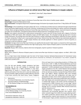
  • 2. Iqra Nehal et al ISRA MEDICAL JOURNAL | Volume 11 - Issue 3 | May – Jun 2019 164 of retinal nerve fiber layer and the macula. Rationale of this study was that myopia is a highly significant problem in Pakistani population3 , not only because of its high prevalence, but also because it can contribute to visual morbidity and increase the risk for vision-threatening conditions. It is important to investigate RNFL changes in myopic patients considering the risk of developing glaucoma with the severity of myopia. This study aids in diagnosing and managing early glaucomatous changes. Hence this study was conducted for the fulfillment of the degree program so glaucomatic parameters like IOP measurement and visual field measurements were the limitations of the study. On the other hand optic neuropathies were excluded and healthy myopic subjects were included due to these limitations. Further the objective of this study was to evaluate the myopic impact on retinal nerve fiber layer thickness in healthy myopic subjects. METHODOLOGY In this prospective observational study, 80 myopic eyes of 40 subjects i.e. both male and female were assessed. This study was conducted at Al-Ibrahim Eye Hospital, Karachi, Pakistan, in the time period of 1st May 2018 to 30th October 2018. The myopic patients were randomly selected attending the general outdoor patient department of Al-Ibrahim Eye Hospital. Mean age was considered as between 20-40 years in both genders. Three groups on the basis of refractive error were made among the respondents were; mild myopia (range= <-3.0 D), moderate myopia (range= -3.0 D to -6.0 D), and high myopia (range= >-6.0 D). Patients were included without any cylindrical component in first two groups, and ≤ 2.0 DC in high myopes. Subjects were excluded having any other ocular pathology such as any ocular media opacity, retinopathy, maculopathy, optic neuropathy, amblyopia, diabetes and hypertension. Subjects having visual acuity after best possible refraction less than 20/30 (6/9) were also excluded. Further, those patients who were not giving consent and those who have age more than 40 were excluded because of increased risk of systemic diseases like hypertension, diabetes mellitus, sarcopenia (age-related degeneration of muscular mass), muscular fibrosis etc. which can affect ocular health. Ethical confirmation of the study has been given by the ethical committee of the institution. An informed approval has been signed by every patient to participate in this study. Non- probability sampling technique was used for sampling. In this study new generation spectral domain Optical Coherrence Tomography used was of Nidek Company (software version was 1.5.5.0). The data was collected by the researcher through the general OPD and investigative department of the hospital. All patients were undergone full ophthalmic examination, starting with visual acuity on Snellen chart, objective refraction, subjective refraction and fundoscopy. After that, scans of SD- OCT were taken on each eye of each patient comprising a single map, i.e. Disc map was taken into consideration to evaluate RNFL thickness, CD ratio, disc area, rim area, RNFL symmetry. Hence conclusively disc map is very important in the interpretation and administration of glaucoma. In objective refraction refractive error was ruled out by Nidek autorefractometer. The mid points of the horizontal and vertical axis were aligned subjectively by the operator to focus centration around the optic nerve head. RNFL thickness was calculated according to 4 types of divisions; average thickness, superior and inferior thickness, four quadrant thickness and clock hour thickness. A 6.0x6.0 mm cube was selected with fine quality to measure the ONH parameters. Scans including artifacts whether central or peripheral, quality index less than 3/5 and signal strength index less than 7/10 were excluded from the study to avoid variations in measurements. Data Analysis: Data inquiry was done by SPSS version 20. Average RNFL thickness and thickness in the four quadrants was compared as according to increasing refractive error in myopes. Statistical significance was set at P < 0.05. RESULTS There were 80 healthy myopic eyes recruited in our study of 40 myopic subjects. Gender distribution was 22(55%) males and 18(54%) females. Average age was calculated as 25.0 ± 5.0 (range= 20 to 40 years). Spherical equivalent was -2.00 ± 0.7 for mild group, -5.00 ± 1.6 for moderate group and -8.00 ± 1.3 for high myopic group. Visual acuity after best possible correction was 20/20 for mild and moderate group and 20/30 for high myopic group. There were 12 (30%) mild, 20(50%) moderate and 08 (20%) highly myopic patients (Table-I). Table-I: Frequency of subjects according to degrees of myopia (N=40) FREQUENCY PERCENT Mild myopia 12 30.0% Moderate myopia 20 50.0% High myopia 8 20.0% Total 40 100.0% Average, superior, inferior and nasal thickness was decreased comparatively in high myopic group other than mild and moderate myopia, but surprisingly inferior quadrant thickness was clinically significant in terms of quantity in high myopic group among all other groups (Figure-1). Average thickness, inferior and nasal quadrant thickness was insignificant among three groups (p-value >0.05) (Figure-1). Decrease in thickness was significant in superior and temporal quadrants between the three groups (p-value <0.05) (Table-I).
  • 3. Iqra Nehal et al ISRA MEDICAL JOURNAL | Volume 11 - Issue 3 | May – Jun 2019 165 Fig-1: Graphical representation of RNFL thickness compared between different degrees of myopia (N=40) DISCUSSION Researchers are striving to identify risk factors for short- sightedness because its epidemic has attracted growing concern worldwide8,9 . The optimal management of the disease and its complications are also being studied rapidly10-12 . Mean of average RNFL thickness was 97.28±8.15 µm conducted by Akhter et al.2 in Islamabad. Correlation analysis among all subjects showed that the average, mean nasal quadrant, upper nasal, and inferonasal sub-quadrant RNFL thickness had positive correlation with spherical equivalent (p=0.045). In this study mean RNFL thickness was 90.85± 11.45 µm. Analysis shows superior and temporal quadrants had a positive correlation with RNFL. In spite of that, some studies13-16 are conducted on axial length correlated with RNFL thickness. Mehboob et al. researched in Pakistan in which they used laser interferometer (IOL master) and SD-OCT to measure axial length and RNFL thickness respectively. Three groups based on axial length were made in this study. A strong negative correlation between axial length and nerve fiber layer thickness of the retina was analyzed (p- value<0.001). They observed that axial length significantly effects measurements of RNFL thickness and on basis of thickness of RNFL; diagnosis of glaucoma should be counted. In this current study axial length was not measured while SD-OCT was used to measure RNFL thickness. Results of our study shows that there was a strong significant correlation in superior and temporal quadrant among 3 groups based on refractive error and average, superior and inferior thickness shows insignificant correlation with the refractive error. Some studies suggest that thickness of nerve fiber layer varies with the type of refractive error and as well as the amount of refractive error. Lee et al.17 studied RNFL thickness in short- sighted, long-sighted and emmetropic children. He found that RNFL thickness was thickest in the hyperopic group followed by myopic and emmetropic group. When the age was adjusted myopes had a thinner RNFL other than two groups while emmetropes and hyperopes had equal RNFL thickness. In our study only, myopic group was considered and mean adult age was taken between 20 to 40 years. We hadn’t adjusted age by any statistical formula because our study aimed to evaluate impact of myopic intensity on RNFL. In contrast studies which discussed about amount of refractive error like Kamath and his collegue18 discussed RNFL thickness in 3600 quadrants of variable degrees of myopic patients. Group III (High myopia) showed statistically significant thinning in 3600 in all quadrants except temporal quadrant compared to group I and II (low and moderate myopia). Results of our study indicate that only superior and temporal quadrant shows statistically significant thinning in high myopes. Amongst studies with SD-OCT similar results were noted by Kang et al.19 , Wang et al.20 , and Savini et al.21 who noted that peri- papillary thickness of RNFL decreases with increments in the dioptric power of myopia. Although Kang et al. and Wang et al. found significant decrease only in superior and inferior and in nasal quadrant while change was insignificant in temporal quadrant. Savini et al also noted decrease in RNFL thickness with weakest correlation of decrease in temporal quadrant. Results of our study represent significant positive correlation in superior and temporal quadrant (p=0.04 and p=0.03 respectively) while significantly negative correlation was found in average (p=0.08), inferior (p=0.10), and nasal quadrants (p=0.28). In contradiction Hoh et al22 suggested that the peri-papillary thickness of RNFL does not alter with degree of myopia or length of the eyeball. These discrepancies can be associated with the different OCT imaging equipment used. They used OCT-1 which obtains only 12 RNFL thickness values from each scan, while rest of the studies, as well as ours, used stratus, Cirrus or SD-OCT. It is likely that the values obtained in OCT-1 scans may be insufficient to pick up differences due to length of the eye ball or degree of refractive error. CONCLUSION Average RNFL thickness was significantly decreased in high myopia as compared to mild myopia while moderate group had slightly thicker thickness than high myopic group. Hence impact of dioptric power on nerve fiber layer thickness in myopic patients is significant. ACKNOWLEDGEMENT 'Thanks to Mr. M. Arslan and Ms. Mahnoor from R and D Department, ATMC, Isra University, Karachi campus for their support'. CONTRIBUTION OF AUTHORS Nehal I: Conceived idea, Literature review, Manuscript writing. Kazi U: Literature review, Manuscript writing Saleem A: Literature review, Manuscript writing 97 114 128 70 67 88 109 114 70 59 87 105 112 80 57 0 20 40 60 80 100 120 140 Total RNFL thickness Superior RNFL thickness Inferior RNFL thickness Temporal RNFL thickness Nasal RNFL thickness RNFLthickness MILD MYOPIA MODERATE MYOPIA HIGH MYOPIA
  • 4. Iqra Nehal et al ISRA MEDICAL JOURNAL | Volume 11 - Issue 3 | May – Jun 2019 166 Disclaimer: None. Conflict of Interest: None. Source of Funding: None. REFERENCES 1. Tai ELM, Ling JL, Gan EH, Adil H, Wan-Hazabbah WH. Comparison Of Peripapillary Retinal Nerve Fiber Layer Thickness Between Myopia Severity Groups And Controls. Int J Ophthalmol 2018;11(2): 274-78 2. Holden BA, Frickle TR, Wilson DA, Jong M, Naidoo KS, Sankaridurg P, et al. Global Prevalence Of Myopia And High Myopia And Temporal Ternds From 2000 Through 2050. Ophthalmol 2016; 123(5): 1036-42 3. Akhtar N, Kausar A, Afzal F, Ali SK, Hamid N. Peripapillary Retinal Nerve Fiber Layer (RNFL) Thickness Measurements by Topcon SD-OCT in Myopic Patients. J of the Coll of Physic & Surg Pak 2017; 28(1): 26-30. 4. Hsu S-Y, Chang M-S, Ko M-L, Harnod T. Retinal Nerve Fiber Layer Thickness And Optic Nerve Head Size Measured In High Myopes By Optical Coherence Tomography. Clini Experimental Optom 2013; 96(1): 373–78 5. Singh D , Mishra SK, Agarwal E ,Sharma R, Bhartiya S, Dada T. Assessment of Retinal Nerve Fiber Layer Changes By Cirrus High-Definition Optical Coherence Tomography In Myopia. J Curr Glaucoma Pract 2017;11(2): 52-57 6. Malakar M, Askari SN, Ashraf H, Waris A, Ahuja A, Asghar A. Optical coherence tomography assisted retinal nerve fibre layer thickness profile in high myopia. J of clini & Diag Res J 2015;9(2): NC01 7. Chowdhary T, Chowdhary DS, Choudhary A. Study of Retinal Nerve Fiber Layer Thickness in Myopic Eye in Rajasthani Population. Int J of Sci & Res 2015; 4(2): 880-81 8. Cuellar‐Partida G, Lu Y, Kho PF, Hewitt AW, Wichmann HE, Yazar S, et al. Assessing the genetic predisposition of education on myopia: a Mendelian randomization study. Genetic Epidemiol. 2016;40(1): 66-72 9. Parssinen O, Kauppinen M. Associations Of Reading Posture, Gaze Angle And Reading Distance With Myopia And Myopic Progression. Acta Ophthalmol 2016; 94(8): 775-79 10. Davidson SL, O'Hara M, Wagner RS. Management of Progressive Myopia. J Pediatric Ophthalmol Strabismus 2016;53(3): 134-36 11. Huang J, Wen D, Wang Q, McAlinden C, Flitcroft I, Chen H, et al. Efficacy comparison of 16 interventions for myopia control in children: a network meta-analysis. Ophthalmol 2016;123(4): 697-08 12. Wolffsohn JS, Calossi A, Cho P, Gifford K, Jones L, Li M, et al. Global trends in myopia management attitudes and strategies in clinical practice. Contact Lens and Anterior Eye 2016;39(2): 106-16 13. Mehboob MA, Islam QU, Yaqub MA. Effect Of Axial Length on Peripapillary Retinal Nerve Fibre Layer Thickness Measured by Spectral Domain Optical Coherence Tomography. Pak Armed Forc Med J 2017; 67(2): 238-42 14. Bae SH, Kang SH, Feng CS, Park J, Jeong JH, Yi K. Influence of myopia on size of optic nerve head and retinal nerve fiber layer thickness measured by spectral domain optical coherence tomography. Korean J Ophthalmol 2016;30(5): 335-43 15. AttaAllah HR, Omar IA, Abdelhalim AS. Evaluation of optic nerve head parameters and retinal nerve fiber layer thickness in axial myopia using SD OCT. Ophthalmol & Therapy. 2017;6(2): 335-41 16. Singh D, Mishra SK, Agarwal E, Sharma R, Bhartiya S, Dada T. Assessment of retinal nerve fiber layer changes by cirrus high-definition optical coherence tomography in myopia. J of Current Glaucoma Pract. 2017;11(2): 52 17. Lee JW, Yau GS, Woo TT, Yick DW, Tam VT, Lai JS. Retinal nerve fiber layer thickness in myopic, emmetropic, and hyperopic children. Medicine 2015; 94(12): e699. 18. Kamath AR, Dudeja L. Peri-papillary retinal nerve fiber layer thickness profile in subjects with myopia measured using optical coherence tomography. J of Clin Ophthalmol & Res 2014;2(3): 131 19. Kang SH, Hong SW, Im SK, Lee SH, Ahn MD. Effect Of Myopia On Thickness Of Retinal Nerve Fiber Layer Measured By Cirrus HD Optical Coherence Tomography. Invest Ophthalmol Vis Sci 2010; 51(1): 4075-83 20. Wang G, Qlu KL, Lu XH, Sun LX, Liao XJ, Chen HL, et al. The Effect Of Myopia On Retinal Nerve Fibre Measurement: A Comparative Study Of Spectral Domain Optical Coherence Tomography And Scanning Laser Polarimetry. Br J Ophthalmol 2011; 95(1): 255-60 21. Savini G, Barboni P, Parisi V, Carbonelli M. The Influence Of Axial Length On Retinal Nerve Fibre Layer Thickness And Optic Disc Size Measurements By Spectral Domain OCT. Br J Ophthalmol 2012;96(1): 57-61 22. Hoh ST, Lim MC, Seah SK, Lim AT, Chew SJ, Foster PJ, et al. Peripapillary Retinal Nerve Fibre Layer Thickness Variations With Myopia. Ophthalmology 2006;113(1): 773-77