SlideShare une entreprise Scribd logo
1  sur  75
1
2
3
3rd
Nerve
PRESENTATION CONTENT
•Origin &
Insertion
•Relation with eye
•Important supply
Embryology
• By the fourth week of development , nuclei
of all cranial nerves are present.
• Parasympathetic (visceral efferent) ganglia
and somatic efferent nuclei are derived
from neural crest cells.
Introduction
• Third cranial nerve.
• It is the largest of the ocular motor nerves.
• It contains some 15,000 axons, including motor fibers and
parasympathetic motor fibers.
• Entirely motor in function.
• Supplies LPS and all extra ocular muscles except lateral
rectus and superior oblique.
• Also supplies the intraocular muscles namely sphincter
pupillae and ciliary muscle.
1 2 3 5
FASCICULUS
NUCLEAR COMPLEX BASILAR
INTRACAVERNOUS
PATHWAY OF OCULAR MOTOR NERVE
4
ORBITAL
Main parts of the Brain
1. Nucleus and cisternal portion
ORIGIN :
The oculomotor nerve originates from the upper part of
the midbrain/Mesencephalon.
The oculomotor nucleus lies in the deep periaqueductal
grey matter at the level of the superior colliculus anterior
to the cerebral aqueduct.
It is a longitudinal column of about 10mm length.
Two Nucleus :
Oculomotor Nucleus
» somatic motor fibers (general somatic
efferents)
Edinger-Westphal nuclei
» visceral motor fibers (general visceral
efferent)
OCULOMOTOR NERVE NUCLEI:
Include two motor nuclei:
 Main Motor Nucleus:
 Site : ventral part of central grey matter "surrounding
the cerebral aqueduct", of midbrain, at the level of
superior colliculus, close to median plane.
 Supply : all extrinsic muscles of the eye except
superior oblique & lateral rectus. The levator palpebrae
superioris muscles of both sides are supplied by single
central group of cells "central caudal nucleus". The
superior rectus muscle is supplied by contralateral
oculomotor nucleus. The remaining muscles are
innervated by ipsilateral oculomotor nucleus.
• Classically, this complex has been described with
four paired subnuclei supplying innervation to the
inferior, medial, and superior rectus muscles and to
the inferior oblique muscle.
• There is a single caudal, dorsal midline nucleus
providing innervation to the levator muscle.
• Innervation of the extraocular muscle from the paired
nuclei is ipsilateral for the inferior and medial rectus
muscles and the inferior oblique muscle.
• However, innervation of the superior rectus is
contralateral.
• The motor fibers to the superior rectus decussate
within the third nerve nucleus and join the fascicle of
the contralateral oculomotor nerve.
• It is composed of subnuclei supplying individual
extraocular muscles as follows :
1. Dorsolateral Nucleus : Ipsilateral Inferior Rectus
2. Intermedial Nucleus : Ipsilateral Inferior Oblique
3. Ventromedial Nucleus : Ipsilateral Medial Rectus
4. Paramedial (Scattered) Nucleus : Contralateral
Superior Rectus
5. Caudal Central Nucleus : Bilateral Levator
Palpebrae Superioris
From To
Uncrossed
fibers
ipsilateral
oculomotor
nucleus
Medial rectus.
Inferior rectus.
Inferior oblique.
Crossed
fibers
Contralateral
oculomotor
nucleus
Superior rectus.
uncrossed
fibers
oculomotor
nuclei
Levator palpebrae
superioris muscle.
Uncrossed
para-
sympathetic
fibers
Ipsilateral
Edinger
Westphal
nucleus
Constrictor pupliae
muscle.
Ciliary muscle.
 Accessory parasympathetic
(EDINGERWESTPHAL) nucleus:
 Site : it lies dorsal to the main motor nucleus.
 Supply : the axons, which are preganglionic
accompany other oculomotor fibres to relay within
ciliary ganglion in orbit and supply sphincter pupillae
& ciliary muscles via short ciliary nerves.
 It consists of a median and two lateral components.
 Perhaps, the cranial half of the nucleus is concerned
with light reflexes and the caudal half with
accommodation.
• In addition to the extraocular muscle motor
fibers, the Edinger-Westphal nuclei supply
the parasympathetic preganglionic neurons
that project to the ciliary ganglion.
• All evidence suggests that these fibers
project ipsilaterally.
Oculomotor
Nerve
Somato motor
Four extraocular
muscle
Superior Rectus
Inferior Rectus
Medial Rectus
Inferior Oblique
Levator palpebral
superiosis
Edinger-Westphal
(visceral motor)
constrictor
pupillae
Ciliary muscles
IMPORTANT SUPPLY TO THE EYE
2. FASCICULUS:
The fibers run through the tegmentum, red
nucleus and medial aspect of the
substantia nigra to emerge from the medial
aspect of the cerebral peduncle.
Emerge from the midbrain and pass into
interpeduncular space
Mid Brain/ Mesencephalon
Periequedoctor grey
matter
2. The Basilar part :
• Starts as a series of 15-20 rootlets in the
interpeduncular fossa.
• These rapidly coalase to form a large medial and a
small lateral root, which unite to form a flattened
nerve, which gets twisted bringing the inferior fibres
superiorly and superior fibers inferiorly; and thus the
nerve becomes a rounded cord.
• The nerve then passes below the posterior cerebral
artery, and above the superior cerebellar artery and
running lateral to & parallel with the posterior
communicating artery to reach the cavernous sinus.
* Starts Upper part of the mid brain
3. Cavernous sinus part:
• It moves anteriorly, passing the nerve pierces
the dura mater. and enters the lateral aspect of
the cavernous sinus superiorly.
• Within the cavernous sinus, it receives sympathetic
branches from the internal carotid plexus.
• These fibres do not combine with the oculomotor
nerve – they merely travel within its sheath.
• In the anterior part of cavernous sinus, the nerve
divides into superior and inferior branches which
enters the orbit through SOF within the annulus of
zinn.
4. Orbital part:
• The nerve enters the orbit through the
superior orbital fissure in the common
tendinous ring which were already divided
into two branches; one superior and one
inferior.
a. Superior branch: Motor innervation to the superior
rectus and levator palpabrae superioris. Sympathetic
fibres run with the superior branch to innervate the
superior tarsal muscle.
b.Inferior branch: Motor innervation to the inferior
rectus, medial rectus and inferior oblique.
Parasympathetic fibres to the ciliary ganglion, which
ultimately innervates the sphincter pupillae and ciliary
muscles.
The larger, inferior division divides into three branches :
1. Nerve To The Medial Rectus passes inferior to the optic nerve.
2. Nerve To Inferior Rectus passes downward and enters the muscle
on its upper aspect, and
3. Nerve To Inferior Oblique (longest of the three branches) passes in
b/w the IR & LR and supplies the IO from its posterior border. It
gives off the motor root to the ciliary ganglion.
Superior Orbital Fissure
Parasympathetic (Motor) Root:
• Carries preganglionic parasympathetic
fibres which arise from cells of Edinger-
Westphal nucleus and run through
oculomotor nerve & its branch to inferior
oblique to reach the ganglion.
• These fibres are relayed in the ganglion
and postganglionic fibres travel in the short
ciliary nerves.
• They carry:
Parasympathetic fibres: supply sphincter
pupillae & ciliary muscles (more than 95% of
these fibres are distributed to ciliary muscle
which is much larger in volume).
BLOOD SUPPLY
• All nerves are supplied with blood from adjacent vessels, which are
usually small and variable.
• The blood supply to the medial aspect of the brainstem is from
vessels directly off the Basilar artery.
• Small perforators off circumflex arteries (posterior cerebral, superior
cerebellar artery) may also supply the fascicular portion of the third
nerve as it courses through the ventral midbrain.
• The vascular supply of 3rd nerve in subarachnoid space is via
vascular twigs from the post. Cerebral A., the sup. Cerebellar A.,
and the tentorial and dorsal meningeal branches of the
meningohypophyseal trunk of the Internal Carotid Artery.
• In the cavernous sinus, the tentorial, dorsal meningeal, and inferior
hypophyseal branches of the meningohypophyseal trunk supply the
nerve along with branches from the ophthalmic artery.
• The blood supply of the oculomotor, trochlear and abducent nerves is
similarly arranged, the nutrient arteries being derived from any adjacent
smaller arteries.
• Though not often noted, it is obvious that deprivation of this blood supply
by spasm, thrombosis or embolism may produce paralysis or paresis of the
muscles supplied.
Small arteries
supply blood
→peripheral
nervous system
CLINICALLY
APPLIED
ASPECTS
ANATOMICAL BASIS OF CLINICAL
FEATURES OF THIRD NERVE PALSY
• A complete & a total third nerve palsy is of
common occurrence.
• May be congenital or acquired.
• Clinical features of complete third nerve
palsy include :
1. Ptosis - paralysis of LPS muscle.
2. Deviation – out, down and intorted – unopposed
action of LR and SO.
4. Pupil is fixed and dilated – paralysis of sphincter
pupillae muscle.
5. Accommodation is completely lost – paralysis of
ciliary muscle.
6. Crossed diplopia – paralytic divergent
squint.
7. Head posture – if the pupillary area is
uncovered, head takes a posture
consistent with the directions of actions of
the paralysed muscles, i.e., head is turned
on the opposite side, tilted towards the
same side and chin is slightly raised.
FEATURES AND CAUSES OF THIRD
NERVE LESIONS AT VARIOUS LEVELS
1. Nuclear lesions
• Lesions involving purely the third nerve nucleus are relatively
uncommon.
• Common causes include ; vascular diseases, demyelination,
primary tumors and metastasis
• Lesions involving nucleus cause an ipsilateral third nerve palsy
with ipsilateral sparing and contralateral weakness of elevation.
• Lesions involving paired medial rectus subnuclei (ventromedial
nucleus) cause a wall-eyed bilateral internuclear ophthalmoplegia (
WEBINO ) characterised by defective convergence and adduction.
2. Fascicular lesions
• Causes are similar to nuclear lesions.
Benedikt syndrome involves the fasciculus as it passes
through the red nucleus and is characterised by
ipsilateral 3rd nerve palsy and contralateral
extrapyramidal signs such as hemitremor.
Weber syndrome involves the fasciculus as it passes
through the cerebral peduncle and is characterised by
ipsilateral 3rd nerve palsy and contralateral hemiparesis.
Nothnagel syndrome involves the fasciculus and the
superior cerebellar peduncle and is characterised by
ipsilateral 3rd nerve palsy and cerebellar ataxia.
Claude syndrome is a combination of Benedikt and
Nothnagel syndromes
COMMON CAUSES OF NUCLEAR AND
FASICULAR THIRD CRANIAL NERVE
CHILDREN
Congenital
- with neurological abnormalities
- with aberrant rennervation
- with cyclic oculomotor spasm
Vascular ( AV
malformations )
Primary tumour
Metastatic tumour
YOUNG
ADULTS
Demyelinating
Vascular
(hemorrhage or
infarction)
Tumor
OLDER
ADULTS
Vascular
(infarction)
Tumour
3. Lesions involving basilar part of the
nerve:
• isolated third nerve palsies are frequently basilar.
Causes :
1. Aneurysms : At the posterior communicating artery cause
isolated third nerve palsy with involvement of pupil.
2. Head trauma: Extradural hematomas which may cause
tentorial pressure cone with downward herniation of the
temporal lobe. This compresses the third nerve as it passes
over the tentorial edge. Initially there occurs fixed, dilated
pupil, which is followed by a total third nerve palsy.
3. Diabetes causes isolated 3rd nerve palsy with sparing of the
pupillary reflexes.
4. Lesions involving intracavernous part
of the nerve:
• Because of its close proximity to other
cranial nerves, intracavernous 3rd nerve
palsies are usually associated with
involvement of the 4th and 6th nerves, and
the 1st division of trigeminal nerve.
• Important Causes :
1. Diabetes may cause vascular palsy.
2. Pituitary apoplexy – may cause a third
nerve palsy (after child birth), if the gland
swells laterally & extension into cavernous
sinus.
3. Intracavernous lesions – aneurysms,
meningiomas, carotid-cavernous fistulae
and Tolosa-Hunt syndrome
(granulomatous inflammation)
5. Lesions of the intraorbital part of
the nerve:
• May cause isolated extraocular muscle
palsies or may involve either superior
division or inferior division or both.
• Causes : orbital tumors, trauma and
vascular diseases.
6. Lesions of pupillomotor fibres
• B/w the brainstem and the cavernous
sinus, pupillomotor fibres are located
superficially in the superior median
quadrant of the nerve.
• They derive the blood supply from the pial
blood vessels whereas the main trunk of
the 3rd nerve is supplied by vasa
nervorum.
• Surgical lesions such as aneurysms,
trauma and uncal herniation
characteristically involve the pupil by
compressing the pial blood vessels and the
superficially located pupillary fibres.
• Medical lesions such as diabetes and
hypertension usually spare the pupil. This
is because the microangiopathy associated
with these diseases involves the vasa
nervorum, causing infarction of the main
trunk, but sparing the superficial pupillary
fibres.
In ISOLATED THIRD NERVE PALSY diabetes
and HTN, is the most common cause of pupil-
sparing 3rd nerve palsy. In most cases
spontaneous recovery occurs within 3 months.
Diabetic 3rd nerve palsy is often assoc. with
periorbital pain and may occasionally be the
presenting feature of diabetes. The presence of
pain is therefore not helpful in differentiating b/w
aneurysmal and diabetic 3rd nerve palsy.
Multiple
choice
Questions
1. Which of the following statements regarding the origin and course
of III is/are correct except?
a) It emerges as a number of rootlets from a nucleus lying level with the superior
colliculus
b) It leaves the brainstem after passing through the red nucleus
c) It runs below and lateral to the free margin of the tentorium cerebelli
d) It pierces the dura midway between the anterior and posterior clinoid processes
e) Its branches cross the same part of the superior orbital fissure as VI
2. A patient suffers damage to the orbit in a road traffic incident
resulting in damage to the third cranial nerve. Which of the
following signs will be present?
a) Pupillary constriction and a medial strabismus
b) Pupillary dilatation and a medial strabismus
c) Pupillary constriction and a lateral strabismus
d) Pupillary dilatation and a lateral strabismus
3. Regarding the distribution of fibres from III
a) Levator palpebrae superioris only receives a motor supply from the superior
division of III
b) It innervates all the muscles at the junction of their middle and posterior thirds
c) The parasympathetic supply to the iris is conveyed via the short ciliary nerves
d) Superior rectus is innervated ipsilaterally
e) Levator palpebrae superioris contralaterally represented at the level of the
oculomotor nuclei
4. Which of the following statements regarding III is correct?
a) The parasympathetic fibres lie centrally in the precavernous sinus portion of the
nerve
b) A third nerve palsy causes the eye down and out.
c) Accommodation is unaffected by such a palsy
d) Compression of the nerve causes a partial ptosis
e) The Edinger-Westphal nucleus lies ventral to the main oculomotor nucleus
5. All of the following are characteristics of oculomotor nerve except:
a) Carries parasympathetic nerve fibers
b) Supplies inferior oblique muscle
c) Enters orbit through the inferior orbital fissure
d) Causes constriction of pupil
6. The following structure(s) synpase(s) in the ciliary ganglion:
a) Nasociliary nerve fibres
b) Parasympathetic nerve fibres from oculomotor nerve
c) Sympathetic nerve fibres from the carotid plexus
d) Parasympathetic nerve fibres form the facial nerve abducent nerve fibres.
Thank You

Contenu connexe

Tendances

EXTRA OCULAR MUSCLES ANATOMY
EXTRA OCULAR MUSCLES ANATOMYEXTRA OCULAR MUSCLES ANATOMY
EXTRA OCULAR MUSCLES ANATOMYRabia Ammer
 
Cranial nerve iii , iv and vi
Cranial nerve iii , iv and viCranial nerve iii , iv and vi
Cranial nerve iii , iv and vifarhan_aq91
 
Visual pathway and lesion
Visual pathway and lesionVisual pathway and lesion
Visual pathway and lesionVikas Khatri
 
Abducens Nerve - Course and relation.
Abducens Nerve - Course and relation.Abducens Nerve - Course and relation.
Abducens Nerve - Course and relation.Izmal Urooj
 
Extraocular muscles
Extraocular muscles Extraocular muscles
Extraocular muscles mgmcri1234
 
Anatomy of lateral geniculate body and visual cortex
Anatomy of lateral geniculate body and visual cortexAnatomy of lateral geniculate body and visual cortex
Anatomy of lateral geniculate body and visual cortexRuturaj Sahoo
 
Ophthalmic artery
Ophthalmic arteryOphthalmic artery
Ophthalmic arteryRawan Ayman
 
Oculomotor nerve
Oculomotor nerveOculomotor nerve
Oculomotor nerveAmr Hassan
 
Anatomy of the orbit (nerves and vessels)
Anatomy of the orbit (nerves and vessels)Anatomy of the orbit (nerves and vessels)
Anatomy of the orbit (nerves and vessels)Dr. Mohammad Mahmoud
 
Anatomy of the lacrimal apparatus
Anatomy of the lacrimal apparatusAnatomy of the lacrimal apparatus
Anatomy of the lacrimal apparatusSSSIHMS-PG
 
Oculomotor nerve
Oculomotor nerveOculomotor nerve
Oculomotor nerveAmyEmtage
 

Tendances (20)

Ciliary ganglion
Ciliary ganglionCiliary ganglion
Ciliary ganglion
 
Orbital anatomy
Orbital anatomyOrbital anatomy
Orbital anatomy
 
EXTRA OCULAR MUSCLES ANATOMY
EXTRA OCULAR MUSCLES ANATOMYEXTRA OCULAR MUSCLES ANATOMY
EXTRA OCULAR MUSCLES ANATOMY
 
Trochlear nerve
Trochlear nerveTrochlear nerve
Trochlear nerve
 
Abducent nerve
Abducent nerveAbducent nerve
Abducent nerve
 
Cavernous sinus
Cavernous sinusCavernous sinus
Cavernous sinus
 
Cranial nerve iii , iv and vi
Cranial nerve iii , iv and viCranial nerve iii , iv and vi
Cranial nerve iii , iv and vi
 
Anatomy of 3rd cranial nerve
Anatomy of 3rd cranial nerveAnatomy of 3rd cranial nerve
Anatomy of 3rd cranial nerve
 
Visual pathway and lesion
Visual pathway and lesionVisual pathway and lesion
Visual pathway and lesion
 
Abducens Nerve - Course and relation.
Abducens Nerve - Course and relation.Abducens Nerve - Course and relation.
Abducens Nerve - Course and relation.
 
Extraocular muscles
Extraocular muscles Extraocular muscles
Extraocular muscles
 
Muscles of the eye
Muscles of the eyeMuscles of the eye
Muscles of the eye
 
Anatomy of lateral geniculate body and visual cortex
Anatomy of lateral geniculate body and visual cortexAnatomy of lateral geniculate body and visual cortex
Anatomy of lateral geniculate body and visual cortex
 
Ophthalmic artery
Ophthalmic arteryOphthalmic artery
Ophthalmic artery
 
Light reflex
Light reflexLight reflex
Light reflex
 
Ophthalmic artery
Ophthalmic arteryOphthalmic artery
Ophthalmic artery
 
Oculomotor nerve
Oculomotor nerveOculomotor nerve
Oculomotor nerve
 
Anatomy of the orbit (nerves and vessels)
Anatomy of the orbit (nerves and vessels)Anatomy of the orbit (nerves and vessels)
Anatomy of the orbit (nerves and vessels)
 
Anatomy of the lacrimal apparatus
Anatomy of the lacrimal apparatusAnatomy of the lacrimal apparatus
Anatomy of the lacrimal apparatus
 
Oculomotor nerve
Oculomotor nerveOculomotor nerve
Oculomotor nerve
 

En vedette

13-03-2017_PORTFOLIO_ACADEMIC_PROFESSIONAL_LOW_RES
13-03-2017_PORTFOLIO_ACADEMIC_PROFESSIONAL_LOW_RES13-03-2017_PORTFOLIO_ACADEMIC_PROFESSIONAL_LOW_RES
13-03-2017_PORTFOLIO_ACADEMIC_PROFESSIONAL_LOW_RESAr. Archana Jaiswal
 
Wheelster hoverboard riding techniques
Wheelster hoverboard riding techniquesWheelster hoverboard riding techniques
Wheelster hoverboard riding techniquesWheelster Inc.
 
Clasificacion de las empresas
Clasificacion de las empresasClasificacion de las empresas
Clasificacion de las empresasManuelsoberanis
 
Reglamento disciplinario para_los_trabajadores
Reglamento disciplinario para_los_trabajadoresReglamento disciplinario para_los_trabajadores
Reglamento disciplinario para_los_trabajadoresSarahi Gonzalez
 
Departamento de producción
Departamento de producciónDepartamento de producción
Departamento de producciónMonserrat Correa
 
Лекция «Точка на карте репрессий. Зюзино». 12.03.2017
Лекция «Точка на карте репрессий. Зюзино». 12.03.2017 Лекция «Точка на карте репрессий. Зюзино». 12.03.2017
Лекция «Точка на карте репрессий. Зюзино». 12.03.2017 zuzino_shtab
 
ISTILAH - ISTILAH DALAM ILMU HADITS
ISTILAH - ISTILAH DALAM ILMU HADITSISTILAH - ISTILAH DALAM ILMU HADITS
ISTILAH - ISTILAH DALAM ILMU HADITSAzzahra Azzahra
 
Report for genius coffee
Report for genius coffeeReport for genius coffee
Report for genius coffeeNgwe Tun
 
Presentation on healthy relationships
Presentation on healthy  relationshipsPresentation on healthy  relationships
Presentation on healthy relationshipscorrieperdok
 

En vedette (10)

13-03-2017_PORTFOLIO_ACADEMIC_PROFESSIONAL_LOW_RES
13-03-2017_PORTFOLIO_ACADEMIC_PROFESSIONAL_LOW_RES13-03-2017_PORTFOLIO_ACADEMIC_PROFESSIONAL_LOW_RES
13-03-2017_PORTFOLIO_ACADEMIC_PROFESSIONAL_LOW_RES
 
Wheelster hoverboard riding techniques
Wheelster hoverboard riding techniquesWheelster hoverboard riding techniques
Wheelster hoverboard riding techniques
 
Clasificacion de las empresas
Clasificacion de las empresasClasificacion de las empresas
Clasificacion de las empresas
 
Reglamento disciplinario para_los_trabajadores
Reglamento disciplinario para_los_trabajadoresReglamento disciplinario para_los_trabajadores
Reglamento disciplinario para_los_trabajadores
 
Departamento de producción
Departamento de producciónDepartamento de producción
Departamento de producción
 
Лекция «Точка на карте репрессий. Зюзино». 12.03.2017
Лекция «Точка на карте репрессий. Зюзино». 12.03.2017 Лекция «Точка на карте репрессий. Зюзино». 12.03.2017
Лекция «Точка на карте репрессий. Зюзино». 12.03.2017
 
ISTILAH - ISTILAH DALAM ILMU HADITS
ISTILAH - ISTILAH DALAM ILMU HADITSISTILAH - ISTILAH DALAM ILMU HADITS
ISTILAH - ISTILAH DALAM ILMU HADITS
 
Proyecto 11
Proyecto 11Proyecto 11
Proyecto 11
 
Report for genius coffee
Report for genius coffeeReport for genius coffee
Report for genius coffee
 
Presentation on healthy relationships
Presentation on healthy  relationshipsPresentation on healthy  relationships
Presentation on healthy relationships
 

Similaire à Oculomotor nerve

Similaire à Oculomotor nerve (20)

Occulo motor nerve dnm
Occulo motor nerve dnmOcculo motor nerve dnm
Occulo motor nerve dnm
 
Anatomy of oculomotor nerve
Anatomy of oculomotor nerveAnatomy of oculomotor nerve
Anatomy of oculomotor nerve
 
3rd, 4th and 6th cranial nerve
3rd, 4th and 6th cranial nerve3rd, 4th and 6th cranial nerve
3rd, 4th and 6th cranial nerve
 
THIRD NERVE - ANATOMY AND APPLIED ASPECT.
THIRD NERVE - ANATOMY AND APPLIED ASPECT.THIRD NERVE - ANATOMY AND APPLIED ASPECT.
THIRD NERVE - ANATOMY AND APPLIED ASPECT.
 
Spinal cord
Spinal cordSpinal cord
Spinal cord
 
Sensory phisiology
Sensory phisiologySensory phisiology
Sensory phisiology
 
2-Anatomy of the Spinal Cord [Autosaved].ppt
2-Anatomy of the Spinal Cord [Autosaved].ppt2-Anatomy of the Spinal Cord [Autosaved].ppt
2-Anatomy of the Spinal Cord [Autosaved].ppt
 
Cranial nerves
Cranial nervesCranial nerves
Cranial nerves
 
Cerebellum
 Cerebellum  Cerebellum
Cerebellum
 
The Brain
The BrainThe Brain
The Brain
 
The Human Brain lectures Brain structures parts functions
The Human Brain lectures Brain structures  parts functionsThe Human Brain lectures Brain structures  parts functions
The Human Brain lectures Brain structures parts functions
 
Spinal cord and spinal nerves
Spinal cord and spinal nervesSpinal cord and spinal nerves
Spinal cord and spinal nerves
 
Ch11 eoc
Ch11 eocCh11 eoc
Ch11 eoc
 
Anatomy & functions of the Brainstem & Cerebellum
Anatomy & functions of the Brainstem & CerebellumAnatomy & functions of the Brainstem & Cerebellum
Anatomy & functions of the Brainstem & Cerebellum
 
Autonomic Nervous System
Autonomic Nervous SystemAutonomic Nervous System
Autonomic Nervous System
 
OCULAR NERVE SUPPLY
OCULAR NERVE SUPPLYOCULAR NERVE SUPPLY
OCULAR NERVE SUPPLY
 
Cerebellum by DR.ARSHAD
Cerebellum by DR.ARSHADCerebellum by DR.ARSHAD
Cerebellum by DR.ARSHAD
 
Anatomy of brain steam midbrain pons and medulla
Anatomy of brain steam midbrain pons and medullaAnatomy of brain steam midbrain pons and medulla
Anatomy of brain steam midbrain pons and medulla
 
DESCENDING-TRACTS.pptx
DESCENDING-TRACTS.pptxDESCENDING-TRACTS.pptx
DESCENDING-TRACTS.pptx
 
11 nerous system1
11 nerous system111 nerous system1
11 nerous system1
 

Dernier

97111 47426 Call Girls In Delhi MUNIRKAA
97111 47426 Call Girls In Delhi MUNIRKAA97111 47426 Call Girls In Delhi MUNIRKAA
97111 47426 Call Girls In Delhi MUNIRKAAjennyeacort
 
Call Girls Thane Just Call 9910780858 Get High Class Call Girls Service
Call Girls Thane Just Call 9910780858 Get High Class Call Girls ServiceCall Girls Thane Just Call 9910780858 Get High Class Call Girls Service
Call Girls Thane Just Call 9910780858 Get High Class Call Girls Servicesonalikaur4
 
Hematology and Immunology - Leukocytes Functions
Hematology and Immunology - Leukocytes FunctionsHematology and Immunology - Leukocytes Functions
Hematology and Immunology - Leukocytes FunctionsMedicoseAcademics
 
Low Rate Call Girls Mumbai Suman 9910780858 Independent Escort Service Mumbai
Low Rate Call Girls Mumbai Suman 9910780858 Independent Escort Service MumbaiLow Rate Call Girls Mumbai Suman 9910780858 Independent Escort Service Mumbai
Low Rate Call Girls Mumbai Suman 9910780858 Independent Escort Service Mumbaisonalikaur4
 
Noida Sector 135 Call Girls ( 9873940964 ) Book Hot And Sexy Girls In A Few C...
Noida Sector 135 Call Girls ( 9873940964 ) Book Hot And Sexy Girls In A Few C...Noida Sector 135 Call Girls ( 9873940964 ) Book Hot And Sexy Girls In A Few C...
Noida Sector 135 Call Girls ( 9873940964 ) Book Hot And Sexy Girls In A Few C...rajnisinghkjn
 
Russian Call Girls Chickpet - 7001305949 Booking and charges genuine rate for...
Russian Call Girls Chickpet - 7001305949 Booking and charges genuine rate for...Russian Call Girls Chickpet - 7001305949 Booking and charges genuine rate for...
Russian Call Girls Chickpet - 7001305949 Booking and charges genuine rate for...narwatsonia7
 
call girls in green park DELHI 🔝 >༒9540349809 🔝 genuine Escort Service 🔝✔️✔️
call girls in green park  DELHI 🔝 >༒9540349809 🔝 genuine Escort Service 🔝✔️✔️call girls in green park  DELHI 🔝 >༒9540349809 🔝 genuine Escort Service 🔝✔️✔️
call girls in green park DELHI 🔝 >༒9540349809 🔝 genuine Escort Service 🔝✔️✔️saminamagar
 
Call Girl Koramangala | 7001305949 At Low Cost Cash Payment Booking
Call Girl Koramangala | 7001305949 At Low Cost Cash Payment BookingCall Girl Koramangala | 7001305949 At Low Cost Cash Payment Booking
Call Girl Koramangala | 7001305949 At Low Cost Cash Payment Bookingnarwatsonia7
 
Call Girl Service Bidadi - For 7001305949 Cheap & Best with original Photos
Call Girl Service Bidadi - For 7001305949 Cheap & Best with original PhotosCall Girl Service Bidadi - For 7001305949 Cheap & Best with original Photos
Call Girl Service Bidadi - For 7001305949 Cheap & Best with original Photosnarwatsonia7
 
Book Call Girls in Kasavanahalli - 7001305949 with real photos and phone numbers
Book Call Girls in Kasavanahalli - 7001305949 with real photos and phone numbersBook Call Girls in Kasavanahalli - 7001305949 with real photos and phone numbers
Book Call Girls in Kasavanahalli - 7001305949 with real photos and phone numbersnarwatsonia7
 
Book Call Girls in Yelahanka - For 7001305949 Cheap & Best with original Photos
Book Call Girls in Yelahanka - For 7001305949 Cheap & Best with original PhotosBook Call Girls in Yelahanka - For 7001305949 Cheap & Best with original Photos
Book Call Girls in Yelahanka - For 7001305949 Cheap & Best with original Photosnarwatsonia7
 
See the 2,456 pharmacies on the National E-Pharmacy Platform
See the 2,456 pharmacies on the National E-Pharmacy PlatformSee the 2,456 pharmacies on the National E-Pharmacy Platform
See the 2,456 pharmacies on the National E-Pharmacy PlatformKweku Zurek
 
Call Girls Service Nandiambakkam | 7001305949 At Low Cost Cash Payment Booking
Call Girls Service Nandiambakkam | 7001305949 At Low Cost Cash Payment BookingCall Girls Service Nandiambakkam | 7001305949 At Low Cost Cash Payment Booking
Call Girls Service Nandiambakkam | 7001305949 At Low Cost Cash Payment BookingNehru place Escorts
 
call girls in munirka DELHI 🔝 >༒9540349809 🔝 genuine Escort Service 🔝✔️✔️
call girls in munirka  DELHI 🔝 >༒9540349809 🔝 genuine Escort Service 🔝✔️✔️call girls in munirka  DELHI 🔝 >༒9540349809 🔝 genuine Escort Service 🔝✔️✔️
call girls in munirka DELHI 🔝 >༒9540349809 🔝 genuine Escort Service 🔝✔️✔️saminamagar
 
Call Girls Hsr Layout Just Call 7001305949 Top Class Call Girl Service Available
Call Girls Hsr Layout Just Call 7001305949 Top Class Call Girl Service AvailableCall Girls Hsr Layout Just Call 7001305949 Top Class Call Girl Service Available
Call Girls Hsr Layout Just Call 7001305949 Top Class Call Girl Service Availablenarwatsonia7
 
Call Girl Nagpur Sia 7001305949 Independent Escort Service Nagpur
Call Girl Nagpur Sia 7001305949 Independent Escort Service NagpurCall Girl Nagpur Sia 7001305949 Independent Escort Service Nagpur
Call Girl Nagpur Sia 7001305949 Independent Escort Service NagpurRiya Pathan
 
Call Girls Service in Bommanahalli - 7001305949 with real photos and phone nu...
Call Girls Service in Bommanahalli - 7001305949 with real photos and phone nu...Call Girls Service in Bommanahalli - 7001305949 with real photos and phone nu...
Call Girls Service in Bommanahalli - 7001305949 with real photos and phone nu...narwatsonia7
 
Call Girl Surat Madhuri 7001305949 Independent Escort Service Surat
Call Girl Surat Madhuri 7001305949 Independent Escort Service SuratCall Girl Surat Madhuri 7001305949 Independent Escort Service Surat
Call Girl Surat Madhuri 7001305949 Independent Escort Service Suratnarwatsonia7
 
College Call Girls Vyasarpadi Whatsapp 7001305949 Independent Escort Service
College Call Girls Vyasarpadi Whatsapp 7001305949 Independent Escort ServiceCollege Call Girls Vyasarpadi Whatsapp 7001305949 Independent Escort Service
College Call Girls Vyasarpadi Whatsapp 7001305949 Independent Escort ServiceNehru place Escorts
 
Call Girls Jp Nagar Just Call 7001305949 Top Class Call Girl Service Available
Call Girls Jp Nagar Just Call 7001305949 Top Class Call Girl Service AvailableCall Girls Jp Nagar Just Call 7001305949 Top Class Call Girl Service Available
Call Girls Jp Nagar Just Call 7001305949 Top Class Call Girl Service Availablenarwatsonia7
 

Dernier (20)

97111 47426 Call Girls In Delhi MUNIRKAA
97111 47426 Call Girls In Delhi MUNIRKAA97111 47426 Call Girls In Delhi MUNIRKAA
97111 47426 Call Girls In Delhi MUNIRKAA
 
Call Girls Thane Just Call 9910780858 Get High Class Call Girls Service
Call Girls Thane Just Call 9910780858 Get High Class Call Girls ServiceCall Girls Thane Just Call 9910780858 Get High Class Call Girls Service
Call Girls Thane Just Call 9910780858 Get High Class Call Girls Service
 
Hematology and Immunology - Leukocytes Functions
Hematology and Immunology - Leukocytes FunctionsHematology and Immunology - Leukocytes Functions
Hematology and Immunology - Leukocytes Functions
 
Low Rate Call Girls Mumbai Suman 9910780858 Independent Escort Service Mumbai
Low Rate Call Girls Mumbai Suman 9910780858 Independent Escort Service MumbaiLow Rate Call Girls Mumbai Suman 9910780858 Independent Escort Service Mumbai
Low Rate Call Girls Mumbai Suman 9910780858 Independent Escort Service Mumbai
 
Noida Sector 135 Call Girls ( 9873940964 ) Book Hot And Sexy Girls In A Few C...
Noida Sector 135 Call Girls ( 9873940964 ) Book Hot And Sexy Girls In A Few C...Noida Sector 135 Call Girls ( 9873940964 ) Book Hot And Sexy Girls In A Few C...
Noida Sector 135 Call Girls ( 9873940964 ) Book Hot And Sexy Girls In A Few C...
 
Russian Call Girls Chickpet - 7001305949 Booking and charges genuine rate for...
Russian Call Girls Chickpet - 7001305949 Booking and charges genuine rate for...Russian Call Girls Chickpet - 7001305949 Booking and charges genuine rate for...
Russian Call Girls Chickpet - 7001305949 Booking and charges genuine rate for...
 
call girls in green park DELHI 🔝 >༒9540349809 🔝 genuine Escort Service 🔝✔️✔️
call girls in green park  DELHI 🔝 >༒9540349809 🔝 genuine Escort Service 🔝✔️✔️call girls in green park  DELHI 🔝 >༒9540349809 🔝 genuine Escort Service 🔝✔️✔️
call girls in green park DELHI 🔝 >༒9540349809 🔝 genuine Escort Service 🔝✔️✔️
 
Call Girl Koramangala | 7001305949 At Low Cost Cash Payment Booking
Call Girl Koramangala | 7001305949 At Low Cost Cash Payment BookingCall Girl Koramangala | 7001305949 At Low Cost Cash Payment Booking
Call Girl Koramangala | 7001305949 At Low Cost Cash Payment Booking
 
Call Girl Service Bidadi - For 7001305949 Cheap & Best with original Photos
Call Girl Service Bidadi - For 7001305949 Cheap & Best with original PhotosCall Girl Service Bidadi - For 7001305949 Cheap & Best with original Photos
Call Girl Service Bidadi - For 7001305949 Cheap & Best with original Photos
 
Book Call Girls in Kasavanahalli - 7001305949 with real photos and phone numbers
Book Call Girls in Kasavanahalli - 7001305949 with real photos and phone numbersBook Call Girls in Kasavanahalli - 7001305949 with real photos and phone numbers
Book Call Girls in Kasavanahalli - 7001305949 with real photos and phone numbers
 
Book Call Girls in Yelahanka - For 7001305949 Cheap & Best with original Photos
Book Call Girls in Yelahanka - For 7001305949 Cheap & Best with original PhotosBook Call Girls in Yelahanka - For 7001305949 Cheap & Best with original Photos
Book Call Girls in Yelahanka - For 7001305949 Cheap & Best with original Photos
 
See the 2,456 pharmacies on the National E-Pharmacy Platform
See the 2,456 pharmacies on the National E-Pharmacy PlatformSee the 2,456 pharmacies on the National E-Pharmacy Platform
See the 2,456 pharmacies on the National E-Pharmacy Platform
 
Call Girls Service Nandiambakkam | 7001305949 At Low Cost Cash Payment Booking
Call Girls Service Nandiambakkam | 7001305949 At Low Cost Cash Payment BookingCall Girls Service Nandiambakkam | 7001305949 At Low Cost Cash Payment Booking
Call Girls Service Nandiambakkam | 7001305949 At Low Cost Cash Payment Booking
 
call girls in munirka DELHI 🔝 >༒9540349809 🔝 genuine Escort Service 🔝✔️✔️
call girls in munirka  DELHI 🔝 >༒9540349809 🔝 genuine Escort Service 🔝✔️✔️call girls in munirka  DELHI 🔝 >༒9540349809 🔝 genuine Escort Service 🔝✔️✔️
call girls in munirka DELHI 🔝 >༒9540349809 🔝 genuine Escort Service 🔝✔️✔️
 
Call Girls Hsr Layout Just Call 7001305949 Top Class Call Girl Service Available
Call Girls Hsr Layout Just Call 7001305949 Top Class Call Girl Service AvailableCall Girls Hsr Layout Just Call 7001305949 Top Class Call Girl Service Available
Call Girls Hsr Layout Just Call 7001305949 Top Class Call Girl Service Available
 
Call Girl Nagpur Sia 7001305949 Independent Escort Service Nagpur
Call Girl Nagpur Sia 7001305949 Independent Escort Service NagpurCall Girl Nagpur Sia 7001305949 Independent Escort Service Nagpur
Call Girl Nagpur Sia 7001305949 Independent Escort Service Nagpur
 
Call Girls Service in Bommanahalli - 7001305949 with real photos and phone nu...
Call Girls Service in Bommanahalli - 7001305949 with real photos and phone nu...Call Girls Service in Bommanahalli - 7001305949 with real photos and phone nu...
Call Girls Service in Bommanahalli - 7001305949 with real photos and phone nu...
 
Call Girl Surat Madhuri 7001305949 Independent Escort Service Surat
Call Girl Surat Madhuri 7001305949 Independent Escort Service SuratCall Girl Surat Madhuri 7001305949 Independent Escort Service Surat
Call Girl Surat Madhuri 7001305949 Independent Escort Service Surat
 
College Call Girls Vyasarpadi Whatsapp 7001305949 Independent Escort Service
College Call Girls Vyasarpadi Whatsapp 7001305949 Independent Escort ServiceCollege Call Girls Vyasarpadi Whatsapp 7001305949 Independent Escort Service
College Call Girls Vyasarpadi Whatsapp 7001305949 Independent Escort Service
 
Call Girls Jp Nagar Just Call 7001305949 Top Class Call Girl Service Available
Call Girls Jp Nagar Just Call 7001305949 Top Class Call Girl Service AvailableCall Girls Jp Nagar Just Call 7001305949 Top Class Call Girl Service Available
Call Girls Jp Nagar Just Call 7001305949 Top Class Call Girl Service Available
 

Oculomotor nerve

  • 1.
  • 3. Embryology • By the fourth week of development , nuclei of all cranial nerves are present. • Parasympathetic (visceral efferent) ganglia and somatic efferent nuclei are derived from neural crest cells.
  • 4.
  • 5. Introduction • Third cranial nerve. • It is the largest of the ocular motor nerves. • It contains some 15,000 axons, including motor fibers and parasympathetic motor fibers. • Entirely motor in function. • Supplies LPS and all extra ocular muscles except lateral rectus and superior oblique. • Also supplies the intraocular muscles namely sphincter pupillae and ciliary muscle.
  • 6.
  • 7.
  • 8. 1 2 3 5 FASCICULUS NUCLEAR COMPLEX BASILAR INTRACAVERNOUS PATHWAY OF OCULAR MOTOR NERVE 4 ORBITAL
  • 9. Main parts of the Brain
  • 10.
  • 11. 1. Nucleus and cisternal portion ORIGIN : The oculomotor nerve originates from the upper part of the midbrain/Mesencephalon. The oculomotor nucleus lies in the deep periaqueductal grey matter at the level of the superior colliculus anterior to the cerebral aqueduct. It is a longitudinal column of about 10mm length. Two Nucleus : Oculomotor Nucleus » somatic motor fibers (general somatic efferents) Edinger-Westphal nuclei » visceral motor fibers (general visceral efferent)
  • 12.
  • 13. OCULOMOTOR NERVE NUCLEI: Include two motor nuclei:  Main Motor Nucleus:  Site : ventral part of central grey matter "surrounding the cerebral aqueduct", of midbrain, at the level of superior colliculus, close to median plane.  Supply : all extrinsic muscles of the eye except superior oblique & lateral rectus. The levator palpebrae superioris muscles of both sides are supplied by single central group of cells "central caudal nucleus". The superior rectus muscle is supplied by contralateral oculomotor nucleus. The remaining muscles are innervated by ipsilateral oculomotor nucleus.
  • 14. • Classically, this complex has been described with four paired subnuclei supplying innervation to the inferior, medial, and superior rectus muscles and to the inferior oblique muscle. • There is a single caudal, dorsal midline nucleus providing innervation to the levator muscle. • Innervation of the extraocular muscle from the paired nuclei is ipsilateral for the inferior and medial rectus muscles and the inferior oblique muscle. • However, innervation of the superior rectus is contralateral. • The motor fibers to the superior rectus decussate within the third nerve nucleus and join the fascicle of the contralateral oculomotor nerve.
  • 15.
  • 16. • It is composed of subnuclei supplying individual extraocular muscles as follows : 1. Dorsolateral Nucleus : Ipsilateral Inferior Rectus 2. Intermedial Nucleus : Ipsilateral Inferior Oblique 3. Ventromedial Nucleus : Ipsilateral Medial Rectus 4. Paramedial (Scattered) Nucleus : Contralateral Superior Rectus 5. Caudal Central Nucleus : Bilateral Levator Palpebrae Superioris
  • 17. From To Uncrossed fibers ipsilateral oculomotor nucleus Medial rectus. Inferior rectus. Inferior oblique. Crossed fibers Contralateral oculomotor nucleus Superior rectus. uncrossed fibers oculomotor nuclei Levator palpebrae superioris muscle. Uncrossed para- sympathetic fibers Ipsilateral Edinger Westphal nucleus Constrictor pupliae muscle. Ciliary muscle.
  • 18.
  • 19.  Accessory parasympathetic (EDINGERWESTPHAL) nucleus:  Site : it lies dorsal to the main motor nucleus.  Supply : the axons, which are preganglionic accompany other oculomotor fibres to relay within ciliary ganglion in orbit and supply sphincter pupillae & ciliary muscles via short ciliary nerves.  It consists of a median and two lateral components.  Perhaps, the cranial half of the nucleus is concerned with light reflexes and the caudal half with accommodation.
  • 20. • In addition to the extraocular muscle motor fibers, the Edinger-Westphal nuclei supply the parasympathetic preganglionic neurons that project to the ciliary ganglion. • All evidence suggests that these fibers project ipsilaterally.
  • 21. Oculomotor Nerve Somato motor Four extraocular muscle Superior Rectus Inferior Rectus Medial Rectus Inferior Oblique Levator palpebral superiosis Edinger-Westphal (visceral motor) constrictor pupillae Ciliary muscles IMPORTANT SUPPLY TO THE EYE
  • 22.
  • 23. 2. FASCICULUS: The fibers run through the tegmentum, red nucleus and medial aspect of the substantia nigra to emerge from the medial aspect of the cerebral peduncle. Emerge from the midbrain and pass into interpeduncular space
  • 25.
  • 26. 2. The Basilar part : • Starts as a series of 15-20 rootlets in the interpeduncular fossa. • These rapidly coalase to form a large medial and a small lateral root, which unite to form a flattened nerve, which gets twisted bringing the inferior fibres superiorly and superior fibers inferiorly; and thus the nerve becomes a rounded cord. • The nerve then passes below the posterior cerebral artery, and above the superior cerebellar artery and running lateral to & parallel with the posterior communicating artery to reach the cavernous sinus.
  • 27.
  • 28.
  • 29. * Starts Upper part of the mid brain
  • 30. 3. Cavernous sinus part: • It moves anteriorly, passing the nerve pierces the dura mater. and enters the lateral aspect of the cavernous sinus superiorly. • Within the cavernous sinus, it receives sympathetic branches from the internal carotid plexus. • These fibres do not combine with the oculomotor nerve – they merely travel within its sheath. • In the anterior part of cavernous sinus, the nerve divides into superior and inferior branches which enters the orbit through SOF within the annulus of zinn.
  • 31.
  • 32.
  • 33.
  • 34.
  • 35. 4. Orbital part: • The nerve enters the orbit through the superior orbital fissure in the common tendinous ring which were already divided into two branches; one superior and one inferior.
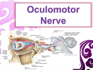
  • 36. a. Superior branch: Motor innervation to the superior rectus and levator palpabrae superioris. Sympathetic fibres run with the superior branch to innervate the superior tarsal muscle. b.Inferior branch: Motor innervation to the inferior rectus, medial rectus and inferior oblique. Parasympathetic fibres to the ciliary ganglion, which ultimately innervates the sphincter pupillae and ciliary muscles. The larger, inferior division divides into three branches : 1. Nerve To The Medial Rectus passes inferior to the optic nerve. 2. Nerve To Inferior Rectus passes downward and enters the muscle on its upper aspect, and 3. Nerve To Inferior Oblique (longest of the three branches) passes in b/w the IR & LR and supplies the IO from its posterior border. It gives off the motor root to the ciliary ganglion.
  • 38.
  • 39.
  • 40. Parasympathetic (Motor) Root: • Carries preganglionic parasympathetic fibres which arise from cells of Edinger- Westphal nucleus and run through oculomotor nerve & its branch to inferior oblique to reach the ganglion. • These fibres are relayed in the ganglion and postganglionic fibres travel in the short ciliary nerves.
  • 41.
  • 42. • They carry: Parasympathetic fibres: supply sphincter pupillae & ciliary muscles (more than 95% of these fibres are distributed to ciliary muscle which is much larger in volume).
  • 44. • All nerves are supplied with blood from adjacent vessels, which are usually small and variable. • The blood supply to the medial aspect of the brainstem is from vessels directly off the Basilar artery. • Small perforators off circumflex arteries (posterior cerebral, superior cerebellar artery) may also supply the fascicular portion of the third nerve as it courses through the ventral midbrain. • The vascular supply of 3rd nerve in subarachnoid space is via vascular twigs from the post. Cerebral A., the sup. Cerebellar A., and the tentorial and dorsal meningeal branches of the meningohypophyseal trunk of the Internal Carotid Artery. • In the cavernous sinus, the tentorial, dorsal meningeal, and inferior hypophyseal branches of the meningohypophyseal trunk supply the nerve along with branches from the ophthalmic artery.
  • 45. • The blood supply of the oculomotor, trochlear and abducent nerves is similarly arranged, the nutrient arteries being derived from any adjacent smaller arteries. • Though not often noted, it is obvious that deprivation of this blood supply by spasm, thrombosis or embolism may produce paralysis or paresis of the muscles supplied.
  • 48. ANATOMICAL BASIS OF CLINICAL FEATURES OF THIRD NERVE PALSY • A complete & a total third nerve palsy is of common occurrence. • May be congenital or acquired. • Clinical features of complete third nerve palsy include :
  • 49. 1. Ptosis - paralysis of LPS muscle. 2. Deviation – out, down and intorted – unopposed action of LR and SO. 4. Pupil is fixed and dilated – paralysis of sphincter pupillae muscle. 5. Accommodation is completely lost – paralysis of ciliary muscle.
  • 50.
  • 51.
  • 52.
  • 53.
  • 54. 6. Crossed diplopia – paralytic divergent squint. 7. Head posture – if the pupillary area is uncovered, head takes a posture consistent with the directions of actions of the paralysed muscles, i.e., head is turned on the opposite side, tilted towards the same side and chin is slightly raised.
  • 55. FEATURES AND CAUSES OF THIRD NERVE LESIONS AT VARIOUS LEVELS 1. Nuclear lesions • Lesions involving purely the third nerve nucleus are relatively uncommon. • Common causes include ; vascular diseases, demyelination, primary tumors and metastasis • Lesions involving nucleus cause an ipsilateral third nerve palsy with ipsilateral sparing and contralateral weakness of elevation. • Lesions involving paired medial rectus subnuclei (ventromedial nucleus) cause a wall-eyed bilateral internuclear ophthalmoplegia ( WEBINO ) characterised by defective convergence and adduction.
  • 56. 2. Fascicular lesions • Causes are similar to nuclear lesions. Benedikt syndrome involves the fasciculus as it passes through the red nucleus and is characterised by ipsilateral 3rd nerve palsy and contralateral extrapyramidal signs such as hemitremor. Weber syndrome involves the fasciculus as it passes through the cerebral peduncle and is characterised by ipsilateral 3rd nerve palsy and contralateral hemiparesis. Nothnagel syndrome involves the fasciculus and the superior cerebellar peduncle and is characterised by ipsilateral 3rd nerve palsy and cerebellar ataxia. Claude syndrome is a combination of Benedikt and Nothnagel syndromes
  • 57.
  • 58. COMMON CAUSES OF NUCLEAR AND FASICULAR THIRD CRANIAL NERVE CHILDREN Congenital - with neurological abnormalities - with aberrant rennervation - with cyclic oculomotor spasm Vascular ( AV malformations ) Primary tumour Metastatic tumour YOUNG ADULTS Demyelinating Vascular (hemorrhage or infarction) Tumor OLDER ADULTS Vascular (infarction) Tumour
  • 59. 3. Lesions involving basilar part of the nerve: • isolated third nerve palsies are frequently basilar. Causes : 1. Aneurysms : At the posterior communicating artery cause isolated third nerve palsy with involvement of pupil. 2. Head trauma: Extradural hematomas which may cause tentorial pressure cone with downward herniation of the temporal lobe. This compresses the third nerve as it passes over the tentorial edge. Initially there occurs fixed, dilated pupil, which is followed by a total third nerve palsy. 3. Diabetes causes isolated 3rd nerve palsy with sparing of the pupillary reflexes.
  • 60.
  • 61.
  • 62.
  • 63. 4. Lesions involving intracavernous part of the nerve: • Because of its close proximity to other cranial nerves, intracavernous 3rd nerve palsies are usually associated with involvement of the 4th and 6th nerves, and the 1st division of trigeminal nerve.
  • 64. • Important Causes : 1. Diabetes may cause vascular palsy. 2. Pituitary apoplexy – may cause a third nerve palsy (after child birth), if the gland swells laterally & extension into cavernous sinus. 3. Intracavernous lesions – aneurysms, meningiomas, carotid-cavernous fistulae and Tolosa-Hunt syndrome (granulomatous inflammation)
  • 65. 5. Lesions of the intraorbital part of the nerve: • May cause isolated extraocular muscle palsies or may involve either superior division or inferior division or both. • Causes : orbital tumors, trauma and vascular diseases.
  • 66. 6. Lesions of pupillomotor fibres • B/w the brainstem and the cavernous sinus, pupillomotor fibres are located superficially in the superior median quadrant of the nerve. • They derive the blood supply from the pial blood vessels whereas the main trunk of the 3rd nerve is supplied by vasa nervorum.
  • 67.
  • 68. • Surgical lesions such as aneurysms, trauma and uncal herniation characteristically involve the pupil by compressing the pial blood vessels and the superficially located pupillary fibres. • Medical lesions such as diabetes and hypertension usually spare the pupil. This is because the microangiopathy associated with these diseases involves the vasa nervorum, causing infarction of the main trunk, but sparing the superficial pupillary fibres.
  • 69. In ISOLATED THIRD NERVE PALSY diabetes and HTN, is the most common cause of pupil- sparing 3rd nerve palsy. In most cases spontaneous recovery occurs within 3 months. Diabetic 3rd nerve palsy is often assoc. with periorbital pain and may occasionally be the presenting feature of diabetes. The presence of pain is therefore not helpful in differentiating b/w aneurysmal and diabetic 3rd nerve palsy.
  • 70.
  • 72. 1. Which of the following statements regarding the origin and course of III is/are correct except? a) It emerges as a number of rootlets from a nucleus lying level with the superior colliculus b) It leaves the brainstem after passing through the red nucleus c) It runs below and lateral to the free margin of the tentorium cerebelli d) It pierces the dura midway between the anterior and posterior clinoid processes e) Its branches cross the same part of the superior orbital fissure as VI 2. A patient suffers damage to the orbit in a road traffic incident resulting in damage to the third cranial nerve. Which of the following signs will be present? a) Pupillary constriction and a medial strabismus b) Pupillary dilatation and a medial strabismus c) Pupillary constriction and a lateral strabismus d) Pupillary dilatation and a lateral strabismus
  • 73. 3. Regarding the distribution of fibres from III a) Levator palpebrae superioris only receives a motor supply from the superior division of III b) It innervates all the muscles at the junction of their middle and posterior thirds c) The parasympathetic supply to the iris is conveyed via the short ciliary nerves d) Superior rectus is innervated ipsilaterally e) Levator palpebrae superioris contralaterally represented at the level of the oculomotor nuclei 4. Which of the following statements regarding III is correct? a) The parasympathetic fibres lie centrally in the precavernous sinus portion of the nerve b) A third nerve palsy causes the eye down and out. c) Accommodation is unaffected by such a palsy d) Compression of the nerve causes a partial ptosis e) The Edinger-Westphal nucleus lies ventral to the main oculomotor nucleus
  • 74. 5. All of the following are characteristics of oculomotor nerve except: a) Carries parasympathetic nerve fibers b) Supplies inferior oblique muscle c) Enters orbit through the inferior orbital fissure d) Causes constriction of pupil 6. The following structure(s) synpase(s) in the ciliary ganglion: a) Nasociliary nerve fibres b) Parasympathetic nerve fibres from oculomotor nerve c) Sympathetic nerve fibres from the carotid plexus d) Parasympathetic nerve fibres form the facial nerve abducent nerve fibres.

Notes de l'éditeur

  1. Craniacaudel sequency
  2. Hemitremor:Tremor affecting the muscles of one side of the body. (lateral part) Hemiparesis: Weakness affecting one side of the body. cerebellar ataxia:failure of muscular coordination due to disease of the cerebellum.