Tissue Definition
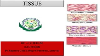
Tissues are groups of cells that have a similar structure and act together to perform a specific function. The word tissue comes from a form of an old French verb meaning “to weave”. There are four different types of tissues in animals: connective, muscle, nervous, and epithelial. In plants, tissues are divided into three types: vascular, ground, and epidermal. Groups of tissues make up organs in the body such as the brain and heart.
Types of Animal Tissues
Connective
Connective tissue connects or separates groups of other tissues. It is found in between all the other tissues and organs in the body. Connective tissue is made up of cells and ground substance, which is a gel that surrounds cells. Most connective tissue, except for lymph and blood, also contains fibers, which are long, narrow proteins. Fibers can be collagenous, which bind bones to tissues; elastic, which allow organs like the lungs to move; or reticular, which provide physical support to cells. Connective tissue also allows oxygen to diffuse from blood vessels into cells.
About 1 in 10 people are have a disorder involving connective tissue. Some connective tissue disorders include sarcomas, Marfan syndrome, lupus, and scurvy, which is a Vitamin C deficiency that leads to fragile connective tissue.
Muscle
Muscle tissue comprises all the muscles in the body, and the specialized nature of the tissue is what allows muscles to contract. There are three types of muscle tissue: skeletal muscle, cardiac muscle, and smooth muscle. Skeletal muscle anchors tendons to bones and allows the body to move. Cardiac muscle is found in the heart and contracts to pump blood. Smooth muscle is found in the intestines, where it helps move food through the digestive tract, and it is also found in other organs like blood vessels, the uterus, and the bladder. Skeletal and cardiac muscles are striated; this means that they contain sarcomeres (a unit of muscle tissue) that are arranged in a uniform pattern. Smooth muscle does not have sarcomeres.
Duchenne muscular dystrophy is an example of a muscle tissue disorder. It is an inherited disorder that causes muscles to atrophy over time. The muscles shorten as they atrophy, which can cause scoliosis and immobile joints. Individuals with the disorder are usually male because the gene responsible for it is found on the X chromosome (of which males have only one).
Nervous
Nervous tissue is found in the brain, spinal cord, and peripheral nerves, which are all parts of the nervous system. It is made up of neurons, which are nerve cells, and neuroglia, which are cells that help nerve impulses travel. Nervous tissue is grouped into four types: gray matter and white matter in the brain, and nerves and ganglia in the peripheral nervous system. The main difference between gray and white matter is that axons of the neurons in gray matter are unmyelinated, while white matter is myelinated. Myelin is a white, fatty substance that insulates neurons and
3. INTRODUCTION
• Tissues are groups of cells
that have a similar structure
and act together to perform a
specific function.
• The word tissue originates
from French, which means
“to weave.”
• Study of tissue is called as
“histology”.
TISSUE
EPITHELIAL CONNECTIVE MUSCULAR NERVOUS
COVERING
SECRETION
ABSORPTION
BINDING
SUPPORT
TRANSPORT
MOVEMENT
LOCOMOTIO
N
CONTROL &
CO-
ORDINATION
4. EPITHELIAL TISSUE
STRUCTURE:-
1. Sheet arrangement
2. Single or multiple layer
3. Closely packed
4. Many junctions
5. Form Covering & Lining throughout the body
6. Not covered by another tissue
FUNCTION:-
1. SELECTIVE BARRIER: - Limit movement of
substances in & out of the body.
2. SECRATORY: - Secretion secreted by cells
released on the free space.
3. PROTECTION :- Protection to the surface
5. BASMENT MEMBRANE
• Thin extracellular layer
• Form a surface
• Restrict passage of large
molecule
• Filtration
6. • Each epithelial tissue is given two names:
The first name indicates the number of layers present:
1. Simple (One)
- Composed of a single cell layer.
- Typically found where absorption and filtration occur and a thin epithelial
barrier is desirable.
2. Stratified (More than one)
- Consist of two or more cell layers stacked one on top of the other
- Common in high-abrasion areas where protection is important, such as the
skin surface and the lining of the mouth.
CLASSIFICATION OF EPITHELIAL TISSUE
7. CONT…
The second name describes the shape of the cells
Three common shapes of epithelial cells: nucleus will be same shape
1. SQUAMOUS CELLS – Flattened and scale like
2. CUBOIDAL CELL- Box like
3. COLUMNAR CELLS- Tall and column shaped
4. CILIATED CELLS- Fibrous shape
8. • Simple epithelia are easy to classify by cell shape because all cells in
the layer usually have the same shape.
• STRATIFIED EPITHELIA:
- Cells shapes usually differ among the different cell layers
- Named according the shape of the cells in the apical layer
CONT…
10. • DESCRIPTION: Single layer of flattened cells with
disc-shaped central nuclei and sparse cytoplasm; the
simplest of the epithelia.
• FUNCTION: Allows passage of materials by
diffusion and filtration in sites where protection is not
important; secretes lubricating substances in serosae.
• LOCATION: Kidney glomeruli. Air sacs of lungs;
lining of heart, blood vessels, and lymphatic vessels;
lining of ventral body cavity (Serosae)
SIMPLE SQUAMOUS EPITHELIUM
19. Description: Gland may consist of a single cell or group of cells.
Specialized cells – secrets substance into duct.
The glands are of 2 types- Exocrine & Endocrine.
Glandular Epithelium
20. Description:
• Ductless gland
• The secretions of endocrine gland enters the interstitial fluid and then
diffuses directly into the blood stream.
• These secretions are called as hormones which regulate the metabolic
and physiological activities of the body in order to maintain
homeostasis.
Location: Pituitary gland, pineal gland, thyroid gland, parathyroid
gland, adrenal glands, pancreas, ovaries, testes and thymus
Function:
• Production of hormones that regulates various metabolic and
physiological activities.
• Pituitary gland secretes human growth hormone responsible for the
normal growth of individuals.
• Pineal gland secrets melatonin hormone responsible for maintaining
the circadian rhythms and seasonal functions.
• Thyroid gland secrets T3 and T4 hormone that are responsible for
maintaining the normal functioning of thyroid gland.
• Pancreas secrets insulin hormone responsible for controlling the blood
sugar level.
Endocrine Gland
21. Description:
Glands with duct.
Secretion of these glands release into ducts that empty
onto the skin surface or the lumen of a hollow organ.
Secretions:- Mucous, sweat, oil, earwax, saliva and
digestive system.
Secretory product release into the duct.
Example- Sudoriferous (sweat) glands, that produces
sweat to help to reduce body temp. and salivary glands
which secrete saliva that keeps the mouth moist.
Location: Sweat gland, sebaceous gland, earwax glands,
salivary glands and pancreas.
Function: Production of sweat, oil, earwax, saliva or
digestive system.
Classification:- Unicellular and multicellular
Exocrine Gland
22. • It is the most abundant and widely distributed tissue system in the
body.
• It binds together, supports and strengthen other body tissue as well
as protect and insulates internal organs.
• It is made up of fibres, cells and ground substances.
Connective Tissues
23. 3 types of fibres are embedded in the extracellular matrix between the cells.
These fibres strengthen and support connective tissues.
Fibres
Collagen Fibres
• These are very strong and
allow tissue flexibility.
• Made up of protein collagen.
• It is the most abundant protein
making up about 25%-35% of
the total body protein.
• They often present in the
parallel bundles.
• It is found in – bone , cartilage,
tendons and ligaments.
Elastic Fibres
• These fibres are smaller in
diameter.
• These are made up of protein
elastin surrounded by a
glycoprotein named fibrillin which
gives strength and stability to
tissue.
• Elastic fibres have ability to return
to its original shape, a property
called as elasticity.
• These are found in- Skin, lungs,
arteries, veins, elastic cartilage,
periodontal ligament and foetal
tissue.
Reticular Fibres
• They consist of collagen
protein arranged in fine
bundles covered with
glycoprotein.
• These are much thinner than
collagen fibres.
• They give support and strength.
• These are found in- Liver, bone
marrow and lymphatic organs
24. • Each cell consist – Fibrinoblast, macrophages, plasma cells, mast cells,
adipocytes and WBC
1. Fibrinoblast: - They are the chief cells of connective tissue.
They are large, flat cells with branching processes.
2. Macrophages: - These cells develop from monocytes, a type, of WBC.
There are 2 types of macrophages.
Play important role in immune response.
a. Fixed macrophages;- Present in particular tissue such as alveolar
macrophages in lungs or spleen macrophages in spleen.
b. Wandering macrophages: - ability to move throughout the tissue and
gather at the site of infection to carry phagocytosis.
Cells
25. 3. Plasma cells:- A small cell that develops from type of white blood cells is
called as beta- lymphocytes. Takes important role in the immune response.
They are present in GIT and respiratory tract , salivary gland, lymph node,
spleen and red bone marrow.
4. Mast cells: - They produce histamine that that dilate the small blood
vessels as a part of the inflammatory responses.
5. Adipocytes: -These are also called as a fat cells or adipose cells. They store
fats. They are found deep to the skin and around heart and kidneys.
6. White blood cells:- In response to inflammatory reaction they migrate
from blood to connective tissue . E.g., Neutrophils gather at the sites of
infection and eosinophils migrate to the sites of allergic response.
Cells
26. Description:
• It is an amorphous gel like substance surrounding the cells.
• In the ground substance, cells and fibres are suspended.
• Ground substance is primarily composed- water ,
glycosaminoglycan (hyaluronan), proteoglycans,
glycoproteins, hyaluronic acid, chondroitin sulphate and
dermatan sulphate.
• The ground substance support cells, binds them together,
stores water and provides a medium through which
substances are exchanged between the blood and cells.
Ground Substance
Function:
• It acts as energy store.
• It provides protection to different body
organs.
• It provide structural framework to the
body.
• It connects different body tissues.
• It connects epithelial tissues to muscle
fibres.
• It supply hormones to all over body
28. • These fibres are loosely woven.
• It has a large proportion of ground
substance.
• They are easily distorted.
• On distortion they become tough
and resist to further deformation.
1. Areolar connective tissue
2. Adipose connective tissue
3. Reticular connective tissue
Loose Connective Tissue
29. Description:- They form a loose network in the intracellular
material and are not arranged in a particular pattern.
Consist:- Fibres- Collagen, Elastic and reticular fibres.
Cell- Fibroblast, macrophages, plasma cells,
adipocytes and mast cells.
Location: - Present below skin, fill the spaces between
muscles, supports blood vessels and nerves in the
alimentary canal.
Yellow elastic fibres are found in arteries and white elastic
fibres are found in kidney and brain.
Function: - It gives strength, elasticity and support to tissue.
Areolar Connective Tissue
30. Description: - It consists of adipocytes which stores fats as a large centrally located
droplet.
Location: - It is present in the subcutaneous layer deep in the skin, around heart,
kidneys and yellow bone marrow.
Function: -
1. It prevents heat loss from body.
2. It acts as a reservoir of energy.
3. It gives shapes to the limbs and body.
4. It protects underlying organ from injury.
Adipose Connective Tissue
31. Description: - It consists of reticular fibres
and reticular cells.
Location: - It is present in the supporting
framework of liver, spleen, lymph nodes, red
bone marrow and is found around the blood
vessels and muscles.
Function: - It forms stroma of organs, binds
together smooth muscle tissue cells, filters
and remove worn-out blood cells in spleen
and microbes in the lymph node.
Reticular Connective Tissue
32. • In this, fibres are densely packed, the fibres content is higher and cell
content is lower as compared to loose connective tissue.
Dense Connective Tissue
1. Dense regular connective tissue
2. Dense irregular connective tissue
3. Elastic connective tissue
33. Description: - Bundles of collagen fibres are arranged in parallel
patterns to provide strength to tissue. Fibrinoblast are appears in rows
between the fibres. It is silvery white in colour and tough in nature.
Location: - It forms tendons (attach muscle to bone) and ligaments
(attach bone to bone).
Function: - It provides strong attachment to structures.
Dense Regular Connective Tissue
34. Description: It contains collagen fibres that are irregularly arranged and
few fibroblasts are appears in rows between the fibres.
Location: It is present in tissue beneath the skin, dermis of skin,
periosteum of bone, membrane capsules around kidneys, liver, testes,
lymph node, pericardium of heart and heart valves.
Function: It provides strength to different organs.
Dense Irregular Connective Tissue
35. Description:
1. It is the hardest connective tissue
2. It has calcified matrix containing
many collagen fibres.
3. It composed of 25% of water, 30% of
organic material and 45%inorganic
salts.
4. It is well vascularised.
5. Bone is arranged in concentric ring
structures called as osteons.
6. At the centre of ring is a structure
called as Haversian canal.
Bone
Haversian canal:
1. Central Haversian channel: It contains
blood vessels and nerves.
2. Lamellae: Surrounding the central canal
concentric plates of bone are present called as
lamellae.
3. Lacunae: It contains mature bone cells called
as osteocytes
4. Canaliculi: Projecting from the lacunae are
canaliculi, network of minute canals
containing the processes of osteocytes.
Location
It present in
compact and
spongy bone
tissue.
Function:
1. To form supporting frame work of the body.
2. To give protection to delicate organs.
3. To form joints essential for locomotion of body.
4. To form RBC in red bone marrow.
5. To provide store of calcium salts.
6. It gives supports and maintains shape.
36. Description:
• It is a connective tissue with liquid extracellular matrix called as blood plasma.
• The blood cells are suspended in the blood plasma.
• It is composed of 55% plasma and 45% of cells.
• Blood plasma is straw coloured liquid in which the blood cells are suspended.
• Plasma is composed 0f 90-92% of water 7% plasma proteins and clotting factors, and 1% of mineral salts, sugar, hormones and
vitamins.
Blood cells are of three types:
1. Erythrocytes(RBC): These cells transport oxygen to body cells and remove carbon dioxide from them.
2. Leucocytes (WBC): These are involved in phagocytosis, immunity and allergic reaction.
3. Thrombocytes (Platelets): Theses cells participates in the blood clotting mechanism.
Location: It is present within blood vessels (arteries, arterioles, capillaries, venules and veins) and within the chambers of heart.
Function:
1. RBCs transport oxygen to body cells and remove carbon dioxide from them.
2. WBCs are involved in phagocytosis, immunity and allergic reaction.
3. Platelets participate in the blood clotting process.
Blood
37. • Muscular tissue consist of
elongated cells called muscle
fibres that can use ATP is generate
force.
• Three type muscular tissues are
present:
1. Skeletal / Striated/ Voluntary
muscle tissue
2. Cardiac mascle
3. Smooth / non-striated/
involuntary muscles tissue
Muscle Tissue
38. Description:
The cells are cylindrical in shape.
The fibres are parallel to each other.
The length of muscle fibres is 30-40 cm.
They have several nuclei located at periphery.
It show alternate dark and light band i. e. striations and hence the name is
straitened muscle.
The muscles are attached to the bones hence called as skeletal muscles.
The activity of fibres is within ones control hence, the name is voluntary
muscle.
Location: It is usually attached to bones by tendons.
Function: It helps to gives motion, posture, heat production and protection.
Skeletal Muscle Tissue
39. Description:
It is present in the myocardium of heart wall.
It is striated but involuntary i.e. the activity of
fibres is beyond once control hence called as
involuntary muscle tissue.
Each fibre is parallel to each other, branched
and multinucleated.
Two cardiac muscle fibres are attached by
thickened plasma membrane called as
intercalated disc.
The intercalated disc contains desmosomes as
well as gap junction.
Location: It is present in the heart wall.
Function: It pumps blood to all parts of the
body, contracts the atria and ventricles of the
heart, causes rhythmic beating of the heart.
Cardiac Muscle Tissue
40. Description:
• A smooth muscle fibre is usually small
• It is thickest in the middle and tapering at the ends.
• It contains single, centrally located nucleus.
• The cells are spindle shaped.
• Alternate light and dark bands are absent hence they are called as smooth/ non
striated.
• Activity of these fibres is beyond once control or wish and hence called as
involuntary.
Location: It is present in the wall of blood vessels, wall of lymph vessels,
alimentary tract, respiratory tract, urinary bladder and uterus.
Function: It gives motion (contraction of blood vessels, airways, propulsion of
food through, GIT, contraction of urinary bladder and gall bladder).
Smooth Muscle Tissue
41. It is made up of two types of nerves cells.
Nervous Tissue
Neurons
Description:
It is made up of cell body, axons, dendrites, and axon terminals.
1. Cell Body: - It contains nucleus and other organelles.
2. Dendrites: - These are input portions of neuron. These are usually short and highly branched forms tree like
structure. Each nerve cell contains many dendrites.
3. Axon: - Each nerve cell contains single axon which is thin, long and cylindrical process. It is major output portion of
a neuron which conducts the signal to effector organs. The axon are surrounded by white, fatty substance called as
myelin sheath. The unmyelinated regions between the myelin segments are called as nodes of ranvier.
Location
It present in the nervous
system
Function
It exhibits sensitivity to various types of stimuli, converts stimuli into nerve impulses
(action potentials), and conducts nerve impulses to other neurons, muscle fibres of glands.