1. Bull. Mater. Sci., Vol. 34, No. 5, August 2011, pp. 1157–1162. c Indian Academy of Sciences.
Preparation of SnO2/C biomorphic materials by biotemplating
from ramie fibres
XIN-HAI HEa,b
, LE-HUA QIa,∗
, JUN-BO WANGb
, MING-QIAN SHENb
, WEI CHANGb
,
CHONG FUb
, MIN-GE YANGb
and XIAO-LEI SUb
aSchool of Mechatronics, Northwestern Polytechnical University, Xi’an 710072, P.R.China
bCollege of Mechanical & Electrical Engineering, Xi’an Polytechnic University, Xi’an 710048, P.R.China
MS received 15 October 2010; revised 31 December 2010
Abstract. A new SnO2/C biomorphic material has been prepared by molding into a composite billet and
carbothermal-reduction under vacuum from ramie fibres/Sn(OH)4 precursors. The phase composition and
microstructure of the prepared materials were characterized. The effects of the carbonization temperature, holding
time and other factors on the crystal structure, morphology and ingredients of the prepared samples were discussed.
The results showed that the sintering temperature and holding time have significant effects on the final products.
When the sintering temperature reached 480◦C, the SnO2/C biomorphic materials were synthesized successfully.
Keywords. Biomorphic materials; carbothermal-reduction; biotemplate.
1. Introduction
Biomorphic materials, as a new kind of porous carbon mate-
rials, are usually fabricated by carbonizing wood or woody
materials impregnated with phenolic resin under vacuum at
an elevated temperature of 300∼2800◦
C. By selective arti-
ficial compounding, biomorphic materials not only maintain
the micro-fine structure of the natural biological materials,
but also is endowed with new features and functions. This
kind of materials is of great application potential in many
industrial areas such as absorbents, sensors, catalyst carrier
materials, self-lubricating materials, biomedical materials,
heat insulating materials and electromagnetic shielding
materials (Griel 2001; Zhang et al 2004; Odeshi et al 2006),
etc. In recent years, a variety of biological materials have
been taken as bio-templates to prepare biomorphic materi-
als, such as wood (Min et al 2006; Ozao et al 2006; Kaul
and Faber 2008), bamboo (Dong 2009), paper (Yang et al
2008), cotton (Amirthan et al 2009), etc. And various oxides
(Dong et al 2007), carbide (Sun et al 2004; Kim et al 2006;
Martinez-Escandell et al 2009) and nitride (Min et al 2008;
Rambo et al 2008) biomorphic materials have been prepared
from biotemplates by sol–gel and carbothermal reduction
method (Qian and Jin 2006), molten metal infiltration (Wang
et al 2006a, b), reactive infiltration of liquid Si (Mallick
et al 2007), etc. Furthermore, the researchers have explored
deeply into the defects of biomorphic materials, such as
poor homogeneity of structure, low mechanical properties,
∗Author for correspondence (qilehua@nwpu.edu.cn)
cracking, etc which lays the theoretical and experimental
basis for application of the materials.
Ramie is a perennial herbaceous plant, which can be annu-
ally harvested in a great amount 2 to 3 times, so the yield of
its natural fibres is very rich. As the prepared template for
biomorphic materials, ramie fibres have good uniformity of
impregnation and controllable texture compared with wood,
bamboo and some other template materials.
In the present work, SnO2/C biomorphic materials have
been fabricated by carbothermal-reduction under vacuum
from ramie fibres biotemplates which has already impreg-
nated with Sn(OH)4 sol using ultrasonic technique. The
phase composition and microstructure of the materials are
analysed and characterized. The mechanism of preparation
has been discussed.
2. Experimental
2.1 Materials preparation
The ramie fibres, crystalline tin tetrachloride (SnCl4·5H2O
AR), polyethylene glycol (PEG AR), ammonia (NH3·H2O
AR) and deionized water were used as raw materials. Firstly,
the PEG (5 g/L) was added to 0·05 mol/L SnCl4 solution
which was prepared by dissolving crystalline SnCl4·5H2O
with deionized water. Secondly, when the solution was
stirred until it was clear and transparent, and then NH3·H2O
was added to the SnCl4 solution slowly, stirring was con-
tinued till pH value reached 2–2·5. Then, it was washed by
deionized water until no chloride ion was found in the preci-
pitation. At last, 6·5 wt% hydrosol of Sn(OH)4 was prepared.
1157
2. 1158 Xin-Hai He et al
To improve the impregnation ratio, ultrasonic vibra-
tion technology was used to impregnate ramie fibres with
Sn(OH)4 hydrosol which was prepared earlier and the treat-
ment process of impregnation-drying was repeated seve-
ral times. Ultimately the ramie fibres/Sn(OH)4 precursor was
obtained after drying it in an oven at 80◦
C for 24 h, and sin-
tering it in a vacuum hot pressing furnace to 480◦
C, 560◦
C
and 630◦
C for 20–60 min in the graphite crucibles. Finally,
the samples of SnO2/C biomorphic materials were prepared
after annealing at 150◦
C for 2·5 h. Fabrication scheme of
SnO2/C biomorphic materials is shown in figure 1. Samples
used in the experiment in different sintering conditions are
shown in table 1.
2.2 Characterization
The differential scanning calorimetry and thermogravime-
try (DSC/TG: TGA/SDTA851e, METTLER TOLEDO,
Switzerland) were used to analyse and characterize the
Figure 1. Fabrication scheme of SnO2/C biomorphic materials.
Table 1. Samples used in experiment in different sintering condi-
tions (weight ratio of ramie/Sn(OH)4 ≈ 1/1·3).
Temperature Heating rate Holding time
Samples (◦C) (◦C/min) (min)
1# 560 5 20
2# 560 5 40
3# 560 5 60
4# 480 5 40
5# 630 5 40
6# 630 8 60
pyrolytic behaviour of ramie fibres, Sn(OH)4 powder and the
ramie fibres/Sn(OH)4 precursor. For crystalline phase identi-
fication, X-ray diffraction was measured on a X-ray diffrac-
tometer (MAXima_X XRD-7000, Shimadzu, Japan) using
Cu radiation produced at 40 kV and 40 mA, at 0·02◦
C step
and at a scanning rate of 10◦
C/min. The microstructure mor-
phology was observed with a scanning electron microscope
(SEM: JSM-6700F, JEOL, Japan) and elemental analysis was
also carried out at several points of the observed area by
using energy dispersive spectroscopy (EDS). FTIR measure-
ments were carried out in the wavenumber range between
4000 and 500 cm−1
by a NICOLET 5700 (Thermo, USA)
spectrophotometer.
3. Results and discussion
3.1 DSC–TG analysis
DSC–TG curves of Sn(OH)4 powder (shown in figure 2)
reveals the pyrolytic behaviour of Sn(OH)4. It shows that
there is a endothermic valley at 70◦
C because the Sn(OH)4
powder is not completely dry and releases some water. The
exothermic peak at 345·7◦
C is related to the heat effect of
decomposition of Sn(OH)4 into SnO2 (Junbo et al 2006).
It can be assumed that the reaction mainly occurs at about
345·7 ◦
C. The reaction involved in this step is
Sn (OH)4 (s)
345·7◦
C
−−−−−→ SnO2 (s) + 2H2O (g) . (1)
Comparing the different thermal weight losses of ramie
fibres/Sn(OH)4 precursors at two kinds of heating rate
(shown in figure 3), it can be found that the heating rate of
5◦
C/min, pyrolysis starts at 189◦
C and ends at 363◦
C. At the
heating rate of 8◦
C/min pyrolysis starts at 217◦
C and ends at
422◦
C. It can be concluded that thermal decomposition curve
shifts to the right, and steepens at high heating rate, which is
attributed to fast decomposition but lagging heat conduction.
Figure 2. DSC–TG curves of Sn(OH)4.
3. Preparation of SnO2/C biomorphic materials by biotemplating from ramie fibres 1159
As a result, pyrolytic reactions lag behind. In the preparation
of biomorphic materials process, it is found that the sample
has obvious cracks and deformation at high heating rate. It
is mainly due to the uneven heat conduction in the sample
which results in a large temperature gradient. Therefore, the
appropriate heating rate is 5◦
C/min.
3.2 XRD analysis
From XRD patterns of samples at different sintering tempe-
ratures (shown in figure 4), it can be seen that the diffrac-
tion peaks of carbon derived from ramie fibres are broad
dispersion peaks when the sintering temperature is 480◦
C
and 560◦
C. Even if the sintering temperature is 630◦
C, there
is no significant change with the peaks. It indicates that the
ramie carbon matrix of SnO2/C biomorphic materials is a
typical amorphous carbon, which is a turbostratic carbon
Figure 3. Different TG of two models of heating to precursors.
Figure 4. XRD patterns of samples prepared at different sintering
temperatures (holding time, 40 min).
material composed of microcrystalline graphite. SnO2 is pro-
duced by the pyrolysis of Sn(OH)4 at 345◦
C, and there is
no significant change in the intensity of diffraction peaks of
SnO2 (JCPDS card No. 41-1445) with increasing carboniza-
tion temperature. But when the temperature reaches 630◦
C
the apparent diffraction peaks of SnO (JCPDS card No.
06-0395) appear, which indicates that the SnO has generated.
Through thermodynamic calculation and analysis, under
the standard state, when the ambient temperature is above
631◦
C and 689·5◦
C, the reduction reactions of SnO2 with
C (2) and (3) will convert into spontaneous reactions from
non-spontaneous ones (Ye 2002).
SnO2 (s) + C (s) → Sn (s,l) + CO2 (g) , (2)
SnO2 (s) + C (s) → SnO (s) + CO (g) . (3)
Figure 5 shows XRD patterns of samples prepared at diffe-
rent holding times sintered at 560◦
C. It can be shown that
the (110), (101) and (200) diffraction peaks of SnO2 become
sharp with the increase of holding time. The diffraction peaks
of SnO and Sn(JCPDS card No. 04-0673) appear when the
holding time reaches 60 min. This can be explained due to the
fact that CO and CO2 gases produced by reactions (2) and (3)
are extracted timely under vacuum, in which way the occur-
rence of the reactions (2) and (3) below 630◦
C are promoted,
and meanwhile the temperature gradient existing in the sam-
ples can also cause localized high temperatures, under which
SnO2 is reduced and decomposed into SnO and Sn by the
effect of strong reducing agent carbon. Moreover, the carbon
derived from ramie fibres can also be further carbonized with
the extension of holding time (Okabe and Saito 1992).
Figure 5. XRD patterns of samples prepared at different holding
times (sintering at 560◦C).
4. 1160 Xin-Hai He et al
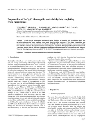
Figure 6. SEM images of carbon biotemplate derived from ramie fibres (a) and carbonized
samples (b).
Figure 7. SEM images and corresponding EDS analysis of carbonized samples. SEM images
of (a) 4# sample, (b) 3# sample, (c) 5# sample, and EDS analysis of (d) 4# sample, (e) 3# sample
and (f) 5# sample.
3.3 SEM analysis
From SEM photographs of carbon biotemplate derived from
ramie fibres (shown in figure 6(a)), it can be seen that
carbon template derived from ramie fibres has a porous
structure, the carbon fibres inherit and maintain the natural
tracheid structure of ramie fibres, and the orientation of the
carbon fibres is random in the template. In the carbon tem-
plate, in addition to the pores among the carbon fibres, there
are about 10 μm∼ 20 μm sized circle-like pores in the
tracheid of ramie fibres. This structure is conducive to the sol
impregnation and the uniform distribution of SnO2 produced
by reactions in the carbon matrix.
Through the microstructure of the specimen (shown in
figure 6(b)), it can be seen that SnO2/C biomorphic materi-
als maintain the structure of carbon template derived from
ramie fibres, and SnO2 distributes in the gap of ramie car-
bon tracheid. However, further studies under high power
(3000-fold) SEM photograph find that the porous structure
of SnO2/C biomorphic materials is not perfectly consistent
with the tracheid structure of ramie fibres, because the ramie
carbon tracheid deforms under the thermal pressure resulting
from the thermal shrinkage of ramie fibres.
Figure 7 shows SEM images and the corresponding EDS
analysis of samples carbonized at 480◦
C, 560◦
C and 630◦
C.
From the SEM image shown in figure 7(a), it is observed that
SnO2 appears as an irregular agglomerate distributing in the
tracheid gap of carbon derived from ramie fibres at a sintering
temperature of 480◦
C. With the increase of sintering tempe-
rature and the extension of holding time, the pellet-like com-
ponents with a diameter of 1 μm∼3 μm and the tile sheet
shaped components with a size of 3 μm∼5 μm appear in the
tracheid gap of carbon derived from ramie fibres (shown in
figure 7(b)). When the sintering temperature reached 630◦
C,
the components are mainly pellet-like with a diameter of
1 μm∼5 μm (shown in figure 7(c)).
5. Preparation of SnO2/C biomorphic materials by biotemplating from ramie fibres 1161
According to EDS analysis of the proportion of Sn and
O (shown in figures 7(d), (e) and (f)), in combination with
Sn–O phase diagram (Li 2002) and the XRD diffraction pat-
terns analysis (shown in figures 4 and 5), it can be ascer-
tained that the irregular agglomerate obtained at 480◦
C is
primarily SnO2, the tile sheet shaped components are mainly
the mixture of SnO2 and SnO and the weight percent-
age of the two ingredients are 40·9% and 59·1%, and the
pellet-like components also contain SnO2 and SnO but their
percentages in weight are 29·1% and 70·9%. The results
suggest that, in the starting phase in the sintering process,
the SnO2 pyrolysed by Sn(OH)4 is shaped as irregular agglo-
merate. But due to the action of strong reducing agent car-
bon, SnO2 changes into the pellet-like or tile sheet shaped
mixture of SnO2 and SnO with increase of sintering tempe-
rature and extension of holding time. And the microstructure
of the mixture changes from the tile sheet to the pellet-like
with the increase of Sn content.
3.4 FTIR analysis
In order to confirm and compare the analysis results of
XRD and SEM testing of all the samples, the SnO2 pow-
der obtained from ramie fibres/Sn(OH)4 precursors with
aerobic sintering at 700◦
C for 40 min has been prepared.
Figure 8 shows FTIR infrared spectra of samples. It is obvi-
ous that samples 1#
–5#
have similar spectrogram distribu-
tion at 4000–750 cm−1
. Except in curve 3#
, the characteristic
absorption peaks of Sn–O–Sn are observed at 750–500 cm−1
,
which is typical of Sn–O asymmetric vibration and symme-
tric vibration peaks.
From the comparison of the samples sintered at 560◦
C,
it can be found that the characteristic absorption peaks of
Sn–O–Sn show width and red-shift with the extension of
Figure 8. FTIR spectral analysis of samples.
holding time. The same results have been found in the pre-
pared samples at the same holding time under different tem-
peratures. However, the red-shift and broadening of peak
is closely related to the crystal structures (Wu et al 1996).
Therefore, the red-shift of peak of Sn–O–Sn bond is due to
the fact that the force generated between ramie carbon and
SnO2 peaks result in the Sn–O bond. In addition, because
the SnO has been generated, the length of Sn–O bond shows
greater distribution, which leads to the broadening of absorp-
tion peak of Sn–O–Sn bond. The results are consistent with
the analysis of XRD and SEM.
4. Conclusions
SnO2/C biomorphic materials were fabricated by
carbothermal-reduction under vacuum from ramie fibres
biotemplates which were already been impregnated with
Sn(OH)4 sol using ultrasonic technique. The thermal decom-
position reaction of ramie fibres mainly concentrate at
about 224·7◦
C and 300·2◦
C. The decomposition of Sn(OH)4
into SnO2 occurs at about 345·7◦
C. Sintering tempera-
ture and holding time have great impact on the final pro-
ducts. The prepared products at the sintering temperature
of 480◦
C are SnO2/C biomorphic materials, and the pre-
pared products at the sintering temperature of 560◦
C and
630◦
C are SnO/SnO2/C multi-phase ceramic materials. The
micro-morphology of SnO/SnO2 mixture is related to the
proportion of SnO and SnO2, and it changes from the tile
sheet to the pellet-like with the increase of Sn content.
However, the preparation of SnO2/C biomorphic materi-
als study is limited to the vacuum environment, the research
work is not still perfect and the reaction mechanism is not
very clear. Therefore, the optimum processing parameters
of the preparation of SnO2/C biomorphic materials, such
as atmosphere, holding time and temperature etc and the
reaction mechanism will be studied in future.
Acknowledgements
The authors wish to express their thanks for the financial
support received from the National Natural Science Founda-
tion of China (No. 51002113), Shaanxi Science and Techno-
logy Research and Development Program (No. 2011K07-10),
the Natural Science Specialized Foundation from Shaanxi
Department of Education (No. 09JK449, 2010JK579), Basic
Research Foundation of Xi’an Polytechnic University (No.
XGJ08010) and Guidance Research Program of China
Textile Industry Association (No. 2008046).
References
Amirthan G, Udayakumar A, BhanuPrasad V V and
Balasubramanian M 2009 Ceram. Int. 35 967
6. 1162 Xin-Hai He et al
Da-Lun Ye 2002 Practical handbook of thermodynamic data of
inorganic metallurgy (Beijing: Industry Press)
Dong Liu 2009 Preparation and charactization of bamboo based
SiC/C composite biological Mimesis Ceramics, M.Sc. thesis,
Beijing Forestry University, Beijing
Dong Qun, Su Huilan, Xu Jiaqiang, Zhang Di and Wang Ruibing
2007 Mater. Lett. 61 2714
Griel P 2001 J. Eur. Ceram. Soc. 21 105
Junbo Wang, Yingmin Li and Minge Yang 2006 Mater. Sci. Eng.
B131 230
Kaul V S and Faber K T 2008 Scr. Mater. 58 886
Kim Jae-Won, Myoung Sang-Won, Kim Hyeon-Cheol, Lee
Je-Hyun, Jung Yeon-Gil and Jo Chang-Yong 2006 Sci. Eng. A434
171
Mallick D, Chakrabarti O P, Majumdar R and Maiti H S 2007
Ceram. Int. 33 217
Martinez-Escandell M, Narciso J and Rodriguez-Reinoso F 2009
Carbon 4 002
Min Luo, Jiqiang Gao and Xiao Zhang 2006 Rare Metal Mater. Eng.
35 133
Min Luo, Jia Cheng and Jing Ma 2008 J. Inorg. Mater. 23 763
Odeshi A G, Mucha H and Wielage B 2006 Carbon 44 1994
Okabe T and Saito K 1992 Meta. Tech. 62 34
Ozao Riko, Nishimoto Yuko and Weiping Pan 2006 Therm. Acta
440 75
Qian Jun-Min and Jin Zhi-Hao 2006 J. Eur. Ceram. Soc. 26 1311
Rambo C R, Sieber H and Genova L A 2008 J. Porous Mater. 15
419
Shang Chien Li 2002 Electro-optical properties of Sb and Ta doped
SnO2 thin films derived from an ultrasonic atomization process,
M.Sc. thesis, National Sun Yat-sen, TaiWan
Sun Binghe, Fan Tongxiang and Zhang Di 2004 Mater. Lett. 58
798
Wang T C, Fan T X, Zhang D and Zhang G D 2006a Mater. Lett. 60
2695
Wang T C, Fan T X, Zhang D and Zhang G D 2006b Carbon 44
900
Xiaohun Wu, Bingsuo Zou, Guilan Zhang, Guoqing Tang, Baolong
Yu and Wenju Chen 1996 Chin. J. Semicond. 6 416
Yang Gangbin, Liu Yinjuan, Qiao Guanjun, Yang Jianfeng and
Wang Hongjie 2008 Mater. Sci. Eng. A492 327
Zhang Di, Sun Binghe and Fan Tongxiang 2004 Sci. China E47 470