SlideShare une entreprise Scribd logo
1  sur  84
Obstetrical Emergencies
1. Cord presentation and cord
prolapse
By Kenbon Seyoum (MSc, Ass’t prof.)
Umbilical cord prolapse
• It is defined as descent of the umbilical cord into the
lower uterine segment.
• There are three clinical types
1.Occult prolapse—The cord is placed by the
side of the presenting part and is not felt by the
fingers on internal examination.
2.Cord presentation (funic presentation)—
The cord is slipped down below the presenting part
and is felt lying in the intact bag of membranes.
3. overt prolapse—The cord is lying inside the
vagina or outside the vulva following rupture of the
membranes.
Types of cord prolapse
.
• INCIDENCE:
–in cephalic presentations is 0.5%,
–frank breech 0.5%,
–complete breech 5%,
–footling breech 15%, and
–transverse lie 20%.
• It is mostly confined to parous women
• Prolapse of the umbilical cord exposes the
cord to intermittent compression
• This compromises fetal circulation
• Depending on the duration and intensity of
compression, it may lead to:
– fetal hypoxia,
– brain damage, and
– death
• In overt cord prolapse, exposure of the umbilical
cord to air causes:-
– irritation and cooling of the cord, resulting in
further vasospasm of the cord vessels.
• perinatal morbidity and mortality because of
intermittent compression of blood flow and
resultant fetal hypoxia.
• The perinatal mortality rate associated with all
cases of overt umbilical cord prolapse approaches
20%.
Causes
• Any obstetric condition that predisposes to poor
application of the fetal presenting part to the
cervix can result in prolapse of the umbilical cord.
The following are associated with cord prolapse.
– Malpresentations
– Contracted pelvis
– Prematurity (< 34 wks' )
– Twins
– Hydramnios
– Placental factor
– Iatrogenic
– Stabilizing induction
• A recent study revealed that obstetric intervention
contributes to nearly half of cases of umbilical cord
prolapse.
• Examples cited include:-
– Amniotomy
– scalp electrode application,
– Intrauterine pressure catheter insertion,
– Attempted ECV
– IPV and
– Expectant management of preterm premature rupture
of membranes.
Clinical Findings
• Overt Cord Prolapse:-can be diagnosed simply by
visualizing the cord protruding from the introitus
or by palpating loops of cord in the vaginal canal.
• Funic Presentation:-made by pelvic examination
if loops of cord are palpated through the
membranes.
• Occult Prolapse:-rarely palpated during pelvic
examination.
• This condition can be inferred only if fetal heart
rate changes
Clinical Findings…
Fetus
• Variable fetal heart rate
• Fetal bradycardia
• Persistent, severe, variable decelerations and
bradycardia  hypoxia, metabolic acidosis,
and eventual damage or death.
• Meconium staining of the amniotic fluid may
be noted at the time of membrane rupture.
Complications
Maternal
– Cesarean section is a major operative procedure
with known anesthetic, hemorrhagic, and operative
complications.
– Laceration of the cervix, vagina, or perineum.
Neonatal
– The neonate at delivery may be hypoxic, acidotic,
or moribund.
Prevention
• Should be treated as high-risk patients.
• Patients with malpresentations or poorly applied
cephalic presentations should be considered for
ultrasonographic examination at the onset of
labor to determine fetal lie and cord position
within the uterine cavity.
• Artificial rupture of membranes should be
avoided until the presenting part is well applied to
the cervix.
• At the time of spontaneous membrane
rupture, a prompt, careful pelvic examination
should be performed to rule out cord prolapse.
• Should amniotomy be required and the
presenting part remains unengaged, careful
needling of the membranes and slow release of
the amniotic fluid can be performed until the
presenting part settles against the cervix.
Management
Overt Cord Prolapse
• An immediate pelvic examination should be
performed.
• Place in the knee–chest position
• Alternatively, 400–700 mL of saline can be
instilled into the bladder in order to elevate the
presenting part.
• Oxygen should be given.
• Abdominal delivery should be accomplished as
rapidly as possible and a pediatric team should be
on standby.
Management…
Occult Cord Prolapse
• If variable decelerations recognized PV should be
performed to rule out overt cord prolapse.
• Place in the lateral Sims or Trendelenburg
position if occult cord prolapse suspected.
• Allow labor to continue if the fetal heart rate
returns to normal
• Administere to oxygen
• Amnioinfusion.
• If the cord compression pattern persists or recurs
to the point of fetal jeopardy  a rapid cesarean
Management
Funic Presentation
• If at term deliver by cesarean section prior to
membrane rupture.
• No consensus on mgt if the fetus is premature.
• The most conservative approach is to
hospitalize the patient on bed rest in the Sims
or Trendelenburg position.
• Serial ultrasonographic to ascertain cord
position, presentation, and gestational age.
.
Route of Delivery
• Vaginal delivery can be successfully accomplished in
cases of overt or occult cord prolapse if, at the time of
prolapse, the cervix is fully dilated, cephalopelvic
disproportion is not anticipated, and an experienced
physician determines that delivery is imminent.
• Cesarean delivery is the preferred route of delivery in
most cases.
• Vaginal delivery is the route of choice for the previable
or dead fetus.
Prognosis
Maternal
• Maternal complications include those related
to:-
–anesthesia,
–blood loss, and
–infection following cesarean section or
operative vaginal delivery.
Prognosis…
Neonatal
• Depending on the degree and duration of
umbilical cord compression occurring
before the diagnosis is made and neonatal
resuscitation is started.
Prognosis…
• If the duration of complete cord occlusion is
less than 5 minutes, the prognosis is good.
• If complete cord occlusion has occurred for
longer than 5 minutes or if intermittent partial
cord occlusion has occurred over a prolonged
period of time, fetal damage or death may
occur.
References
• Current Diagnosis & Treatment Obstetrics &
Gynecology, Tenth Edition
• Dc Duttas’ Textbook Of Obstetrics Seventh Edition
???
Amniotic Fluid Embolism(AFE)
• Also called Liquor Amnii Embolism
• It is rare
• Catastrophic and fatal complication of
pregnancy.
• Major causes of mortality in modern
obstetrics.
• Mechanism: Liquor amnii is forced into the
maternal circulation either through a rent in the
membranes or placenta.
• Thromboplastin rich liquor amnii containing
the debris, blocks the pulmonary arteries and
triggers the complex coagulation mechanism
leading to DIC.
• There is massive fibrin deposition distributed
throughout the entire pulmonary vascular tree.
• If the patient survives from the severe
cardiopulmonary embarrassment which
stimulates thromboembolic phenomenon, there
will be severe clotting defect with profuse
bleeding per vaginam or through the veno-
puncture sites due to consumption of
coagulation factors.
• From the damaged endothelium of the pulmonary
arteries  massive fibrinolytic activators are
produced  excite the fibrinolytic system converting
the plasminogen to plasmin  produces lysis of
fibrin, fibrinogen and even the factor V and factor
VIII.
• Thus, there is secondary fibrinolysis on top of
primary fibrinogen depletion arising out of DIC.
Incidence and Risk Factors
• exact incidence is not known.
• However, in one study utilizing a
computerized database from over 300 hospitals
in California, the authors calculated frequency
of amniotic fluid embolism of approximately 1
per 20,500 deliveries.
Possible or Reported Risk Factors Associated
with Amniotic Fluid Embolism
Tumultuous labor
(placenta abruption)
Prolonged labor
Induction/augmentation
of labor
Trauma
Cesarean delivery
Multiparity
Advanced maternal age
Operative vaginal
delivery
Hydramnios
Uterine rupture
Multifetal gestation
Male fetal sex
Eclampsia
Allergy history
Diagnosis
• The diagnosis of AFE is primarily clinical,
based on a high degree of suspicion.
– Sudden complain of difficulty breathing,
– Becomes hypotensive,
– Gasps for air, and
– Develops seizures.
– Fetal bradycardia, and
– The pt. is noted to be oozing from her intravenous
site.
– Hypoxia which may soon be followed by
circulatory collapse and cardiopulmonary arrest
DX…
• There are few tests that can be utilized to
confirm the diagnosis of AFE.
• In the presence of an overt coagulopathy,
– The serum fibrinogen level often will be quite
low and
– The fibrin degradation products elevated.
• The platelet count also may be decreased but
generally not much below 100,000.
• An arterial blood gas will often reveal a
decreased Po2.
TREATMENT
• PREVENTIVE:
• The responsible factors in prevention are the
changes in the trends of obstetric management:
– Abruptio placentae
– Early institution of appropriate therapy in shock
– IUD
– Infusion of polymolecular gelatin as plasma expander
– Emptying the uterus and controlling the infection early
with antibiotics.
– Avoiding instillation of hypertonic saline for induction
of abortion.
• Adjuvant therapies (Vitamin K)
CURATIVE
• Delivery of the fetus brings the resolution of
coagulopathy.
• The other part of the management is to
achieve:
– a platelet count > 50,000/µL and
– a fibrinogen level > 100 mg/dl.
ACTUAL MANAGEMENT
– Volume replacement •
– Blood component therapy
– Heparin
– Fibrinolytic inhibitors
References
• Dc Duttas’ Textbook Of Obstetrics Seventh Edition
• Danforth's Obstetrics and Gynecology, 10th Edition.
??? Please!
What do you think this picture tell us?
UTERINE RUPTURE
• DEFINITION: Disruption in the continuity of the all
uterine layers (endometrium, myometrium and
serosa) any time beyond 28 weeks of pregnancy
is called rupture of the uterus.
• classified as:-
– Complete:-all layers of the uterine wall are separated
– Incomplete:- uterine muscle is separated but the
visceral peritoneum is intact.
• Incomplete rupture is also commonly referred to
as uterine dehiscence.
• Morbidity and mortality rates are appreciably
greater when rupture is complete.
• The greatest risk factor for either form of
rupture is prior cesarean delivery.
• ETIOLOGY: broadly divided into:-
♠Spontaneous
♠Scar Rupture
♠Iatrogenic
SPONTANEOUS
During pregnancy: It is rare for an apparently
uninjured uterus
causes:-
֍Previous damage to the uterine walls
֍Rarely in grand multiparae due to thin uterine
walls
֍Congenital malformation of the uterus-rare
possibility
֍In Couvelaire uterus
During labor: it is due to:
♠ Obstructive rupture—This is the end result
of an obstructed labor.
♠ Non-obstructive rupture —Grand multiparae
are usually affected and rupture usually occurs
in early labor.
• The rupture usually involves the fundal area
and is complete.
SCAR RUPTURE
• Primary cesarean section, scar rupture
constitutes significantly to the overall
incidence of uterine rupture.
• The incidence of lower segment scar rupture is
about 1–2%, while that following classical one
is 5–10 times higher.
• Uterine scar following hysterotomy behaves
like that of a classical scar and is of growing
concern.
SCAR RUPTURE
• During pregnancy: Classical cesarean or
hysterotomy scar is likely to give way during later
months of pregnancy.
• Lower segment scar rarely ruptures during
pregnancy.
• During labor: The classical or hysterotomy scar
is more vulnerable to rupture during labor.
• Although rare, lower segment scar
predominantly ruptures during labor.
IATROGENIC OR TRAUMATIC
During pregnancy:
Injudicious administration of oxytocin
Use of prostaglandins for induction of
abortion or labor
Forcible external version
Fall or blow on the abdomen.
During labor:
Internal podalic version
Destructive operation.
Manual removal of placenta.
Application of forceps or breech extraction
through incompletely dilated cervix
Injudicious administration of oxytocin for
augmentation of labor.
PATHOLOGY
TYPES:
• In incomplete rupture, the peritoneum remains
intact.
• It usually results from:-
♣ rupture of the lower segment scar or
♣ extension of a cervical tear into the lower segment
with formation of a broad ligament hematoma.
Pathology…
• Complete rupture usually occurs following
disruption of the scar in upper segment.
• It may also be due to spontaneous rupture of
both obstructive and non-obstructive type.
Pathology…
• SITES:
♣ Spontaneous non-obstructive rupture usually
involves the upper segment and often involves
the fundus.
♣ In obstructive type, the rupture involves the
anterior lower segment transversely and often
extends upwards along the lateral uterine wall.
♣ The margins are ragged and necrosed.
• The posterior wall may be involved due to
friction with the sacral promontory.
• Not infrequently, the tear extends downwards to
involve the cervix and the vaginal wall
(colporrhexis).
• The bladder may be involved, at times.
• Rupture over the previous scar is:-
ᴥ Almost always located at the site of the scar.
ᴥ The margins of the ruptured cesarean scar are usually
clean and look fibrosed.
ᴥ May extend to one or both the sides to involve the
major branches of uterine vessels
Dehiscence and scar rupture
Scar dehiscence—
(a) disruption of part of scar and not the
entire length
(b)fetal membranes remain intact and
(c) bleeding is almost nil or minimal.
Dehiscence and scar rupture …
Scar rupture—
a) Disruption of the entire length of the scar
b) Complete separation of all the uterine layers
including serosa
c) Rupture of the membranes with
a) Varying amount of bleeding from the margins or from
its extension
d) Uterine cavity and peritoneal cavity become
continuous.
FETUS AND PLACENTA
In incomplete rupture,
Both the fetus and placenta remain inside the
uterine cavity or part of the fetus may occupy in
between the layers of broad ligament.
In complete rupture,
The fetus with or without the placenta usually
escapes out of the uterus.
The uterus remains contracted.
Blood loss is not much unless major vessels are
affected.
PROGNOSIS:
• Lower segment scar rupture gives a
comparatively better prognosis.
• But, rupture following obstructed labor either
spontaneous or due to instrumentation gives a
maternal death rate of about 20% or more.
• The major causes of death are hemorrhage,
shock and sepsis.
• Late sequelae include intestinal obstruction and
scar rupture in subsequent pregnancies if the
uterine rent has been repaired.
DIAGNOSIS OF RUPTURE UTERUS
• No specific pattern that presages uterine rupture.
• Before hypovolemic shock develops, symptoms and
physical findings in women with uterine rupture may
appear bizarre unless the possibility is kept in mind.
• For example, hemoperitoneum from a ruptured uterus
may result in diaphragmatic irritation with pain
referred to the chest—directing one to a diagnosis of
pulmonary or amnionic fluid embolism instead of
uterine rupture.
DX…
+ Non-reassuring fetal heart rate
+ Cessation of contractions
+ The appearance of uterine rupture is identical
to that of placental abruption.
+ Little appreciable pain or tenderness.
+ Pain and tenderness may not be readily
apparent.
+ Maternal hypovolemia from concealed
hemorrhage.
+ If the fetal presenting part has entered the pelvis
with labor, loss of station may be detected by
pelvic examination.
+ fetus is partly or totally extruded from the uterine
rupture site.
+ Abdominal palpation or vaginal examination may
be helpful to identify the presenting part, which
will have moved away from the pelvic inlet.
+ A firm contracted uterus may at times be felt
alongside the fetus.
MANAGEMENT OF RUPTURE UTERUS
PROPHYLAXIS:The at-risk mothers, likely to rupture,
should have mandatory hospital delivery. These
are—
– Contracted pelvis
– Previous history of cesarean section, hysterotomy or
myomectomy
– Uncorrected transverse lie
– Grand multiparity
– Known case of hydrocephalus.
– Etc…
MANAGEMENT…
TREATMENT:
– Resuscitation
– Laparotomy
• Laparotomy: Any of the three procedures
may be adopted following laparotomy.
Hysterectomy
Repair
Repair and sterilization
Decision-to-Delivery
• Time With rupture and expulsion of the fetus
into the peritoneal cavity, the chances for
intact fetal survival are dismal, and reported
mortality rates range from 50 to 75 percent.
• Fetal condition depends on the degree to
which the placental implantation remains
intact, although this can change within
minutes
• With rupture, the only chance of fetal survival
is afforded by immediate delivery—most
often by laparotomy—otherwise, hypoxia is
inevitable.
• If rupture is followed by immediate total
placental separation, then very few intact
fetuses will be salvaged.
• Thus, even in the best of circumstances, fetal
salvage will be impaired.
• Of the 35 laboring patients with a uterine
rupture, the decision-to-delivery time was <
18 minutes in 17, and none of these infants
had an adverse neurological outcome.
• Of the 18 born > 18 minutes from decision
time, the three infants with long-term
neurological impairments were delivered at
31, 40, and 42 minutes.
References
• Dc Duttas’ Textbook Of Obstetrics Seventh
Edition
• Williams OBSTETRICS 24TH EDITION
SHOULDER DYSTOCIA
• Shoulder dystocia is difficult delivery of the
shoulders after delivery of the fetal head.
• occurs when either the anterior or the posterior
(rare) fetal shoulder impacts on the maternal
symphysis or on the sacral promontory.
• Overall incidence varies between 0.2 and 1
percent.
• Consensus regarding a specific definition of
shoulder dystocia is lacking.
• However, the diagnosis continues to rely on
the clinical perception that the normal
downward traction needed for fetal shoulder
delivery is ineffective.
• Because of these differing definitions, the
incidence of shoulder dystocia varies.
factors:
Predisposing
‡ Fetal macrosomia
‡ Obesity
‡ Diabetes
‡ Postmaturity
‡ Multiparity
‡ Anencephaly
‡ Fetal ascites
Complications
Fetal:
– Asphyxia
– brachial plexus injury (plexopathy) due to stretch,
Erb, Klumpke palsy
– fracture humerus,
– clavicle or sternomastoid hematoma during
delivery.
– Perinatal morbidity and mortality are high.
Maternal:
–PPH,
–cervical, vaginal, perineal tears,
–Increased operative delivery and morbidity.
Prediction and Prevention
The American College of Obstetricians and
Gynecologists (2012b) reviewed studies and
concluded that:-
– 1. Most cases of shoulder dystocia cannot be
accurately predicted or prevented.
– 2. Elective induction of labor or elective cesarean
delivery for all women suspected of having a
macrosomic fetus is not appropriate.
– 3.Planned cesarean delivery may be considered
for the nondiabetic woman with a fetus whose
estimated fetal weight is > 5000 g or for the
diabetic woman whose fetus is estimated to weigh
> 4500 g.
Prediction and Prevention…
• Prevention of shoulder dystocia is not possible
accurately
Birthweight
• Commonly cited maternal characteristics associated
with increased fetal birthweight are:-
 obesity,
 postterm pregnancy,
 multiparity,
 diabetes, and
 gestational diabetes.
• There is universal agreement that increasing
birthweight is associated with an increasing incidence
of shoulder dystocia.
Prediction and Prevention…
Intrapartum Factors
• Some labor characteristics have been
associated with an increased shoulder
dystocia risk and include:-
–prolonged second stage labor,
–operative vaginal delivery, and
–prior shoulder dystocia
Shoulder dystocia cont…
Diagnosis
Definite recoil of the head back against the
perineum (turtle-neck sign)
Inadequate spontaneous restitution
Fetal face becomes plethoric.
Shoulder dystocia cont…
Management
• Gentle traction:-An initial gentle attempt at
traction, assisted by maternal expulsive
efforts, is recommended.
• After gentle traction, various techniques can
be used to free the anterior shoulder from its
impacted position behind the symphysis
pubis.
The following maneuvers can be used
1. suprapubic pressure
2. McRoberts maneuver:- removing the legs
from the stirrups and sharply flexing them up
onto the abdomen
3. Delivery of the posterior shoulder:-, consists
of carefully sweeping the posterior arm of the
fetus across its chest, followed by delivery of the
arm.
.
4. Woods corkscrew maneuver
• progressively rotating the posterior shoulder
180 degrees in a corkscrew fashion, the
impacted anterior shoulder could be released.
5. fracture of the anterior clavicle;-Deliberate
fracture of the anterior clavicle by using the thumb
to press it toward and against the pubic ramus can
be attempted to free the shoulder impaction.
6. Cleidotomy: One or both clavicles may be cut
with scissors to reduce the shoulder girth.
This is applicable to a living anencephalic baby as a
first choice or in a dead fetus.
7. The second Rubin maneuver.
A. The shoulder-to-shoulder diameter is aligned
vertically.
B. The more easily accessible fetal shoulder is
pushed toward the anterior chest wall of the
fetus.
• Most often, this results in abduction of both
shoulders, which reduces the shoulder-to-
shoulder diameter and frees the impacted
anterior shoulder
8. Zavanelli maneuver:- cephalic replacement
into the pelvis followed by cesarean delivery.
9. Symphysiotomy:-intervening symphyseal
cartilage and much of its ligamentous support is
cut to widen the symphysis pubis
References
• Dc Duttas’ Textbook Of Obstetrics Seventh
Edition
• Williams OBSTETRICS 24TH EDITION

Contenu connexe

Similaire à Obstetrical emergencies.pptx

CORD PROLAPSE
CORD PROLAPSECORD PROLAPSE
CORD PROLAPSEsony arun
 
Obstetrical Emergency & Management.pptx
Obstetrical Emergency & Management.pptxObstetrical Emergency & Management.pptx
Obstetrical Emergency & Management.pptxPreetiChouhan6
 
OBSTETRIC EMERGENCIES.pptx
OBSTETRIC EMERGENCIES.pptxOBSTETRIC EMERGENCIES.pptx
OBSTETRIC EMERGENCIES.pptxIram Chaudhry
 
Obg emergency DR. UDAY PRATAP SINGH , M.L.B. M.C. JHANSI
Obg emergency   DR. UDAY PRATAP SINGH , M.L.B. M.C. JHANSIObg emergency   DR. UDAY PRATAP SINGH , M.L.B. M.C. JHANSI
Obg emergency DR. UDAY PRATAP SINGH , M.L.B. M.C. JHANSIDrUday Pratap Singh
 
cordprolapse-190701165943 (1).pdf
cordprolapse-190701165943 (1).pdfcordprolapse-190701165943 (1).pdf
cordprolapse-190701165943 (1).pdfPreetiChouhan6
 
Late pregnancy bleeding
Late pregnancy bleedingLate pregnancy bleeding
Late pregnancy bleedingEneutron
 
Late pregnancy bleeding Ver2
Late pregnancy bleeding Ver2Late pregnancy bleeding Ver2
Late pregnancy bleeding Ver2Musa Abusabha
 
cordprolapse-190701165943 (1).pptx
cordprolapse-190701165943 (1).pptxcordprolapse-190701165943 (1).pptx
cordprolapse-190701165943 (1).pptxPreetiChouhan6
 
Cranial Hemorrhage of The Newborn
Cranial Hemorrhage  of  The Newborn Cranial Hemorrhage  of  The Newborn
Cranial Hemorrhage of The Newborn Syed Kamrul Hasan
 
Antepartum & Postpartum.ppt
Antepartum & Postpartum.pptAntepartum & Postpartum.ppt
Antepartum & Postpartum.pptraishemali
 
ABRUPTIO PLACENTAE ppt.pptx
ABRUPTIO PLACENTAE ppt.pptxABRUPTIO PLACENTAE ppt.pptx
ABRUPTIO PLACENTAE ppt.pptxYuganshChouhan
 
amniotic fluid disorder
amniotic fluid disorderamniotic fluid disorder
amniotic fluid disordernastehokedir
 
Late Pregnancy Bleeding.pptx
Late Pregnancy Bleeding.pptxLate Pregnancy Bleeding.pptx
Late Pregnancy Bleeding.pptxLara Masri
 
Umbilical Cord prolapse
Umbilical Cord prolapseUmbilical Cord prolapse
Umbilical Cord prolapseDeepa Mishra
 

Similaire à Obstetrical emergencies.pptx (20)

CORD PROLAPSE
CORD PROLAPSECORD PROLAPSE
CORD PROLAPSE
 
Obstetric emergencies
Obstetric  emergencies Obstetric  emergencies
Obstetric emergencies
 
Obstetrical Emergency & Management.pptx
Obstetrical Emergency & Management.pptxObstetrical Emergency & Management.pptx
Obstetrical Emergency & Management.pptx
 
postpartum hemorrhage
postpartum hemorrhagepostpartum hemorrhage
postpartum hemorrhage
 
OBSTETRIC EMERGENCIES.pptx
OBSTETRIC EMERGENCIES.pptxOBSTETRIC EMERGENCIES.pptx
OBSTETRIC EMERGENCIES.pptx
 
Obg emergency DR. UDAY PRATAP SINGH , M.L.B. M.C. JHANSI
Obg emergency   DR. UDAY PRATAP SINGH , M.L.B. M.C. JHANSIObg emergency   DR. UDAY PRATAP SINGH , M.L.B. M.C. JHANSI
Obg emergency DR. UDAY PRATAP SINGH , M.L.B. M.C. JHANSI
 
cordprolapse-190701165943 (1).pdf
cordprolapse-190701165943 (1).pdfcordprolapse-190701165943 (1).pdf
cordprolapse-190701165943 (1).pdf
 
Cord prolapse
Cord prolapseCord prolapse
Cord prolapse
 
NCM 109 WEEK 8
NCM 109 WEEK 8NCM 109 WEEK 8
NCM 109 WEEK 8
 
Late pregnancy bleeding
Late pregnancy bleedingLate pregnancy bleeding
Late pregnancy bleeding
 
Late pregnancy bleeding Ver2
Late pregnancy bleeding Ver2Late pregnancy bleeding Ver2
Late pregnancy bleeding Ver2
 
cordprolapse-190701165943 (1).pptx
cordprolapse-190701165943 (1).pptxcordprolapse-190701165943 (1).pptx
cordprolapse-190701165943 (1).pptx
 
Cranial Hemorrhage of The Newborn
Cranial Hemorrhage  of  The Newborn Cranial Hemorrhage  of  The Newborn
Cranial Hemorrhage of The Newborn
 
Antepartum & Postpartum.ppt
Antepartum & Postpartum.pptAntepartum & Postpartum.ppt
Antepartum & Postpartum.ppt
 
ABRUPTIO PLACENTAE ppt.pptx
ABRUPTIO PLACENTAE ppt.pptxABRUPTIO PLACENTAE ppt.pptx
ABRUPTIO PLACENTAE ppt.pptx
 
Obstetrical emergencies
Obstetrical emergenciesObstetrical emergencies
Obstetrical emergencies
 
amniotic fluid disorder
amniotic fluid disorderamniotic fluid disorder
amniotic fluid disorder
 
Late Pregnancy Bleeding.pptx
Late Pregnancy Bleeding.pptxLate Pregnancy Bleeding.pptx
Late Pregnancy Bleeding.pptx
 
Umbilical Cord prolapse
Umbilical Cord prolapseUmbilical Cord prolapse
Umbilical Cord prolapse
 
obstetric emergency
 obstetric emergency obstetric emergency
obstetric emergency
 

Dernier

Big Data Analysis Suggests COVID Vaccination Increases Excess Mortality Of ...
Big Data Analysis Suggests COVID  Vaccination Increases Excess Mortality Of  ...Big Data Analysis Suggests COVID  Vaccination Increases Excess Mortality Of  ...
Big Data Analysis Suggests COVID Vaccination Increases Excess Mortality Of ...sdateam0
 
SYNDESMOTIC INJURY- ANATOMICAL REPAIR.pptx
SYNDESMOTIC INJURY- ANATOMICAL REPAIR.pptxSYNDESMOTIC INJURY- ANATOMICAL REPAIR.pptx
SYNDESMOTIC INJURY- ANATOMICAL REPAIR.pptxdrashraf369
 
epilepsy and status epilepticus for undergraduate.pptx
epilepsy and status epilepticus  for undergraduate.pptxepilepsy and status epilepticus  for undergraduate.pptx
epilepsy and status epilepticus for undergraduate.pptxMohamed Rizk Khodair
 
Case Report Peripartum Cardiomyopathy.pptx
Case Report Peripartum Cardiomyopathy.pptxCase Report Peripartum Cardiomyopathy.pptx
Case Report Peripartum Cardiomyopathy.pptxNiranjan Chavan
 
COVID-19 (NOVEL CORONA VIRUS DISEASE PANDEMIC ).pptx
COVID-19  (NOVEL CORONA  VIRUS DISEASE PANDEMIC ).pptxCOVID-19  (NOVEL CORONA  VIRUS DISEASE PANDEMIC ).pptx
COVID-19 (NOVEL CORONA VIRUS DISEASE PANDEMIC ).pptxBibekananda shah
 
SGK HÓA SINH NĂNG LƯỢNG SINH HỌC 2006.pdf
SGK HÓA SINH NĂNG LƯỢNG SINH HỌC 2006.pdfSGK HÓA SINH NĂNG LƯỢNG SINH HỌC 2006.pdf
SGK HÓA SINH NĂNG LƯỢNG SINH HỌC 2006.pdfHongBiThi1
 
Culture and Health Disorders Social change.pptx
Culture and Health Disorders Social change.pptxCulture and Health Disorders Social change.pptx
Culture and Health Disorders Social change.pptxDr. Dheeraj Kumar
 
METHODS OF ACQUIRING KNOWLEDGE IN NURSING.pptx by navdeep kaur
METHODS OF ACQUIRING KNOWLEDGE IN NURSING.pptx by navdeep kaurMETHODS OF ACQUIRING KNOWLEDGE IN NURSING.pptx by navdeep kaur
METHODS OF ACQUIRING KNOWLEDGE IN NURSING.pptx by navdeep kaurNavdeep Kaur
 
MedDRA-A-Comprehensive-Guide-to-Standardized-Medical-Terminology.pdf
MedDRA-A-Comprehensive-Guide-to-Standardized-Medical-Terminology.pdfMedDRA-A-Comprehensive-Guide-to-Standardized-Medical-Terminology.pdf
MedDRA-A-Comprehensive-Guide-to-Standardized-Medical-Terminology.pdfSasikiranMarri
 
Apiculture Chapter 1. Introduction 2.ppt
Apiculture Chapter 1. Introduction 2.pptApiculture Chapter 1. Introduction 2.ppt
Apiculture Chapter 1. Introduction 2.pptkedirjemalharun
 
PNEUMOTHORAX AND ITS MANAGEMENTS.pdf
PNEUMOTHORAX   AND  ITS  MANAGEMENTS.pdfPNEUMOTHORAX   AND  ITS  MANAGEMENTS.pdf
PNEUMOTHORAX AND ITS MANAGEMENTS.pdfDolisha Warbi
 
PERFECT BUT PAINFUL TKR -ROLE OF SYNOVECTOMY.pptx
PERFECT BUT PAINFUL TKR -ROLE OF SYNOVECTOMY.pptxPERFECT BUT PAINFUL TKR -ROLE OF SYNOVECTOMY.pptx
PERFECT BUT PAINFUL TKR -ROLE OF SYNOVECTOMY.pptxdrashraf369
 
The next social challenge to public health: the information environment.pptx
The next social challenge to public health:  the information environment.pptxThe next social challenge to public health:  the information environment.pptx
The next social challenge to public health: the information environment.pptxTina Purnat
 
Presentation for Bella Mahl 2024-03-28-24-MW-Overview-Bella.pptx
Presentation for Bella Mahl 2024-03-28-24-MW-Overview-Bella.pptxPresentation for Bella Mahl 2024-03-28-24-MW-Overview-Bella.pptx
Presentation for Bella Mahl 2024-03-28-24-MW-Overview-Bella.pptxpdamico1
 
PULMONARY EDEMA AND ITS MANAGEMENT.pdf
PULMONARY EDEMA AND  ITS  MANAGEMENT.pdfPULMONARY EDEMA AND  ITS  MANAGEMENT.pdf
PULMONARY EDEMA AND ITS MANAGEMENT.pdfDolisha Warbi
 
History and Development of Pharmacovigilence.pdf
History and Development of Pharmacovigilence.pdfHistory and Development of Pharmacovigilence.pdf
History and Development of Pharmacovigilence.pdfSasikiranMarri
 
Radiation Dosimetry Parameters and Isodose Curves.pptx
Radiation Dosimetry Parameters and Isodose Curves.pptxRadiation Dosimetry Parameters and Isodose Curves.pptx
Radiation Dosimetry Parameters and Isodose Curves.pptxDr. Dheeraj Kumar
 
97111 47426 Call Girls In Delhi MUNIRKAA
97111 47426 Call Girls In Delhi MUNIRKAA97111 47426 Call Girls In Delhi MUNIRKAA
97111 47426 Call Girls In Delhi MUNIRKAAjennyeacort
 
Presentation on General Anesthetics pdf.
Presentation on General Anesthetics pdf.Presentation on General Anesthetics pdf.
Presentation on General Anesthetics pdf.Prerana Jadhav
 
maternal mortality and its causes and how to reduce maternal mortality
maternal mortality and its causes and how to reduce maternal mortalitymaternal mortality and its causes and how to reduce maternal mortality
maternal mortality and its causes and how to reduce maternal mortalityhardikdabas3
 

Dernier (20)

Big Data Analysis Suggests COVID Vaccination Increases Excess Mortality Of ...
Big Data Analysis Suggests COVID  Vaccination Increases Excess Mortality Of  ...Big Data Analysis Suggests COVID  Vaccination Increases Excess Mortality Of  ...
Big Data Analysis Suggests COVID Vaccination Increases Excess Mortality Of ...
 
SYNDESMOTIC INJURY- ANATOMICAL REPAIR.pptx
SYNDESMOTIC INJURY- ANATOMICAL REPAIR.pptxSYNDESMOTIC INJURY- ANATOMICAL REPAIR.pptx
SYNDESMOTIC INJURY- ANATOMICAL REPAIR.pptx
 
epilepsy and status epilepticus for undergraduate.pptx
epilepsy and status epilepticus  for undergraduate.pptxepilepsy and status epilepticus  for undergraduate.pptx
epilepsy and status epilepticus for undergraduate.pptx
 
Case Report Peripartum Cardiomyopathy.pptx
Case Report Peripartum Cardiomyopathy.pptxCase Report Peripartum Cardiomyopathy.pptx
Case Report Peripartum Cardiomyopathy.pptx
 
COVID-19 (NOVEL CORONA VIRUS DISEASE PANDEMIC ).pptx
COVID-19  (NOVEL CORONA  VIRUS DISEASE PANDEMIC ).pptxCOVID-19  (NOVEL CORONA  VIRUS DISEASE PANDEMIC ).pptx
COVID-19 (NOVEL CORONA VIRUS DISEASE PANDEMIC ).pptx
 
SGK HÓA SINH NĂNG LƯỢNG SINH HỌC 2006.pdf
SGK HÓA SINH NĂNG LƯỢNG SINH HỌC 2006.pdfSGK HÓA SINH NĂNG LƯỢNG SINH HỌC 2006.pdf
SGK HÓA SINH NĂNG LƯỢNG SINH HỌC 2006.pdf
 
Culture and Health Disorders Social change.pptx
Culture and Health Disorders Social change.pptxCulture and Health Disorders Social change.pptx
Culture and Health Disorders Social change.pptx
 
METHODS OF ACQUIRING KNOWLEDGE IN NURSING.pptx by navdeep kaur
METHODS OF ACQUIRING KNOWLEDGE IN NURSING.pptx by navdeep kaurMETHODS OF ACQUIRING KNOWLEDGE IN NURSING.pptx by navdeep kaur
METHODS OF ACQUIRING KNOWLEDGE IN NURSING.pptx by navdeep kaur
 
MedDRA-A-Comprehensive-Guide-to-Standardized-Medical-Terminology.pdf
MedDRA-A-Comprehensive-Guide-to-Standardized-Medical-Terminology.pdfMedDRA-A-Comprehensive-Guide-to-Standardized-Medical-Terminology.pdf
MedDRA-A-Comprehensive-Guide-to-Standardized-Medical-Terminology.pdf
 
Apiculture Chapter 1. Introduction 2.ppt
Apiculture Chapter 1. Introduction 2.pptApiculture Chapter 1. Introduction 2.ppt
Apiculture Chapter 1. Introduction 2.ppt
 
PNEUMOTHORAX AND ITS MANAGEMENTS.pdf
PNEUMOTHORAX   AND  ITS  MANAGEMENTS.pdfPNEUMOTHORAX   AND  ITS  MANAGEMENTS.pdf
PNEUMOTHORAX AND ITS MANAGEMENTS.pdf
 
PERFECT BUT PAINFUL TKR -ROLE OF SYNOVECTOMY.pptx
PERFECT BUT PAINFUL TKR -ROLE OF SYNOVECTOMY.pptxPERFECT BUT PAINFUL TKR -ROLE OF SYNOVECTOMY.pptx
PERFECT BUT PAINFUL TKR -ROLE OF SYNOVECTOMY.pptx
 
The next social challenge to public health: the information environment.pptx
The next social challenge to public health:  the information environment.pptxThe next social challenge to public health:  the information environment.pptx
The next social challenge to public health: the information environment.pptx
 
Presentation for Bella Mahl 2024-03-28-24-MW-Overview-Bella.pptx
Presentation for Bella Mahl 2024-03-28-24-MW-Overview-Bella.pptxPresentation for Bella Mahl 2024-03-28-24-MW-Overview-Bella.pptx
Presentation for Bella Mahl 2024-03-28-24-MW-Overview-Bella.pptx
 
PULMONARY EDEMA AND ITS MANAGEMENT.pdf
PULMONARY EDEMA AND  ITS  MANAGEMENT.pdfPULMONARY EDEMA AND  ITS  MANAGEMENT.pdf
PULMONARY EDEMA AND ITS MANAGEMENT.pdf
 
History and Development of Pharmacovigilence.pdf
History and Development of Pharmacovigilence.pdfHistory and Development of Pharmacovigilence.pdf
History and Development of Pharmacovigilence.pdf
 
Radiation Dosimetry Parameters and Isodose Curves.pptx
Radiation Dosimetry Parameters and Isodose Curves.pptxRadiation Dosimetry Parameters and Isodose Curves.pptx
Radiation Dosimetry Parameters and Isodose Curves.pptx
 
97111 47426 Call Girls In Delhi MUNIRKAA
97111 47426 Call Girls In Delhi MUNIRKAA97111 47426 Call Girls In Delhi MUNIRKAA
97111 47426 Call Girls In Delhi MUNIRKAA
 
Presentation on General Anesthetics pdf.
Presentation on General Anesthetics pdf.Presentation on General Anesthetics pdf.
Presentation on General Anesthetics pdf.
 
maternal mortality and its causes and how to reduce maternal mortality
maternal mortality and its causes and how to reduce maternal mortalitymaternal mortality and its causes and how to reduce maternal mortality
maternal mortality and its causes and how to reduce maternal mortality
 

Obstetrical emergencies.pptx

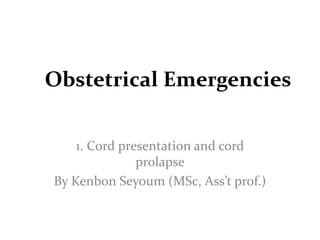
  • 1. Obstetrical Emergencies 1. Cord presentation and cord prolapse By Kenbon Seyoum (MSc, Ass’t prof.)
  • 2. Umbilical cord prolapse • It is defined as descent of the umbilical cord into the lower uterine segment. • There are three clinical types 1.Occult prolapse—The cord is placed by the side of the presenting part and is not felt by the fingers on internal examination. 2.Cord presentation (funic presentation)— The cord is slipped down below the presenting part and is felt lying in the intact bag of membranes. 3. overt prolapse—The cord is lying inside the vagina or outside the vulva following rupture of the membranes.
  • 3. Types of cord prolapse
  • 4. .
  • 5. • INCIDENCE: –in cephalic presentations is 0.5%, –frank breech 0.5%, –complete breech 5%, –footling breech 15%, and –transverse lie 20%. • It is mostly confined to parous women
  • 6. • Prolapse of the umbilical cord exposes the cord to intermittent compression • This compromises fetal circulation • Depending on the duration and intensity of compression, it may lead to: – fetal hypoxia, – brain damage, and – death
  • 7. • In overt cord prolapse, exposure of the umbilical cord to air causes:- – irritation and cooling of the cord, resulting in further vasospasm of the cord vessels. • perinatal morbidity and mortality because of intermittent compression of blood flow and resultant fetal hypoxia. • The perinatal mortality rate associated with all cases of overt umbilical cord prolapse approaches 20%.
  • 8. Causes • Any obstetric condition that predisposes to poor application of the fetal presenting part to the cervix can result in prolapse of the umbilical cord. The following are associated with cord prolapse. – Malpresentations – Contracted pelvis – Prematurity (< 34 wks' ) – Twins – Hydramnios – Placental factor – Iatrogenic – Stabilizing induction
  • 9. • A recent study revealed that obstetric intervention contributes to nearly half of cases of umbilical cord prolapse. • Examples cited include:- – Amniotomy – scalp electrode application, – Intrauterine pressure catheter insertion, – Attempted ECV – IPV and – Expectant management of preterm premature rupture of membranes.
  • 10. Clinical Findings • Overt Cord Prolapse:-can be diagnosed simply by visualizing the cord protruding from the introitus or by palpating loops of cord in the vaginal canal. • Funic Presentation:-made by pelvic examination if loops of cord are palpated through the membranes. • Occult Prolapse:-rarely palpated during pelvic examination. • This condition can be inferred only if fetal heart rate changes
  • 11. Clinical Findings… Fetus • Variable fetal heart rate • Fetal bradycardia • Persistent, severe, variable decelerations and bradycardia  hypoxia, metabolic acidosis, and eventual damage or death. • Meconium staining of the amniotic fluid may be noted at the time of membrane rupture.
  • 12. Complications Maternal – Cesarean section is a major operative procedure with known anesthetic, hemorrhagic, and operative complications. – Laceration of the cervix, vagina, or perineum. Neonatal – The neonate at delivery may be hypoxic, acidotic, or moribund.
  • 13. Prevention • Should be treated as high-risk patients. • Patients with malpresentations or poorly applied cephalic presentations should be considered for ultrasonographic examination at the onset of labor to determine fetal lie and cord position within the uterine cavity. • Artificial rupture of membranes should be avoided until the presenting part is well applied to the cervix.
  • 14. • At the time of spontaneous membrane rupture, a prompt, careful pelvic examination should be performed to rule out cord prolapse. • Should amniotomy be required and the presenting part remains unengaged, careful needling of the membranes and slow release of the amniotic fluid can be performed until the presenting part settles against the cervix.
  • 15. Management Overt Cord Prolapse • An immediate pelvic examination should be performed. • Place in the knee–chest position • Alternatively, 400–700 mL of saline can be instilled into the bladder in order to elevate the presenting part. • Oxygen should be given. • Abdominal delivery should be accomplished as rapidly as possible and a pediatric team should be on standby.
  • 16. Management… Occult Cord Prolapse • If variable decelerations recognized PV should be performed to rule out overt cord prolapse. • Place in the lateral Sims or Trendelenburg position if occult cord prolapse suspected. • Allow labor to continue if the fetal heart rate returns to normal • Administere to oxygen • Amnioinfusion. • If the cord compression pattern persists or recurs to the point of fetal jeopardy  a rapid cesarean
  • 17. Management Funic Presentation • If at term deliver by cesarean section prior to membrane rupture. • No consensus on mgt if the fetus is premature. • The most conservative approach is to hospitalize the patient on bed rest in the Sims or Trendelenburg position. • Serial ultrasonographic to ascertain cord position, presentation, and gestational age.
  • 18. .
  • 19. Route of Delivery • Vaginal delivery can be successfully accomplished in cases of overt or occult cord prolapse if, at the time of prolapse, the cervix is fully dilated, cephalopelvic disproportion is not anticipated, and an experienced physician determines that delivery is imminent. • Cesarean delivery is the preferred route of delivery in most cases. • Vaginal delivery is the route of choice for the previable or dead fetus.
  • 20. Prognosis Maternal • Maternal complications include those related to:- –anesthesia, –blood loss, and –infection following cesarean section or operative vaginal delivery.
  • 21. Prognosis… Neonatal • Depending on the degree and duration of umbilical cord compression occurring before the diagnosis is made and neonatal resuscitation is started.
  • 22. Prognosis… • If the duration of complete cord occlusion is less than 5 minutes, the prognosis is good. • If complete cord occlusion has occurred for longer than 5 minutes or if intermittent partial cord occlusion has occurred over a prolonged period of time, fetal damage or death may occur.
  • 23. References • Current Diagnosis & Treatment Obstetrics & Gynecology, Tenth Edition • Dc Duttas’ Textbook Of Obstetrics Seventh Edition
  • 24. ???
  • 25. Amniotic Fluid Embolism(AFE) • Also called Liquor Amnii Embolism • It is rare • Catastrophic and fatal complication of pregnancy. • Major causes of mortality in modern obstetrics.
  • 26. • Mechanism: Liquor amnii is forced into the maternal circulation either through a rent in the membranes or placenta. • Thromboplastin rich liquor amnii containing the debris, blocks the pulmonary arteries and triggers the complex coagulation mechanism leading to DIC. • There is massive fibrin deposition distributed throughout the entire pulmonary vascular tree.
  • 27. • If the patient survives from the severe cardiopulmonary embarrassment which stimulates thromboembolic phenomenon, there will be severe clotting defect with profuse bleeding per vaginam or through the veno- puncture sites due to consumption of coagulation factors.
  • 28. • From the damaged endothelium of the pulmonary arteries  massive fibrinolytic activators are produced  excite the fibrinolytic system converting the plasminogen to plasmin  produces lysis of fibrin, fibrinogen and even the factor V and factor VIII. • Thus, there is secondary fibrinolysis on top of primary fibrinogen depletion arising out of DIC.
  • 29. Incidence and Risk Factors • exact incidence is not known. • However, in one study utilizing a computerized database from over 300 hospitals in California, the authors calculated frequency of amniotic fluid embolism of approximately 1 per 20,500 deliveries.
  • 30. Possible or Reported Risk Factors Associated with Amniotic Fluid Embolism Tumultuous labor (placenta abruption) Prolonged labor Induction/augmentation of labor Trauma Cesarean delivery Multiparity Advanced maternal age Operative vaginal delivery Hydramnios Uterine rupture Multifetal gestation Male fetal sex Eclampsia Allergy history
  • 31. Diagnosis • The diagnosis of AFE is primarily clinical, based on a high degree of suspicion. – Sudden complain of difficulty breathing, – Becomes hypotensive, – Gasps for air, and – Develops seizures. – Fetal bradycardia, and – The pt. is noted to be oozing from her intravenous site. – Hypoxia which may soon be followed by circulatory collapse and cardiopulmonary arrest
  • 32. DX… • There are few tests that can be utilized to confirm the diagnosis of AFE. • In the presence of an overt coagulopathy, – The serum fibrinogen level often will be quite low and – The fibrin degradation products elevated. • The platelet count also may be decreased but generally not much below 100,000. • An arterial blood gas will often reveal a decreased Po2.
  • 33. TREATMENT • PREVENTIVE: • The responsible factors in prevention are the changes in the trends of obstetric management: – Abruptio placentae – Early institution of appropriate therapy in shock – IUD – Infusion of polymolecular gelatin as plasma expander – Emptying the uterus and controlling the infection early with antibiotics. – Avoiding instillation of hypertonic saline for induction of abortion. • Adjuvant therapies (Vitamin K)
  • 34. CURATIVE • Delivery of the fetus brings the resolution of coagulopathy. • The other part of the management is to achieve: – a platelet count > 50,000/µL and – a fibrinogen level > 100 mg/dl.
  • 35. ACTUAL MANAGEMENT – Volume replacement • – Blood component therapy – Heparin – Fibrinolytic inhibitors
  • 36. References • Dc Duttas’ Textbook Of Obstetrics Seventh Edition • Danforth's Obstetrics and Gynecology, 10th Edition. ??? Please!
  • 37. What do you think this picture tell us?
  • 38. UTERINE RUPTURE • DEFINITION: Disruption in the continuity of the all uterine layers (endometrium, myometrium and serosa) any time beyond 28 weeks of pregnancy is called rupture of the uterus. • classified as:- – Complete:-all layers of the uterine wall are separated – Incomplete:- uterine muscle is separated but the visceral peritoneum is intact. • Incomplete rupture is also commonly referred to as uterine dehiscence.
  • 39. • Morbidity and mortality rates are appreciably greater when rupture is complete. • The greatest risk factor for either form of rupture is prior cesarean delivery. • ETIOLOGY: broadly divided into:- ♠Spontaneous ♠Scar Rupture ♠Iatrogenic
  • 40. SPONTANEOUS During pregnancy: It is rare for an apparently uninjured uterus causes:- ֍Previous damage to the uterine walls ֍Rarely in grand multiparae due to thin uterine walls ֍Congenital malformation of the uterus-rare possibility ֍In Couvelaire uterus
  • 41. During labor: it is due to: ♠ Obstructive rupture—This is the end result of an obstructed labor. ♠ Non-obstructive rupture —Grand multiparae are usually affected and rupture usually occurs in early labor. • The rupture usually involves the fundal area and is complete.
  • 42. SCAR RUPTURE • Primary cesarean section, scar rupture constitutes significantly to the overall incidence of uterine rupture. • The incidence of lower segment scar rupture is about 1–2%, while that following classical one is 5–10 times higher. • Uterine scar following hysterotomy behaves like that of a classical scar and is of growing concern.
  • 43. SCAR RUPTURE • During pregnancy: Classical cesarean or hysterotomy scar is likely to give way during later months of pregnancy. • Lower segment scar rarely ruptures during pregnancy. • During labor: The classical or hysterotomy scar is more vulnerable to rupture during labor. • Although rare, lower segment scar predominantly ruptures during labor.
  • 44. IATROGENIC OR TRAUMATIC During pregnancy: Injudicious administration of oxytocin Use of prostaglandins for induction of abortion or labor Forcible external version Fall or blow on the abdomen.
  • 45. During labor: Internal podalic version Destructive operation. Manual removal of placenta. Application of forceps or breech extraction through incompletely dilated cervix Injudicious administration of oxytocin for augmentation of labor.
  • 46.
  • 47. PATHOLOGY TYPES: • In incomplete rupture, the peritoneum remains intact. • It usually results from:- ♣ rupture of the lower segment scar or ♣ extension of a cervical tear into the lower segment with formation of a broad ligament hematoma.
  • 48. Pathology… • Complete rupture usually occurs following disruption of the scar in upper segment. • It may also be due to spontaneous rupture of both obstructive and non-obstructive type.
  • 49. Pathology… • SITES: ♣ Spontaneous non-obstructive rupture usually involves the upper segment and often involves the fundus. ♣ In obstructive type, the rupture involves the anterior lower segment transversely and often extends upwards along the lateral uterine wall. ♣ The margins are ragged and necrosed. • The posterior wall may be involved due to friction with the sacral promontory.
  • 50. • Not infrequently, the tear extends downwards to involve the cervix and the vaginal wall (colporrhexis). • The bladder may be involved, at times. • Rupture over the previous scar is:- ᴥ Almost always located at the site of the scar. ᴥ The margins of the ruptured cesarean scar are usually clean and look fibrosed. ᴥ May extend to one or both the sides to involve the major branches of uterine vessels
  • 51. Dehiscence and scar rupture Scar dehiscence— (a) disruption of part of scar and not the entire length (b)fetal membranes remain intact and (c) bleeding is almost nil or minimal.
  • 52. Dehiscence and scar rupture … Scar rupture— a) Disruption of the entire length of the scar b) Complete separation of all the uterine layers including serosa c) Rupture of the membranes with a) Varying amount of bleeding from the margins or from its extension d) Uterine cavity and peritoneal cavity become continuous.
  • 53. FETUS AND PLACENTA In incomplete rupture, Both the fetus and placenta remain inside the uterine cavity or part of the fetus may occupy in between the layers of broad ligament. In complete rupture, The fetus with or without the placenta usually escapes out of the uterus. The uterus remains contracted. Blood loss is not much unless major vessels are affected.
  • 54. PROGNOSIS: • Lower segment scar rupture gives a comparatively better prognosis. • But, rupture following obstructed labor either spontaneous or due to instrumentation gives a maternal death rate of about 20% or more. • The major causes of death are hemorrhage, shock and sepsis. • Late sequelae include intestinal obstruction and scar rupture in subsequent pregnancies if the uterine rent has been repaired.
  • 55. DIAGNOSIS OF RUPTURE UTERUS • No specific pattern that presages uterine rupture. • Before hypovolemic shock develops, symptoms and physical findings in women with uterine rupture may appear bizarre unless the possibility is kept in mind. • For example, hemoperitoneum from a ruptured uterus may result in diaphragmatic irritation with pain referred to the chest—directing one to a diagnosis of pulmonary or amnionic fluid embolism instead of uterine rupture.
  • 56. DX… + Non-reassuring fetal heart rate + Cessation of contractions + The appearance of uterine rupture is identical to that of placental abruption. + Little appreciable pain or tenderness. + Pain and tenderness may not be readily apparent. + Maternal hypovolemia from concealed hemorrhage.
  • 57. + If the fetal presenting part has entered the pelvis with labor, loss of station may be detected by pelvic examination. + fetus is partly or totally extruded from the uterine rupture site. + Abdominal palpation or vaginal examination may be helpful to identify the presenting part, which will have moved away from the pelvic inlet. + A firm contracted uterus may at times be felt alongside the fetus.
  • 58. MANAGEMENT OF RUPTURE UTERUS PROPHYLAXIS:The at-risk mothers, likely to rupture, should have mandatory hospital delivery. These are— – Contracted pelvis – Previous history of cesarean section, hysterotomy or myomectomy – Uncorrected transverse lie – Grand multiparity – Known case of hydrocephalus. – Etc…
  • 59. MANAGEMENT… TREATMENT: – Resuscitation – Laparotomy • Laparotomy: Any of the three procedures may be adopted following laparotomy. Hysterectomy Repair Repair and sterilization
  • 60. Decision-to-Delivery • Time With rupture and expulsion of the fetus into the peritoneal cavity, the chances for intact fetal survival are dismal, and reported mortality rates range from 50 to 75 percent. • Fetal condition depends on the degree to which the placental implantation remains intact, although this can change within minutes
  • 61. • With rupture, the only chance of fetal survival is afforded by immediate delivery—most often by laparotomy—otherwise, hypoxia is inevitable. • If rupture is followed by immediate total placental separation, then very few intact fetuses will be salvaged. • Thus, even in the best of circumstances, fetal salvage will be impaired.
  • 62. • Of the 35 laboring patients with a uterine rupture, the decision-to-delivery time was < 18 minutes in 17, and none of these infants had an adverse neurological outcome. • Of the 18 born > 18 minutes from decision time, the three infants with long-term neurological impairments were delivered at 31, 40, and 42 minutes.
  • 63.
  • 64. References • Dc Duttas’ Textbook Of Obstetrics Seventh Edition • Williams OBSTETRICS 24TH EDITION
  • 65. SHOULDER DYSTOCIA • Shoulder dystocia is difficult delivery of the shoulders after delivery of the fetal head. • occurs when either the anterior or the posterior (rare) fetal shoulder impacts on the maternal symphysis or on the sacral promontory. • Overall incidence varies between 0.2 and 1 percent.
  • 66. • Consensus regarding a specific definition of shoulder dystocia is lacking. • However, the diagnosis continues to rely on the clinical perception that the normal downward traction needed for fetal shoulder delivery is ineffective. • Because of these differing definitions, the incidence of shoulder dystocia varies.
  • 67. factors: Predisposing ‡ Fetal macrosomia ‡ Obesity ‡ Diabetes ‡ Postmaturity ‡ Multiparity ‡ Anencephaly ‡ Fetal ascites
  • 68. Complications Fetal: – Asphyxia – brachial plexus injury (plexopathy) due to stretch, Erb, Klumpke palsy – fracture humerus, – clavicle or sternomastoid hematoma during delivery. – Perinatal morbidity and mortality are high.
  • 69. Maternal: –PPH, –cervical, vaginal, perineal tears, –Increased operative delivery and morbidity.
  • 70. Prediction and Prevention The American College of Obstetricians and Gynecologists (2012b) reviewed studies and concluded that:- – 1. Most cases of shoulder dystocia cannot be accurately predicted or prevented. – 2. Elective induction of labor or elective cesarean delivery for all women suspected of having a macrosomic fetus is not appropriate. – 3.Planned cesarean delivery may be considered for the nondiabetic woman with a fetus whose estimated fetal weight is > 5000 g or for the diabetic woman whose fetus is estimated to weigh > 4500 g.
  • 71. Prediction and Prevention… • Prevention of shoulder dystocia is not possible accurately Birthweight • Commonly cited maternal characteristics associated with increased fetal birthweight are:-  obesity,  postterm pregnancy,  multiparity,  diabetes, and  gestational diabetes. • There is universal agreement that increasing birthweight is associated with an increasing incidence of shoulder dystocia.
  • 72. Prediction and Prevention… Intrapartum Factors • Some labor characteristics have been associated with an increased shoulder dystocia risk and include:- –prolonged second stage labor, –operative vaginal delivery, and –prior shoulder dystocia
  • 73. Shoulder dystocia cont… Diagnosis Definite recoil of the head back against the perineum (turtle-neck sign) Inadequate spontaneous restitution Fetal face becomes plethoric.
  • 74. Shoulder dystocia cont… Management • Gentle traction:-An initial gentle attempt at traction, assisted by maternal expulsive efforts, is recommended. • After gentle traction, various techniques can be used to free the anterior shoulder from its impacted position behind the symphysis pubis.
  • 75. The following maneuvers can be used 1. suprapubic pressure 2. McRoberts maneuver:- removing the legs from the stirrups and sharply flexing them up onto the abdomen
  • 76.
  • 77. 3. Delivery of the posterior shoulder:-, consists of carefully sweeping the posterior arm of the fetus across its chest, followed by delivery of the arm.
  • 78.
  • 79. . 4. Woods corkscrew maneuver • progressively rotating the posterior shoulder 180 degrees in a corkscrew fashion, the impacted anterior shoulder could be released.
  • 80. 5. fracture of the anterior clavicle;-Deliberate fracture of the anterior clavicle by using the thumb to press it toward and against the pubic ramus can be attempted to free the shoulder impaction. 6. Cleidotomy: One or both clavicles may be cut with scissors to reduce the shoulder girth. This is applicable to a living anencephalic baby as a first choice or in a dead fetus.
  • 81. 7. The second Rubin maneuver. A. The shoulder-to-shoulder diameter is aligned vertically. B. The more easily accessible fetal shoulder is pushed toward the anterior chest wall of the fetus. • Most often, this results in abduction of both shoulders, which reduces the shoulder-to- shoulder diameter and frees the impacted anterior shoulder
  • 82.
  • 83. 8. Zavanelli maneuver:- cephalic replacement into the pelvis followed by cesarean delivery. 9. Symphysiotomy:-intervening symphyseal cartilage and much of its ligamentous support is cut to widen the symphysis pubis
  • 84. References • Dc Duttas’ Textbook Of Obstetrics Seventh Edition • Williams OBSTETRICS 24TH EDITION

Notes de l'éditeur

  1. Placental factor—minor degree placenta praevia with marginal insertion of the cord or long cord
  2. Adjuvant therapies (Vitamin K)—The vitamin K dependent factors II, VII, IX, X are consumed in DIC. 5-10 mg of Inj Vit K given (IM), can help to replenish these procoagulants.