Recommandé

Recommandé
Contenu connexe
Tendances
Tendances (20)
En vedette
En vedette (7)
Similaire à "Time course evaluation & treatment of post-TBI brain tumor with corresponding visual field loss" [Poster, American Academy of Optometry – October 2013]
Similaire à "Time course evaluation & treatment of post-TBI brain tumor with corresponding visual field loss" [Poster, American Academy of Optometry – October 2013] (20)
Dernier
Dernier (20)
"Time course evaluation & treatment of post-TBI brain tumor with corresponding visual field loss" [Poster, American Academy of Optometry – October 2013]
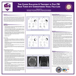
- 1. Time Course Evaluation & Treatment of Post-TBI Brain Tumor with Corresponding Visual Field Loss Maggie Jan, O.D. Southern California College of Optometry at Marshall B. Ketchum University VA Long Beach – Traumatic Brain Injury & Low Vision Rehabilitation Residency Background In recent research, traumatic brain injury (TBI) has been theorized as a possible risk factor contributing to the development of brain tumors.1 In this case, a patient with no previous history of brain tumors and no family predisposition for tumors experiences a TBI. Seven years after sustaining IED blast-related military brain injuries, the patient presents with symptoms consistent with a cranial neoplasm. This study tracks the patient’s symptoms and visual field defect juxtaposed with corresponding MRI images identified during the time course of tumor treatment. Treatment & Management • Medical team starts the patient on Parlodel® (bromocriptine) drug therapy aimed at reducing the prolactin-secreting tumor size. • If tumor fails to respond to drug therapy, invasive surgical pituitary tumor resection remains an option but will require hormone replacement therapy. • Testosterone therapy for decreased libido, weight gain, gynecomastia. • Initiated mental care, psychiatry for depression and anger. Mood-enhancing drug Wellbutrin® (bupropion HCl) and trazadone prescribed for insomnia and mood changes. • The visual fields are taken concurrently as tumor treatment is initiated. During the time course of the patient’s treatment, a series of four 24-2 Humphery threshold visual fields are taken over one year (Figure 1b). Challenges acquiring visual fields from TBI patients exist – decreased visual attention, PTSD and claustrophobia make it difficult to test. • No prescription needed for distance vision. Sun wear with polarization prescribed for protection against glare and symptomatic photophobia secondary to TBI. Rx reading glasses for accommodative dysfunction. References 1. McKinney, P A. “Brain Tumours: Incidence, Survival, and Aetiology.” Journal of Neurology, Neurosurgery & Psychiatry. 75, 2004. 2. Mumenthaler, Marco, and Heinrich Mattle. “Visual Field Defects.” Fundamentals of Neurology: An Illustrated Guide. Stuttgart: Thieme, 2006. 3. Kaufman, David M. “Visual Field Loss.” Clinical Neurology for Psychiatrists. Philadelphia: Saunders/Elsevier, 2007. 4. Silverstone B, Lang MA, Rosenthal BP, Faye EE, editors. “Signs and Symptoms of Chiasmal Lesions.” The Lighthouse Handbook on Vision Impairment and Vision Rehabilitation. Vol. 2. New York: Oxford University Press. p 188-190. 5. Center for Disease Control and Prevention (CDC). National Center for Injury Prevention and Control. Report to Congress on traumatic brain injury in the United States. Atlanta (GA): Centers for Disease Control and Prevention. 1999, Dec. 6. Center for Disease Control and Prevention (CDC). National Center for Injury Prevention and Control. Report to Congress on Traumatic Brain Injury in the United States: Understanding the Public Health Problem among Current and Former Military Personnel. Atlanta (GA): Centers for Disease Control and Prevention. 2013, Aug. 7. Elder, Gregory A, and Adrian Cristian. “Blast-related Mild Traumatic Brain Injury: Mechanisms of Injury and Impact on Clinical Care.” Mount Sinai Journal of Medicine: a Journal of Translational and Personalized Medicine. 76(2), 2009: 111-118. Conclusions • Addition of visual efficiency training or vision rehabilitation could be potentially valuable in the future for helping the patient compensate for field loss. • Growths + minute changes to mass/volume in confined space of sella turcica can affect integrity of surrounding structures such as optic nerve, visual function, and hormone secretions. • Tracking changes to visual field not only helps radiologists gauge potential treatment efficacy but serves dual purpose of helping the patient visualize health status while continuing to undergo the remaining time course of scheduled treatment with slow recovery. • Patient’s brain tumor development following acquisition of TBI could be coincidental. Although neuronal disruption and inflammation processes has been hypothesized as a possible risk factor in epidemiological studies1, no link is proven currently. Further research into patients that have both TBIs and tumors to see if tumors do develop over time. The study must control for and rule out genetic predisposition and subjects with existing tumors. • 10% to 20% of veterans returning from Iraq and Afghanistan operations are estimated to have suffered a traumatic brain injury.7 In addition, according to CDC statistics the incidence of TBIs in current and former military personnel has tripled in the six years between 2005 and 20116. High incidence and quantifiable impact on quality of life necessitates the need to manage and treat patients with TBI. Case Report Patient History • 37-year-old Caucasian male, active duty military soldier complaining of visual changes, difficulty with peripheral vision, difficulty seeing objects or people who “suddenly appear” to his right or left. • Vision loss is accompanied with symptoms of severe headaches worse in the morning, poor balance and dizziness, decreased attention, personality changes such as increased irritability and anger, problems with memory and word retrieval, insomnia, weight gain, and gynecomastia. • Med Hx: TBI secondary to fall with loss of consciousness from explosions accompanied with impact in Iraq 7 years ago. Frontal lobe contusion with encephalomalacia. MRIs taken at the time of the acquired brain injury revealed no cranial lesions or masses. No family history of tumors. • Ocular Hx: Unremarkable • Medications: None Exam Findings • Ocular findings are unremarkable, with exception of mild decrease in accommodative amplitudes, photophobia, and abnormal confrontation visual field with findings of neglect in the superior temporal and inferior temporal fields of both eyes. • Baseline Humphery Threshold Visual Field 24-2 Sita Fast: temporal visual field loss detected OD & OS (Figure 1a). Field loss is repeatable and displays pattern of bitemporal hemaniopsia. • Lab studies: Blood serum analysis reveals high levels of ACTH, elevated prolactin, low levels of testosterone. • Radiology studies: MRI imaging reveals a mass in the right side of the pituitary gland measuring approximately 7x11x11mm which invades the sphenoid sinus (Figure 2). Differential Diagnosis & Discussion • Differential Diagnoses (DDx) for Visual field loss: Pituitary adenoma (primary DDx), mid chiasm neoplasm, craniopharyngiomas, meningiomas, anterior carotid artery aneurysm.2 • Cranial neoplasm located in the pituitary determined to be the cause of patient’s bitemporal visual loss, hyperprolactinemia, and hypogonadism.4 • Visual Field Loss & Visual PathwayAnalysis: Nerve fibers originating in the nasal retina of each eye converge at the optic chiasm before crossing over to opposite sides of the brain for higher level processing.2 Thus a compression of the optic chiasm affects the visual impulse from both nasal retina which corresponds to loss of temporal visual field in both eyes.3 • Symptoms of a moderate TBI could impact speech, sensory, vision and cognitive symptoms5. Patient’s symptoms of: headaches, vertigo, trouble with memory, attention, concentration deficits, irritability related to acquired brain injury.6 Of these TBI-related symptoms some may closely resemble characteristics of a brain tumor. • Visual symptoms most commonly experienced by patients with TBI include: accommodative dysfunction, noncompensating vergences, large phorias or tropias, and photophobia. • In this case symptoms of headaches, inattention, and irritability overlap in both conditions; fortuitously, addressing these common symptoms via treatment of the brain tumor will not exacerbate the underlying TBI. • The patient’s TBI is categorized as “moderate” due to loss of consciousness greater than half an hour but less than six hours, without open intracranial wound5. Even with identification and diagnosis of the traumatic brain injury, there exists no method of reversal for physical damage resulting from brain injury. Current TBI care is centered around providing treatment of symptoms and functional rehabilitation rather elimination of the condition. Figure 2: T1 Post-‐Gadolinium MRI Images AXIAL CORONAL Acknowledgements to Major Jennifer Stoecklin Stowe, O.D.