1. Passive avoidance training decreases synapse density in
the hippocampus of the domestic chick
A. M. Nikolakopoulou,1,2
H. A. Davies1
and M. G. Stewart1
1
The Open University, Biological Sciences, Walton Hall, Milton Keynes MK7 6AA, UK
2
University of California, Irvine, Neurobiology and Behavior, 2205 McGaugh Hall, Irvine, CA 92697, USA
Keywords: axo-spinous density, dorsal, learning, stress, ventral
Abstract
The bird hippocampus (Hp), although lacking the cellular lamination of the mammalian Hp, possesses comparable roles in spatial
orientation and is implicated in passive avoidance learning. As in rodents it can be divided into dorsal and ventral regions based on
immunocytochemical, tracing and electrophysiological studies. To study the effects of passive avoidance learning on synapse
morphometry in the Hp, spine and shaft synapse densities of 1-day-old domestic chicks were determined in dorsal and ventral Hp of
each hemisphere by electron microscopy, 6 and 24 h following training to avoid pecking at a bead coated with a bitter-tasting
substance, methyl anthranilate (MeA). The density of asymmetric spine and shaft synapses in MeA-trained birds at 6 h post-training
was significantly lower in the dorsal and ventral Hp of the right hemisphere relative to control (untrained) chicks, but by 24 h this
difference was absent. A hemispheric asymmetry was apparent in the ventral Hp where the water-trained group showed enhanced
shaft and spine synapse density in the left hemisphere, whilst in the MeA-trained group only asymmetric shaft synapses follow the
same pattern in relation to the right hemisphere. There were no differences in asymmetric shaft synapses in the dorsal Hp at 6 h post-
training, but at 24 h post-training there was a reduction in the density of shaft synapses in the right hemisphere in MeA compared with
control birds. These data are discussed in relation to the pruning effects of stress and learning on synapse density in chick Hp.
Introduction
Alterations in connectivity via changes in synaptic efficacy are
believed to underlie learning and memory storage (Hebb, 1949; Bliss
& Collingridge, 1993). Changes in synaptic morphology can be very
rapid, with alterations in shape occurring within seconds (Hering &
Sheng, 2001). A number of studies have focused on synaptogenesis in
the chick brain mainly after passive avoidance learning (PAL) where
the aversive experience is exposure to a bitter-tasting substance,
methyl anthranilate (MeA) (Stewart et al., 1987), and in filial (Horn
et al., 1985; Horn, 2004) and acoustic imprinting (Thode et al., 2005).
Increases in spine and synapse density have been demonstrated in the
left medial striatum (MSt) (Stewart et al., 1987; Lowndes & Stewart,
1994) and intermediate medial mesopallium (IMM) (Patel & Stewart,
1988) at 24 h after PAL in MeA-trained chicks compared with water
control birds.
The chick hippocampus (Hp) has been suggested to be homologous
to the mammalian Hp (Kallen, 1962; Erichsen et al., 1991; Atoji et al.,
2002). Previous studies have shown that as in mammals the avian Hp
plays a key role in spatial memory (Bingman et al., 1990; Regolin &
Rose, 1999; Kahn & Bingman, 2004) and demonstrates synaptic
plasticity with some similarities to mammalian long-term potentiation
(LTP) (Margrie et al., 1998). This region has also been implicated in
the passive avoidance paradigm (Sandi et al., 1992). Unal et al. (2002)
demonstrated an increase in density of shaft and spine synapses with
time after PAL in the dorsolateral Hp of chicks, with an increase at
both 24 and 48 h after training in the density of shaft synapses in
MeA- compared with water-trained birds.
In the present study we have examined the effects of PAL on
synaptic morphometry in the ventral and dorsal Hp of the domestic
chick, the divisions based on the published findings (Casini et al.,
1986; Krebs et al., 1991; Szekely, 1999). Each area was studied
individually, because previous studies from our group have demon-
strated synapse density reduction in the dorsal Hp after ischaemia
(Horner et al., 1996), and in rat brain the dorsal and ventral Hp have
been shown to play different roles in learning tasks (Moser et al.,
1993; Hock & Bunsey, 1998; Moser & Moser, 1998). Our studies
were conducted at 6 h and then 24 h after PAL and included two
control groups, a completely untrained control group and birds trained
to peck a bead identical to that used with the aversive tasting MeA, but
coated with water. The 6 h time-point was chosen because a protein
cascade takes place by this time that enables the structural changes
necessary for short-term memory to be consolidated into long-term
memory (Rose, 1991, 1995a,b), and by 24 h this process is well
advanced (Rose & Stewart, 1999). Each hemisphere was studied
separately as chicks show evidence of hemispheric asymmetry
(Stewart et al., 1987; Sandi et al., 1993; Gagliardo et al., 2001).
Materials and methods
Animals and training
Commercially obtained Ross Chunky eggs (domestic chick Gallus
domesticus) were incubated and hatched in our own brooders until
18 ± 6 h old. Chicks were placed in pairs in small aluminium pens
illuminated by red bulbs at a temperature of 25–30 °C. The animals were
Correspondence: Dr A.M. Nikolakopoulou, as above.2
E-mail: anikolak@uci.edu
Received 5 August 2005, revised 7 December 2005, accepted 10 December 2005
European Journal of Neuroscience, Vol. 23, pp. 1054–1062, 2006 doi:10.1111/j.1460-9568.2006.04619.x
ª The Authors (2006). Journal Compilation ª Federation of European Neuroscience Societies and Blackwell Publishing Ltd
2. separated into three groups, naı¨ve (undisturbed), water-trained (W) and
methyl anthranilate-trained (MeA). Chicks were first pretrained by
being presented a small white bead 3 mm in diameter three times with
intervals of 5 min between presentations, essentially as described
previously (Mileusnic et al., 2000; Dermon et al., 2002). Animals that
successfully pecked the bead were noted as ‘peck’, while those that did
not peck were noted as ‘no peck’. In order for the chicks to be included in
the study, they should have pecked the white bead three times. Ten
minutes after the last presentation of the white bead, a chrome bead
4 mm in diameter dipped in either water or MeA was presented to the
animals (the training session). Chicks peck once, and having tasted MeA
they exhibit a disgust response by shaking their heads, emitting stress
sounds and beating their beaks on the ground. Chicks were tested 6 h
(naı¨ve group n ¼ 7, water group n ¼ 5, MeA group n ¼ 5) or 24 h
(naı¨ve group n ¼ 6, water group n ¼ 6, MeA group n ¼ 5) after
training by the presentation of a dry chrome bead. Chicks in the water
group pecked the dry bead whilst those in the MeA group exhibited
recall of the bitter taste and avoided the chrome bead. Chicks that failed
to give the correct response to the task within 20 s, and pecked at the dry
bead although previously exposed to MeA, were excluded from the
study. All experimental procedures took place under UK Home Licence
and were also in agreement with the European Communities directive
(86 ⁄ 609 ⁄ EEC) for the care and use of laboratory animals.
Tissue fixation
Animals were anaesthetized by intraperitoneal (i.p.) injection with
0.2 mL of sodium pentobarbital immediately after testing. They were
then transcardially perfused with heparin in 0.9% saline followed by
3.75% acrolein (TAAB, UK) in 2% paraformaldehyde in 0.1 m
phosphate buffer (PB), pH 7.4, followed by 2% paraformaldehyde in
PB. Brains were removed from the skull and postfixed at 4 °C
overnight in the latter fixative.
Tissue processing
The left hemisphere was marked and the brains were cut at 100 lm
thickness with a vibrating microtome (Leica, UK); sections of the
antero-posterior level A 8.2 were used for this study (Kuenzel &
Masson, 1988) (Fig. 1). The sections were postfixed with 2% osmium
tetroxide in 0.1 m PB for 1 h, washed in 0.1 m PB dehydrated through
a graded ethanol series followed by propylene oxide and Epon (Agar
Scientific, Stansted, UK) before being flat-embedded between two
sheets of Aclar (Agar Scientific, Stansted, UK). Sections were
polymerized at 60 °C for 48 h. With the use of a stereoscope to view
the sections, the Hp was divided into ventral and dorsal parts (Szekely
& Krebs, 1996; Szekely, 1999), as indicated in Fig. 1.
Preparation for electron microscopy
The tissue blocks were coded and all subsequent procedures were
performed blind. Blocks were cut on a UCT ultramicrotome (Leica),
and ribbons of sections 70 nm thick were collected on formvar ⁄ car-
bon films on copper slot grids. The sections were counterstained in
uranyl acetate and Reynolds lead citrate before examination in a JEOL
1010 electron microscope. Digital images were collected at a
magnification of 12 000 using a Gatan BioScan CCDTV.
Electron microscopy and synapse counting
Synapse identification
Synapses were identified on the basis of a thickening of the pre-
and postsynaptic membranes and the presence of vesicles in close
proximity to the presynaptic zone. Synapses with a prominent
postsynaptic density (PSD) are termed asymmetric and most
commonly occur on dendritic spines, though some may be found
on dendritic shafts (Fig. 2A–D). Synapses where the pre- and
postsynaptic membranes are of equal thickness are termed sym-
metric and are typically found on dendritic shafts (Fig. 2B).
Symmetric synapses overall comprised less than 2% of the total and
were too infrequent for a statistical analysis and are therefore not
included in the present analyses. Similarly, perforated synapses,
which are those with a split PSD, were not taken into account due
to their rarity.
Synapse counting
The physical dissector was used for estimation of synapse density
(Sterio, 1984), where synapses are counted if seen in one section
(the nominated) and not in the other (reference section) (Fig. 3).
The mathematical formula for calculation of the numerical density
is: Nsynapse ¼ SQ–
syn ⁄ tA, where SQ–
syn is the total number of counted
synapses only in the nominated sections, t is the section thickness
(distance between the two sections) and A is the area of the
counting frame. Section thickness was determined as described in
earlier studies (de Groot & Bierman, 1986; De Groot, 1988).
Briefly, the relative electron transmission (PET) was applied to
determine the thickness of a section by using the thickness
estimation diagram that occurs from the application of Small’s
minimal fold technique (de Groot & Bierman, 1986; De Groot,
1988; Tigges et al., 1996). Synapses are not counted if they touch
the forbidden lines (left side and bottom lines).
Synapse height
Synapse height (Hsyn, which is a measure of the size of the PSD) was
estimated using parameters derived from the dissector method as
Hsyn ¼ ð
X
Qsyn=
X
QÀ
synÞ Â t
where SQ–
syn is the total number of counted synapses only in the
nominated sections and SQsyn is the total number of synapses in both
the nominated and reference fields.
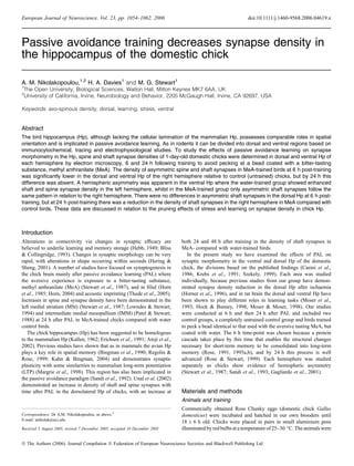
Fig. 1. Coronal sections of the chick brain showing the location of the ventral
and dorsal hippocampus (vHp and dHp). AA, anterior arcopallium; AD, dorsal
arcopallium; CPi, piriform cortex; GP, globus pallidus; LSt, lateral striatum;
M, mesopallium; N, nidopallium; SL, lateral septal nucleus; SM, medial septal
nucleus; TnA, nucleus taeniae of amygdala; black dots A14, dopaminergic
neurons in the paraventricular nucleus (PVN).
Effects of PAL on the chick hippocampus 6 and 24 h after training 1055
ª The Authors (2006). Journal Compilation ª Federation of European Neuroscience Societies and Blackwell Publishing Ltd
European Journal of Neuroscience, 23, 1054–1062
3. Statistics
Four-way analysis of variance (anova) for hemisphere (right, left), time
of study (6 h, 24 h), brain area (dorsal, ventral) and training (control,
water-trained, MeA-trained) was used to check statistically significant
differences between the groups tested, and values P < 0.05 were taken
as significant. When a P-value was not significant for the main factors
but only for their interaction, a three-way anova was conducted for the
factors in the interaction to determine any significant differences. If the
P-value was significant, a Fisher least significant difference (LSD)
post hoc test was performed in order to find specific differences.
Results
Synapse density alterations in shaft and spine synapses (Fig. 2A–D)
were examined in both the ventral and dorsal subdivisions of the chick
Hp 6 and 24 h after PAL. Although the borders of ventral and dorsal
Hp in chicks have not been clearly defined by previous studies, the
dorsal Hp in this study was taken to correspond to that described as
area 3 and 4 by Erichsen et al. (1991) (dorsomedial Hp and part of
dorsolateral as in Szekely & Krebs, 1996). This region has been
suggested to be homologous to the dentate gyrus and hilus,
respectively, whereas the ventral Hp in chicks is related to area 2,
which shows homology with Ammon’s horn (CA).
Synapse ultrastructure
As described above, two different types of synapses were examined;
asymmetric dendritic shaft and asymmetric spine (also termed
asymmetric axo-dendritic and axo-spinous, respectively) (Fig. 2A
and D).
Fig. 2. Photomicrographs of synaptic contacts in the chick Hp. (A–D). (A) An asymmetric shaft synapse (on dendrites) in dorsal 1-day-old chick Hp indicated with a
black asterisk. The presynaptic part can be clearly distinguished by the presence of vesicles (ves). (B) Representative symmetric shaft synapse marked with an
arrowhead from the dorsal Hp of the MeA-trained group. Asterisk indicates spine synapses. (C) Representation of asymmetric spine synapses marked with black
asterisks from the right hemisphere of the ventral Hp of the water-trained group. (D) Two dendrites in the ventral Hp of a control bird 24 h post-training receiving two
asymmetric synapses from a presynaptic axon terminal (At) resulting in axo-dendritic synapses. At, axon terminal; Den, dendrite; mit, mitochondrion; sp, spine. Scale
bars, 200 nm.
1056 A.M. Nikolakopoulou et al.
ª The Authors (2006). Journal Compilation ª Federation of European Neuroscience Societies and Blackwell Publishing Ltd
European Journal of Neuroscience, 23, 1054–1062
4. Effects of PAL 6 h post-training
Two sets of data are presented in Fig. 4, for synapse density 6 h after
PAL in ventral and dorsal Hp from each hemisphere.
Asymmetric shaft density
Data for asymmetric shaft synapses in the ventral and dorsal Hp 6 h
post-training for control, water and MeA birds are presented in
Fig. 4A and B. Four-way anova showed statistical differences for
brain regions examined (ventral and dorsal Hp) (F1,109 ¼ 4.648,
P ¼ 0.033), and the interaction between hemisphere and area of study
(F1,109 ¼ 5.546, P ¼ 0.02). There is a 48% difference in the
asymmetric axo-dendritic (asym shaft) synapses in the left hemisphere
of the ventral Hp of the water-trained group compared with the right
hemisphere. LSD post hoc analysis demonstrated that 6 h post-
training the ventral Hp of the right hemisphere shows significantly
lower synapse density (25%) in relation to the left hemisphere of the
same area (P ¼ 0.045) in the water-trained group.
Asymmetric spine density
Mean data for asymmetric spine synapse density in left and right dorsal
and ventral Hp of the three bird groups are shown in Fig. 4A and B. Four-
way anova revealed statistically significant differences for the inter-
action of time after training and training group (F2,109 ¼ 3.377,
P ¼ 0.038), but not for single factors. Therefore, a three-way anova
only for the 6 h time course (training group, hemisphere, area) was
performed, which revealed a statistically significant difference between
the training groups (F2,52 ¼ 3.856, P ¼ 0.027) and hemisphere.
Post hoc tests showed that 6 h post-training the dorsal Hp of the right
hemisphere of the MeA-trained group has significantly fewer asym-
metric spine synapses (35% less) in comparison to control birds
(P ¼ 0.00084). It also has fewer of this type of synapse than the ventral
part of the right hemisphere of controls (P ¼ 0.04). Furthermore, the
water- and MeA-trained groups exhibit significant hemispheric differ-
ences 6 h after training, with the right ventral Hp having fewer
asymmetric spine synapses in comparison to the left hemisphere (44%
for water, P ¼ 0.017; 33% for MeA-trained birds, P ¼ 0.04).
Effects of PAL 24 h post-training
Asymmetric shaft density
Data for asymmetric axo-dendritic synapses in control, water- and
MeA-trained birds 24 h post-training are shown in Fig. 5A and B.
A four-way anova shows statistically significant differences for brain
regions examined (ventral vs. dorsal Hp) (F1,109 ¼ 4.648, P ¼ 0.033),
and the interaction between hemisphere and region examined
(F1,109 ¼ 5.546, P ¼ 0.02). Fisher LSD post hoc tests showed that
in the dorsal Hp of the right hemisphere there is a 33% decrease in
synapse density in the MeA-trained group in comparison with control
birds (P ¼ 0.038; Fig. 5A), whilst the density of axo-dendritic
synapses (asym shaft) in the right hemisphere of the dorsal Hp of
control birds is 29% greater than in the left hemisphere (P ¼ 0.031).
Differences in synaptic density in Hp between 6 h and 24 h
post-training groups
Asymmetric shaft density
After a Fisher post hoc test, a 48% increase was confirmed in
asymmetric shaft synapses of water-trained chicks in the ventral Hp of
the right hemisphere (P ¼ 0.012) at 24 h in comparison to 6 h post-
training.
Asymmetric spine density
Post hoc tests showed a contrasting pattern between the control and
the water-trained groups in the dorsal and ventral Hp. At 6 h there
were significantly more asymmetric spine synapses (1.61 vs. 1.31 per
Fig. 3. Example of two images used for synapse density estimation with the dissector method. The image on the right is the ‘nominated or look-up’ image, whilst
the left is the ‘reference’ image. Only synapses that are located within the borders of the lines are counted. The dashed lines are the forbidden lines; a synapse
touching the dashed lines is not counted. An asymmetric spine on a dendrite is marked with an asterisk in both images and it is not counted. The black arrow in the
look-up section indicates an asymmetric synapse onto a dendrite, which is counted as it does not appear in the reference section. The black arrow indicates a
symmetric synapse onto a spine (look-up section). The black arrowheads show an asymmetric synapse onto a spine in both images and therefore are not counted. In
the reference image the star indicates a symmetric axo-dendritic synapse. In this case three synapses would have been counted, one symmetric axo-dendritic and one
asymmetric axo-dendritic synapse in the reference section, and one symmetric axo-spinous in the look-up section.
Effects of PAL on the chick hippocampus 6 and 24 h after training 1057
ª The Authors (2006). Journal Compilation ª Federation of European Neuroscience Societies and Blackwell Publishing Ltd
European Journal of Neuroscience, 23, 1054–1062
5. lm3
, a 23% increase) in the dorsal Hp of the right hemisphere of the
control group (P ¼ 0.033) than in the right ventral Hp; however, this
difference disappeared by 24 h. In contrast, in the right ventral Hp of
the water-trained group 6 h after training there were fewer asymmetric
spine synapses (35% less) in comparison to the 24 h water-trained
group (P ¼ 0.0024). No other changes were observed.
Synaptic height
Asymmetric shaft synapse height
Mean data for asymmetric shaft synaptic height (Hsyn) in the three
chick groups at 6 and 24 h post-training are shown in Table 1.
Four-way anova showed significant differences for the interaction
of time after training and the Hp area examined (F1,108 ¼ 5.88,
P ¼ 0.017).
Post hoc tests at 6 h post-training show that in the ventral Hp of the
left hemisphere, Hsyn in control birds is greater than in water-trained
chicks (P ¼ 0.0096), but is reduced relative to MeA-trained birds
(P ¼ 0.036). Additionally, the MeA-trained group demonstrates
increased Hsyn relative to water-trained chicks (P > 0.0001). In the
dorsal part of the right hemisphere Hsyn is higher in the MeA-trained
group than in water (P ¼ 0.0001) or control birds (P ¼ 0.005).
At 24 h post-training there are no differences in Hsyn between the
groups in either hemisphere of ventral or dorsal Hp. However,
comparison between values at 6 and 24 h shows that Hsyn in the MeA-
trained group is greater at 6 than at 24 h in the left ventral Hp
(P ¼ 0.0059), whilst Hsyn in the right dorsal Hp of the water-trained
group is greater than at 6 h (P ¼ 0.036).
Asymmetric spine synaptic height
The data for the height (Hsyn) of the major class of synapses examined,
asymmetric spine synapses, are presented in Table 2. Four-way anova
showed no differences in spine synaptic height for any of the factors
examined.
Discussion
Our data reveal marked alterations in the density of asymmetric
synapses (both on spines and dendritic shafts) in the chick Hp after
passive avoidance training. In general, training results in a decrease
rather than an increase in synapse density. In the ventral Hp of the
right hemisphere of the water-trained group at 6 h post-training,
asymmetric shaft synaptic density was reduced in comparison to the
Fig. 4. Histograms showing asymmetric synapse
densities (Nv ⁄ lm3
) in the dorsal (A) and ventral
(B) hippocampus (Hp) of the right and left hemi-
sphere of chicks 6 h after avoidance training, and
in water-trained and naı¨ve controls (control n ¼ 6,
water n ¼ 5, MeA n ¼ 5). Vertical bars on the
histogram blocks represent means ± SEM.
(A) The asterisks indicate significant reductions in
asymmetric spine synapse density in the dorsal Hp
of the right hemisphere of MeA-trained chicks
(P ¼ 0.0008). (B) Significant differences are in-
dicated between the right and left hemisphere in
the water-trained group (n ¼ 5) (P ¼ 0.045) for
asymmetric shaft synapses (à), whilst and +
show differences for asymmetric spine synapse
densities between right and left hemisphere of
water and MeA-trained groups (P ¼ 0.017 and
P ¼ 0.04, respectively).
1058 A.M. Nikolakopoulou et al.
ª The Authors (2006). Journal Compilation ª Federation of European Neuroscience Societies and Blackwell Publishing Ltd
European Journal of Neuroscience, 23, 1054–1062
6. left hemisphere, and asymmetric axo-spinous synapse density was also
reduced in the right hemisphere of both the water- and MeA-trained
groups. Similarly, in the dorsal Hp of the right hemisphere, there was a
decline in synaptic density of asymmetric axo-spinous synapses 6 h
after training in the MeA-trained group in comparison to the control
group. Our data differ from that of Unal et al. (2002), where an
increase in the density of shaft synapses was found in Hp at both 24
and 48 h post-training, with small decreases in spine synapse density
at 24 h post-MeA training. A reason for the differences may be the
dissimilarity in the Hp regions examined (Unal et al., 2002 studied the
dorsolateral Hp only), and also the control group in the Unal et al.
study did not include naı¨ve (completely untrained) animals. Further-
more, unlike the findings in the study by Unal et al. (2002), our data
indicate that synaptic changes occur without alteration in PSD size
Fig. 5. Histograms showing asymmetric synapse
densities (Nv ⁄ lm3
) in the dorsal (A) and ventral
(B) hippocampus (Hp) in the right and left hemi-
sphere of chicks 24 h after avoidance training, and
in water-trained and naı¨ve controls (control n ¼ 6,
water n ¼ 6, MeA n ¼ 6). Vertical bars on the
histogram blocks represent means ± SEM.
(A) There are significantly fewer (à) asymmetric
shaft synapses in the dorsal Hp of the left hemis-
phere of control animals, compared with the right
hemisphere (P ¼ 0.031). Also the MeA-trained
group has significantly fewer asymmetric shaft
synapses (*) in comparison to the control group
(P ¼ 0.038). (B) None of the differences in the
ventral Hp between the groups or hemispheres at
24 h for either asymmetric shaft or spine synapses
are significant.
Table 1. Asymmetric shaft synapse height
Asymmetric shaft synapse height 6 h after training (in lm) Asymmetric shaft synapse height 24 h after training (in lm)
Ventral hippocampus Dorsal hippocampus Ventral hippocampus Dorsal hippocampus
R L R L R L R L
Control 0.12 ± 0.006 0.12 ± 0.008 0.11 ± 0.1 0.12 ± 0.009 0.12 ± 0.008 0.12 ± 0.006 0.12 ± 0.01 0.13 ± 0.01
Water 0.13 ± 0.01 0.10 ± 0.008** 0.095 ± 0.01 0.11 ± 0.007 0.11 ± 0.006 0.11 ± 0.006 0.13 ± 0.009à 0.12 ± 0.006
MeA 0.12 ± 0.01 0.14 ± 0.016*,
0.13 ± 0.01* 0.11 ± 0.01 0.12 ± 0.01 0.10 ± 0.006 0.12 ± 0.01 0.13 ± 0.02
Data are presented as means (± SEM) showing asymmetric shaft synaptic height (Hsyn) in right (R) and left hemisphere (L) of the three chick groups 6 and 24 h after
passive avoidance training. *P < 0.05 and **P < 0.01, comparing control and trained birds at 6 h after training, but there were no significant differences in Hsyn
between the groups in either hemisphere of ventral or dorsal hippocampus.
MeA-trained group 6 h Ventral (R) > MeA-trained group 24 h Ventral (R)
(P ¼ 0.0059). à
Water-trained group 6h Dorsal (R) < water-trained group 24 h Dorsal (R) (P ¼ 0.036).
Effects of PAL on the chick hippocampus 6 and 24 h after training 1059
ª The Authors (2006). Journal Compilation ª Federation of European Neuroscience Societies and Blackwell Publishing Ltd
European Journal of Neuroscience, 23, 1054–1062
7. (Hsyn), at least in asymmetric axo-spinous synapses, which are the
major class of synapses in chick Hp (three times more numerous than
asymmetric shaft). There are alterations in the size of asymmetric shaft
synapses at 6 h post-training, with Hsyn larger in the left hemisphere of
MeA birds than in water or control birds, but these differences
disappear by 24 h post-training.
The synapse reduction in both the dorsal and ventral Hp would
appear to suggest that the training process per se may have
contributed to a large extent to the decrease in synapse density in
the chick Hp, as it occurs both in the water- and MeA-trained groups.
Conversely, in the rat, the passive avoidance task (O’Malley et al.,
1998) or water maze training (Moser et al., 1994) results in an
increase in dendritic spine density in Hp dentate gyrus and CA1,
respectively, whilst the enriched environment has similar effects in
CA3 (Altschuler, 1979). One may argue that in chicks passive
avoidance training is not a spatial task and therefore the Hp is affected
differently from mammals, resulting in reduced synaptic connectivity
in the MeA-trained group. However, recent studies in rat Hp have also
identified synapse density alterations after non-spatial tasks such as
olfactory learning (Knafo et al., 2004). They emphasize that an
increase in axo-spinous synapse density in CA1 apical dendrites
occurs only if odour exposure is accompanied by olfactory learning.
Although chicks associate the intense smelling MeA with learning
(Marples & Roper, 1997; Richard & Davies, 2000; Dermon et al.,
2002), no synapse density enhancement has been monitored in our
studies. On the other hand, studies in the IMM 1 h after PAL have
shown increases in spine density in the right hemisphere in relation to
the left in the MeA-trained group as well as in comparison to
untrained animals (Doubell & Stewart, 1993). It is important to note
that at 1 h post-training the biochemical cascade for memory
formation is at an early stage, and is different than at 6 h (Rose,
1995a; Rose & Stewart, 1999), as cell adhesion molecules, which are
essential for memory formation, are activated only 5–8 h post-training
(Scholey et al., 1993, 1995). The c-fos and c-jun proteins show a peak
in expression 2 h after imprinting (McCabe & Horn, 1994; Amba-
lavanar et al., 1999; Suge & McCabe, 2004), and 1–2 h after passive
avoidance training (Freeman & Rose, 1995) in IMM. Consequently,
6 h may be the time-point when the procedures for long-term memory
formation start to take place, which may be reflected by increased
synaptogenesis. In contrast to these studies, auditory filial imprinting
in the chick has been detected to cause spine density reduction in the
dorsocaudal nidopallium (Bock & Braun, 1999) and mediorostral
nidopallium ⁄ mesopallium (Bock & Braun, 1998), implying that
learning is not always accompanied by synapse density increases,
which is consistent with our results from the trained groups reported
here. One possible explanation for these findings is that the synapse
reduction and the likely synaptic remodelling may relate to the
process of selective stabilization of synapses, meaning that the
training process prunes any overexpression. The functional conse-
quences are likely to be pathways that are more specified, but we
cannot make further assumptions about this on the basis of our
information alone. However, synapse elimination or pruning has been
suggested to be a natural procedure occurring during neuronal
activation and synaptic remodelling (Goda & Davis, 2003).
Twenty-four hours post-training, a reduction in asymmetric shaft
synapses in the MeA-trained group was demonstrated in the dorsal
part of the right Hp. One hypothesis may be that shaft synapses
develop into spine synapses as there is no difference at 24 h in spine
synaptic density between the untrained (control) and MeA-trained
birds. However, the overall synaptic density is not notably elevated in
the MeA-trained group as this significant reduction of shaft synapses
is not compensated by substantial increases in spine synapse density.
The decline 6 h after training in axo-spinous synapse density in
MeA chicks in the dorsal hemisphere of the right hemisphere could be
explained firstly either by late spine formation, or secondly by branch
or synaptic elimination. In the first case, it is known from mammalian
studies that axo-dendritic synapses appear first and give rise to
dendritic spines (Mates & Lund, 1983; Fiala et al., 1998). The present
data, however, have not shown any differences in the number of shaft
synapses 6 h post-training in the MeA-trained group in relation to
controls. The second hypothesis could be that branch elimination
results in decreased dendritic spine formation. Several explanations
could be given for this phenomenon; apoptosis may occur to eliminate
the dendritic tree together with synaptic connections. Thus, apoptosis
may modulate synaptic remodelling by inducing cell death of old or
newly formed neurons after training so that new contacts take place to
transform short- to long-term memory, keeping the synaptic balance in
the chick brain.
Another more plausible explanation based on prior data could be
that the passive avoidance training is a stressful experience. Sandi &
Rose (1997) have demonstrated that plasma corticosterone levels
increase 5 min after MeA tasting, but return to basal levels by 15 min,
whilst recent studies from our lab (Nikolakopoulou, 2005) have shown
that the levels of cortisol are higher in the chick Hp of the MeA-
trained group in relation to controls 20 min after passive avoidance
training. Although in the Sandi & Rose (1997) study it was shown that
the levels of corticosterone return to normal, the elevated corticoster-
one levels may affect synaptic plasticity by acting on brain-derived
neurotrophic factor (BDNF), which has reduced expression after stress
(Smith et al., 1995; Ueyama et al., 1997), thus influencing synaptic
dismantling (Hu et al., 2005). BDNF has also been shown to mediate
synaptic plasticity, as levels are increased after LTP (Castren et al.,
1993) and regulate axonal remodelling and branching (Inoue & Sanes,
1997; Lom & Cohen-Cory, 1999; McAllister et al., 1999), synapse
Table 2. Asymmetric spine synapse height
Asymmetric spine synapse height 6 h after training (in lm) Asymmetric spine synapse height 24 h after training (in lm)
Ventral hippocampus Dorsal hippocampus Ventral hippocampus Dorsal hippocampus
R L R L R L R L
Control 0.11 ± 0.005 0.13 ± 0.007 0.1 ± 0.009 0.12 ± 0.007 0.12 ± 0.007 0.12 ± 0.004 0.12 ± 0.006 0.13 ± 0.008
Water 0.13 ± 0.01 0.1 ± 0.008 0.1 ± 0.02 0.11 ± 0.004 0.12 ± 0.004 0.12 ± 0.009 0.12 ± 0.005 0.13 ± 0.009
MeA 0.11 ± 0.004 0.13 ± 0.006 0.13 ± 0.01 0.11 ± 0.005 0.11 ± 0.01 0.11 ± 0.004 0.11 ± 0.01 0.12 ± 0.007
Data are presented as means (± SEM) of asymmetric spine Hsyn in the three chick groups 6 and 24 h after passive avoidance training. There were no significant
differences in Hsyn at either 6 or 24 h after training in either left (L) or right (R) hemispheres of any of the three chick groups.
1060 A.M. Nikolakopoulou et al.
ª The Authors (2006). Journal Compilation ª Federation of European Neuroscience Societies and Blackwell Publishing Ltd
European Journal of Neuroscience, 23, 1054–1062
8. formation and stability (Poo, 2001) and synaptic transmission
(Boulanger & Poo, 1999). Stress reduces neurogenesis (Gould &
Tanapat, 1999), causes axon degeneration by Ca2+
excitotoxicity
(Choi, 1995; Rothstein et al., 1996) and synapse reduction in CA3
(Sandi et al., 2003; Stewart et al., 2005). Thus, stress-induced
apoptosis may cause dendritic atrophy, resulting in reduced spine
density.
However, the fact that other chick brain areas, notably the IMM and
striatal regions (MSt), are affected positively in terms of increased
synapse formation after PAL (Rose et al., 1980; Rose & Csillag, 1985;
Stewart et al., 1987; Stewart & Rusakov, 1995) may indicate that
although Hp participates in PAL, its role is in the early stages of
memory acquisition (Sandi et al., 1992) but not the longer term
memory storage stages, which in PAL are centred in the mesopallium
and medial striatum (Rose & Stewart, 1999).
Acknowledgements
The authors would like to thank Mrs Frances Colyer for technical support,
Dr Jose Julio Rodriguez for helpful comments, Dr Mark Gardener for his help
with the statistical analyses, and the personnel of the animal unit for animal
welfare.
Abbreviations
BDNF, brain-derived neurotrophic factor; CA, Ammon’s horn; Hp, hippocam-
pus; IMM, intermediate medial mesopallium; LSD, least significant difference;
LTP, long-term potentiation; MeA, methyl anthranilate; MSt, medial striatum;
PAL, passive avoidance learning; PB, phosphate buffer; PSD, postsynaptic
density.
References
Altschuler, R.A. (1979) Morphometry of the effect of increased experience and
training on synaptic density in area CA3 of the rat hippocampus.
J. Histochem. Cytochem., 27, 1548–1550.
Ambalavanar, R., McCabe, B.J., Potter, K.N. & Horn, G. (1999) Learning-
related fos-like immunoreactivity in the chick brain: time-course and
co-localization with GABA and parvalbumin. Neuroscience, 93, 1515–1524.
Atoji, Y., Wild, J.M., Yamamoto, Y. & Suzuki, Y. (2002) Intratelencephalic
connections of the hippocampus in pigeons (Columba livia). J. Comp.
Neurol., 447, 177–199.
Bingman, V.P., Ioale, P., Casini, G. & Bagnoli, P. (1990) The avian
hippocampus: evidence for a role in the development of the homing pigeon
navigational map. Behav. Neurosci., 104, 906–911.
Bliss, T.V. & Collingridge, G.L. (1993) A synaptic model of memory: long-
term potentiation in the hippocampus. Nature, 361, 31–39.
Bock, J. & Braun, K. (1998) Differential emotional experience leads to pruning of
dendritic spines in the forebrain of domestic chicks. Neural Plast., 6, 17–27.
Bock, J. & Braun, K. (1999) Filial imprinting in domestic chicks is associated
with spine pruning in the associative area, dorsocaudal neostriatum. Eur. J.
Neurosci., 11, 2566–2570.
Boulanger, L. & Poo, M.M. (1999) Presynaptic depolarization facilitates
neurotrophin-induced synaptic potentiation. Nat. Neurosci., 2, 346–351.
Casini, G., Bingman, V.P. & Bagnoli, P. (1986) Connections of the pigeon
dorsomedial forebrain studied with WGA-HRP and 3H-proline. J. Comp.
Neurol., 245, 454–470.
Castren, E., Pitkanen, M., Sirvio, J., Parsadanian, A., Lindholm, D., Thoenen,
H. & Riekkinen, P.J. (1993) The induction of LTP increases BDNF and NGF
mRNA but decreases NT-3 mRNA in the dentate gyrus. Neuroreport, 4,
895–898.
Choi, D.W. (1995) Calcium: still center-stage in hypoxic-ischemic neuronal
death. Trends Neurosci., 18, 58–60.
De Groot, D.M. (1988) Comparison of methods for the estimation of the
thickness of ultrathin tissue sections. J. Microsc., 151, 23–42.
Dermon, C.R., Zikopoulos, B., Panagis, L., Harrison, E., Lancashire, C.L.,
Mileusnic, R. & Stewart, M.G. (2002) Passive avoidance training enhances
cell proliferation in 1-day-old chicks. Eur. J. Neurosci., 16, 1267–1274.
Doubell, T.P. & Stewart, M.G. (1993) Short-term changes in the numerical
density of synapses in the intermediate and medial hyperstriatum ventrale
following one-trial passive avoidance training in the chick. J. Neurosci., 13,
2230–2236.
Erichsen, J.T., Bingman, V.P. & Krebs, J.R. (1991) The distribution of
neuropeptides in the dorsomedial telencephalon of the pigeon (Columba
livia): a basis for regional subdivisions. J. Comp. Neurol., 314, 478–492.
Fiala, J.C., Feinberg, M., Popov, V. & Harris, K.M. (1998) Synaptogenesis via
dendritic filopodia in developing hippocampal area CA1. J. Neurosci., 18,
8900–8911.
Freeman, F.M. & Rose, S.P. (1995) MK-801 blockade of Fos and Jun
expression following passive avoidance training in the chick. Eur. J.
Neurosci., 7, 563–569.
Gagliardo, A., Ioale, P., Odetti, F., Bingman, V.P., Siegel, J.J. & Vallortigara, G.
(2001) Hippocampus and homing in pigeons: left and right hemispheric
differences in navigational map learning. Eur. J. Neurosci., 13, 1617–1624.
Goda, Y. & Davis, G.W. (2003) Mechanisms of synapse assembly and
disassembly. Neuron, 40, 243–264.
Gould, E. & Tanapat, P. (1999) Stress and hippocampal neurogenesis. Biol.
Psychiatry, 46, 1472–1479.
de Groot, D.M. & Bierman, E.P. (1986) A critical evaluation of methods for
estimating the numerical density of synapses. J. Neurosci. Meth., 18, 79–101.
Hebb, D.O. (1949) The Organization of Behavior. A Neuropsychological
Theory. John Wiley, New York.
Hering, H. & Sheng, M. (2001) Dendritic spines: structure, dynamics and
regulation. Nat. Rev. Neurosci., 2, 880–888.
Hock, B.J. Jr & Bunsey, M.D. (1998) Differential effects of dorsal and ventral
hippocampal lesions. J. Neurosci., 18, 7027–7032.
Horn, G. (2004) Pathways of the past: the imprint of memory. Nat. Rev.
Neurosci., 5, 108–120.
Horn, G., Bradley, P. & McCabe, B.J. (1985) Changes in the structure of
synapses associated with learning. J. Neurosci., 5, 3161–3168.
Horner, C.H., Davies, H.A., Brown, J. & Stewart, M.G. (1996) Reduction in
numerical synapse density in chick (Gallus domesticus) dorsal hippocampus
following transient cerebral ischaemia. Brain Res., 735, 354–359.
Hu, B., Nikolakopoulou, A.M. & Cohen-Cory, S. (2005) BDNF stabilizes
synapses and maintains the structural complexity of optic axons in vivo.
Development, 132, 4285–4298.
Inoue, A. & Sanes, J.R. (1997) Lamina-specific connectivity in the brain:
regulation by N-cadherin, neurotrophins, and glycoconjugates. Science, 276,
1428–1431.
Kahn, M.C. & Bingman, V.P. (2004) Lateralization of spatial learning in the
avian hippocampal formation. Behav. Neurosci., 118, 333–344.
Kallen, B. (1962) II. Embryogenesis of brain nuclei in the chick telencephalon.
Ergeb Anat Entwicklungsgesch, 36, 62–82.
Knafo, S., Ariav, G., Barkai, E. & Libersat, F. (2004) Olfactory learning-
induced increase in spine density along the apical dendrites of CA1
hippocampal neurons. Hippocampus, 14, 819–825.
Krebs, J.R., Erichsen, J.T. & Bingman, V.P. (1991) The distribution of
neurotransmitters and neurotransmitter-related enzymes in the dorsomedial
telencephalon of the pigeon (Columba livia). J. Comp. Neurol., 314, 467–
477.
Kuenzel, W. & Masson, M. (1988) A Stereotaxic Atlas of the Brain of the Chick
(Gallus Domesticus). The Johns Hopkins University Press, Maryland.
Lom, B. & Cohen-Cory, S. (1999) Brain-derived neurotrophic factor
differentially regulates retinal ganglion cell dendritic and axonal arborization
in vivo. J. Neurosci., 19, 9928–9938.
Lowndes, M. & Stewart, M.G. (1994) Dendritic spine density in the lobus
parolfactorius of the domestic chick is increased 24 h after one-trial passive
avoidance training. Brain Res., 654, 129–136.
Margrie, T.W., Rostas, J.A. & Sah, P. (1998) Long-term potentiation of synaptic
transmission in the avian hippocampus. J. Neurosci., 18, 1207–1216.
Marples, N.M. & Roper, T.J. (1997) Response of domestic chicks to methyl
anthranilate odour. Anim Behav., 53, 1263–1270.
Mates, S.L. & Lund, J.S. (1983) Spine formation and maturation of type 1
synapses on spiny stellate neurons in primate visual cortex. J. Comp.
Neurol., 221, 91–97.
McAllister, A.K., Katz, L.C. & Lo, D.C. (1999) Neurotrophins and synaptic
plasticity. Annu. Rev. Neurosci., 22, 295–318.
McCabe, B.J. & Horn, G. (1994) Learning-related changes in Fos-like
immunoreactivity in the chick forebrain after imprinting. Proc. Natl Acad.
Sci. USA, 91, 11417–11421.
Mileusnic, R., Lancashire, C.L., Johnston, A.N. & Rose, S.P. (2000) APP is
required during an early phase of memory formation. Eur. J. Neurosci., 12,
4487–4495.
Moser, M.B. & Moser, E.I. (1998) Functional differentiation in the
hippocampus. Hippocampus, 8, 608–619.
Effects of PAL on the chick hippocampus 6 and 24 h after training 1061
ª The Authors (2006). Journal Compilation ª Federation of European Neuroscience Societies and Blackwell Publishing Ltd
European Journal of Neuroscience, 23, 1054–1062
9. Moser, E., Moser, M.B. & Andersen, P. (1993) Spatial learning impairment
parallels the magnitude of dorsal hippocampal lesions, but is hardly present
following ventral lesions. J. Neurosci., 13, 3916–3925.
Moser, M.B., Trommald, M. & Andersen, P. (1994) An increase in dendritic
spine density on hippocampal CA1 pyramidal cells following spatial learning
in adult rats suggests the formation of new synapses. Proc. Natl Acad. Sci.
USA, 91, 12673–12675.
Nikolakopoulou, A.M. (2005) Neural and synaptic plasticity in the chick brain
after passive avoidance training. PhD Thesis, The Open University,
Biological Sciences, Milton Keynes.
O’Malley, A., O’Connell, C. & Regan, C.M. (1998) Ultrastructural analysis
reveals avoidance conditioning to induce a transient increase in hippocampal
dentate spine density in the 6 hour post-training period of consolidation.
Neuroscience, 87, 607–613.
Patel, S.N. & Stewart, M.G. (1988) Changes in the number and structure of
dendritic spines 25 hours after passive avoidance training in the domestic
chick, Gallus domesticus. Brain Res., 449, 34–46.
Poo, M.M. (2001) Neurotrophins as synaptic modulators. Nat. Rev. Neurosci.,
2, 24–32.
Regolin, L. & Rose, S.P. (1999) Long-term memory for a spatial task in young
chicks. Anim. Behav., 57, 1185–1191.
Richard, S. & Davies, D.C. (2000) Comparison of methyl anthranilate and
denatonium benzoate as aversants for learning in chicks. Physiol. Behav., 70,
521–525.
Rose, S.P. (1991) How chicks make memories: the cellular cascade from c-fos
to dendritic remodelling. Trends Neurosci., 14, 390–397.
Rose, S.P. (1995a) Cell-adhesion molecules, glucocorticoids and long-term-
memory formation. Trends Neurosci., 18, 502–506.
Rose, S.P. (1995b) Glycoproteins and memory formation. Behav. Brain Res.,
66, 73–78.
Rose, S.P. & Csillag, A. (1985) Passive avoidance training results in lasting
changes in deoxyglucose metabolism in left hemisphere regions of chick
brain. Behav. Neural Biol., 44, 315–324.
Rose, S.P., Gibbs, M.E. & Hambley, J. (1980) Transient increase in forebrain
muscarinic cholinergic receptor binding following passive avoidance
learning in the young chick. Neuroscience, 5, 169–178.
Rose, S.P. & Stewart, M.G. (1999) Cellular correlates of stages of memory
formation in the chick following passive avoidance training. Behav. Brain
Res., 98, 237–243.
Rothstein, J.D., Dykes-Hoberg, M., Pardo, C.A., Bristol, L.A., Jin, L., Kuncl,
R.W., Kanai, Y., Hediger, M.A., Wang, Y., Schielke, J.P. & Welty, D.F.
(1996) Knockout of glutamate transporters reveals a major role for astroglial
transport in excitotoxicity and clearance of glutamate. Neuron, 16, 675–686.
Sandi, C., Davies, H.A., Cordero, M.I., Rodriguez, J.J., Popov, V.I. & Stewart,
M.G. (2003) Rapid reversal of stress induced loss of synapses in CA3 of rat
hippocampus following water maze training. Eur. J. Neurosci., 17, 2447–
2456.
Sandi, C., Patterson, T.A. & Rose, S.P. (1993) Visual input and lateralization of
brain function in learning in the chick. Neuroscience, 52, 393–401.
Sandi, C. & Rose, S.P. (1997) Training-dependent biphasic effects of
corticosterone in memory formation for a passive avoidance task in chicks.
Psychopharmacology (Berl.), 133, 152–160.
Sandi, C., Rose, S.P. & Patterson, T.A. (1992) Unilateral hippocampal lesions
prevent recall of a passive avoidance task in day-old chicks. Neurosci. Lett.,
141, 255–258.
Scholey, A.B., Mileusnic, R., Schachner, M. & Rose, S.P. (1995) A role for
a chicken homolog of the neural cell adhesion molecule L1 in consolida-
tion of memory for a passive avoidance task in the chick. Learn. Mem., 2,
17–25.
Scholey, A.B., Rose, S.P., Zamani, M.R., Bock, E. & Schachner, M. (1993) A
role for the neural cell adhesion molecule in a late, consolidating phase of
glycoprotein synthesis six hours following passive avoidance training of the
young chick. Neuroscience, 55, 499–509.
Smith, M.A., Makino, S., Kvetnansky, R. & Post, R.M. (1995) Stress and
glucocorticoids affect the expression of brain-derived neurotrophic factor
and neurotrophin-3 mRNAs in the hippocampus. J. Neurosci., 15, 1768–
1777.
Sterio, D.C. (1984) The unbiased estimation of number and sizes of arbitrary
particles using the disector. J. Microsc., 134, 127–136.
Stewart, M.G., Csillag, A. & Rose, S.P. (1987) Alterations in synaptic structure
in the paleostriatal complex of the domestic chick, Gallus domesticus,
following passive avoidance training. Brain Res., 426, 69–81.
Stewart, M.G., Davies, H.A., Sandi, C., Kraev, I.V., Rogachevsky, V.V., Peddie,
C.J., Rodriguez, J.J., Cordero, M.I., Donohue, H.S., Gabbott, P.L. & Popov,
V.I. (2005) Stress suppresses and learning induces plasticity in CA3 of rat
hippocampus: a three-dimensional ultrastructural study of thorny excres-
cences and their postsynaptic densities. Neuroscience, 131, 43–54.
Stewart, M.G. & Rusakov, D.A. (1995) Morphological changes associated with
stages of memory formation in the chick following passive avoidance
training. Behav. Brain Res., 66, 21–28.
Suge, R. & McCabe, B.J. (2004) Early stages of memory formation in filial
imprinting: Fos-like immunoreactivity and behavior in the domestic chick.
Neuroscience, 123, 847–856.
Szekely, A.D. (1999) The avian hippocampal formation: subdivisions and
connectivity. Behav. Brain Res., 98, 219–225.
Szekely, A.D. & Krebs, J.R. (1996) Efferent connectivity of the hippocampal
formation of the zebra finch (Taenopygia guttata): an anterograde pathway
tracing study using Phaseolus vulgaris leucoagglutinin. J. Comp. Neurol.,
368, 198–214.
Thode, C., Bock, J., Braun, K. & Darlison, M.G. (2005) The chicken
immediate-early gene ZENK is expressed in the medio-rostral
neostriatum ⁄ hyperstriatum ventrale, a brain region involved in acoustic
imprinting, and is up-regulated after exposure to an auditory stimulus.
Neuroscience, 130, 611–617.
Tigges, J., Herndon, J.G. & Rosene, D.L. (1996) Preservation into old age of
synaptic number and size in the supragranular layer of the dentate gyrus in
rhesus monkeys. Acta Anat. (Basel), 157, 63–72.
Ueyama, T., Kawai, Y., Nemoto, K., Sekimoto, M., Tone, S. & Senba, E.
(1997) Immobilization stress reduced the expression of neurotrophins and
their receptors in the rat brain. Neurosci. Res., 28, 103–110.
Unal, B., Bradley, P.M., Sahin, B., Canan, S., Aslan, H. & Kaplan, S. (2002)
Estimation of numerical density and mean synaptic height in chick
hippocampus 24 and 48 hours after passive avoidance training. Brain Res.
Dev. Brain Res., 136, 135–144.
1062 A.M. Nikolakopoulou et al.
ª The Authors (2006). Journal Compilation ª Federation of European Neuroscience Societies and Blackwell Publishing Ltd
European Journal of Neuroscience, 23, 1054–1062