The immune system recognizes foreign organisms through pattern recognition receptors (PRRs) that detect pathogen-associated molecular patterns (PAMPs) shared by pathogens. The innate immune system responds first through phagocytic cells like granulocytes and antigen-presenting cells that engulf and kill pathogens. It also activates the adaptive immune system. Adaptive immunity recognizes pathogens through highly specific B and T cell receptors generated through genetic recombination, ensuring recognition of virtually any pathogen. Activated T cells then stimulate B cells and other T cells to eliminate the pathogen through targeted antibody production and cell-mediated responses.
1. MARK BOTIRIUS
P a g e 1 | 10
Describe how the immune system recognizes foreign organisms,
reacts to the foreign organisms, and then immobilizes and kills them.
The immune systemisahighlyspecializedandcomplexentitythatutilizesavarietyof
strategiesforthe recognition,immobilization,anddestructionof foreignorganisms. Althoughitis
complex,the strategiesof the immune systemcanbe dividedintotwogeneral categories:those
strategiesemployedbythe innate system, andthose employedbythe adaptive system. Dividingthe
immune systemintothese twogeneral categorieshasthe addedbenefitof bestowinga
chronological componenttothe immune system, whichprovidesanotherwaytoorganize andmake
sense of itsextremelycomplicatedstructure because,generallyspeaking,the innate systemreacts
first,followedbythe adaptive system. Those elementsthatworktoeliminate pathogensthatdonot
involve anycellularparticipationwill be excluded,simplybecause Idonot perceive themtobe
relevanttothe answerthe questionislookingfor. Specifically,those partsof the immune system
relatedtoanatomical barrierstoinfection(e.g.the skin),non-specificphysical andchemical
defenses(e.g.mucous,saliva,antimicrobial peptidesfoundinsweatand/ortearsetc.) andthe
complementsystem(atotallypassivesystemthatdoesnotactivelyrecognize orreactto microbes,
but doessopassively) willnotbe discussed. 1
In orderto describe howthe innate systemreacts,itisnecessarytofirstdescribe whatis
doingthe reacting(inotherwords,whoare the “players”of the system?) followedbyhow the
systemreacts. Therefore,whatistofollow will be adescriptionof the following“players”of the
innate system: granulocytes(neutrophils,basophils,mastcells,andeosinophils),myeloidantigen
presentingcells(macrophagesanddendriticcells),andNK(natural killer) cells. The discussionof
some of these cells(forexample,dendriticcells) will overlapintothe adaptive systembecause,of
course,activationof the innate systemleadstothe activationof the adaptive system.
Granulocytes
The common definingcharacteristicof granulocytesis, asthe name indicates,the presence
of granulesthatare releasedtokill anypathogensthattheyencounter. Inaddition,granulocytes
exhibitoddshaped,multi-lobednucleiincontrasttothe roundnuclei foundinlymphocytes. While
all granulocytespossessgranules,onlyneutrophilsandeosinophilsare phagocytic. There are many
differenttypesof granulessecretedbygranulocytes. Some examplesare:proteases(elastase,
collagenase)antimicrobialproteins(defensins,lysozyme),histamine,cytokines, andchemokines.
Histamines,cytokines,andchemokinesare particularlyimportant. Cytokinesupregulate the
immune response while chemokinesare achemical signal thatattracts more white bloodcells.
Histaminesare importantbecause theypromotevasodilationandinflammationthatbringsmore
bloodto the site of infection. The effectsof these three proteinsaddtogether,toresultinrecruiting
more cellstothe site of infectioninordertocombatthe invadingpathogens.Lastly,another
importantchemokine secretedbygranulocytesare cell adhesionmolecules(CAM’s) whichcause
1 I began to includethe complement system in my original draftand itbecame increasingly clearto me that,
although the complement system plays a major rolein the immune response, it nonetheless plays a pa ssive,
cooperative rolewith the innate and adaptivesystems, not an activeone. As a result,it didn’tadd much to the
answer and I deemed itto be ancillary to what the question was asking.
2. MARK BOTIRIUS
P a g e 2 | 10
newlyarrivedwhite bloodcellstoslowly“roll”alongthe bloodvesselsatthe site of infectionand
extravasate intothe infectedtissues. (Owen,2013, p. 168)
Myeloid Antigen Presenting Cells
The principal importance of myeloidantigenpresentingcellsistotake inthe antigens
releasedbypathogensandthenmigrate tothe secondarylymphorganstopresentthese antigensto
activate the T cellsandB-cellsof the adaptive system. There are several waysthatAPC’scan
accomplishthis. Macrophagesare phagocyticand are notonlyable to provide afirstline of defense
againstinvadingmicrobes,butare alsoable tointernalizeandprocessinvadersintoantigensfor
presentationtoTcells. Dendriticcellsare the mosthighlyspecializedandeffective APC’sthat
continuallyscanforantigenstotake inby phagocytoses,pinocytosis,orreceptormediated
endocytosis. Once theyhave theirantigen,dendriticcellsmigrate intothe secondarylymphoid
organs (suchas the lymphnodes,toname one) topresenttheirantigentoT cellsandB-cells. (Owen,
2013, p.33)
Natural Killer (NK) cells and NKT cells
Althoughnatural killercellsare lymphoidinlineage theyare consideredtobe part of the
innate systemnonethelessbecause theydonotexhibitantigenspecificreceptors,whichisone of
the definingcharacteristicsof the adaptive system. Explainingwhythese cellsare partof the innate
systemprovidesme anexcellentsegue intoanimportantdistinctionbetweenthe innateand
adaptive systems. Thatisthe difference inhow the twosystemsrecognizepathogens. The innate
systemisthe firstresponder whenitcomestoinfection. The questionis,how doesthe system
recognize somethingthatithasneverseenbefore? The
answeristhat a great deal of invadingmicrobesismostly
comprisedof fourgeneral types:viruses,fungi,parasites,
and bacteria. Fortunately,thesefourtypesof pathogens
have a great deal incommon,whichhasprovided
evolutionaryselective pressure forimmune systemsto
developwaystocombatthese microbesbyrecognizing
whattheyhave in common. In the language of
immunology, these commoncharacteristicssharedbya
particulargroupof microbesare called“Pathogen
AssociatedMolecularPatterns”(PAMPs). Cellsof the
innate systemhave evolvedreceptorsthatrecognize these
commonpatterns. These receptorsare called“Pattern
RecognitionReceptors”(PRRs). (Iwasa,2016, p. 663) How
effectiveare PRRs? Considerthatacrossall bacteriathat
possessacell wall (whichisalmostall thatwe know of)
there are over 100 differenttypesof peptidoglycan. Even
withall of thisdiversity,theyare all nonethelesscomposed
of N-acetylglucosamine andN-acetylmuramicacidandare
connectedwithaβ-1,4 linkage. (Madigan,2012, p. 58)
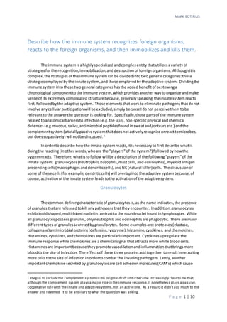
Witha single PRR(i.e.TLR-2),the immune systemcan
identifyanybacteriawithapeptidoglycancell wall(whichis
TLR's Ligands
TLR1 Triacyl lipopeptides
TLR2 Peptidoglycans
GPI-Linked proteins
Lipoproteins
Zymosan
Phosphatidlyserine
TLR3 Double stranded RNA
TLR4 LPS
F-Protein
Mannans
TLR5 Flagellin
TLR6 Diacyl lipopolypeptides
Zymosan
TLR7 Single stranded RNA
TLR8 Single stranded RNA
TLR9 CpG unmethylated dinucleotides
Dinucleotides
Herpes Virus components
Hemozoin
Table 1. This is just a small sample of only
one categoryof PRR’s known as “TollLike
Receptors”. (Owen, 2013, p. 155)
3. MARK BOTIRIUS
P a g e 3 | 10
almostall of them),andthisis just oneexample. Because PRRshave evolvedtorecognizealarge
portionof invadingmicrobesthatshare a commoncharacteristic,itwould be counterproductive for
themto be adaptive andchanging. Therefore,PRRsare germline encodedandinvariable whichis
not the case withB-cell andT-cell receptorswhichare variable andconstructedviasomaticDNA
recombination. The reasonnatural killercellsare partof the innate system, isbecause theyalso
identifytheirtargetsusinganinvariable strategythatisnotadaptive innature. In the case of NK
cells,theyprimarilyidentifytheirtargetsviathe absence of MHC classI proteinsthat isexpressedon
almostall cells. Inthe case of NKT cellstheydohave T-cell receptors,howevertheyare notvery
diverse,andtheyare limitedinwhattheyrecognize(theyonlyrecognize particularlipidsand
glycolipids). Inotherwords,theirreceptorscanbe classifiedasTCR’s,althoughtheyare notreally
the same as the receptorsfoundonactual T-cells. (Owen,2013, p. 40) NKTsare therefore
consideredtobe a hybrid,spanningboththe innate andadaptive categories. Lastly,like
granulocytes,NKandNKTcellskill theirtargetsbyreleasinggranulescontainingantimicrobial
proteins.Havingdescribedthe playersinvolvedinthe innate response andhow theyrecognize
foreignorganisms,Icannow describe how the innate systemreactsandkillsforeignorganisms.
Leukocytes(mostlyneutrophilswhichcomprise50% to 70%) (Owen,2013, p. 33)routinely
travel aroundthe circulatorysystemandbodytissuessearchingforpathogens. Supposesomeone
cuts themselves. PAMP’sof invadingpathogenssuchasbacteriaare recognizedbythe PRRsof
leukocytesresidinginthe areaand are destroyedusingmethodsthatdependonthe type of cell.
Neutrophilsandeosinophilswill beginphagocytizingandusingtheirgranules,whilebasophilsand
mast cellswill attackusingtheirgranulesaswell. Inadditiontophagocytizingandreleasing
granules,these cellswillalsorelease chemokines(suchasIL-8) thatbring more leukocytesintothe
battle andcytokines(suchasIL-4, IL-10, and IL-13) that alertotherleukocytesof the invasion,
therebyupregulatingtheiractivity (Owen,2013, p. 35). Histidine andCAMproteinsare also
releasedtoaidinthe fightby increasingbloodflow,causinginflammation,andsignalingwhere
arrivingleukocyteswillbegintoroll alongthe bloodvessel epitheliumeventuallycomingtoastop
and transmigratingintothe infectedtissuetojointhe fight. Meanwhile,APC’sinthe areasuchas
dendriticcellsandmacrophagesare takingupantigensreleasedinthe battle andare migratingto
secondarylymphtissuessuchasthe lymphnodes(orthe spleeninthe case of bloodborne
pathogens) topresentthese antigenstoT-cells. Notice thatB-cellsandT-cellsare entirelyabsentin
the fight. In fact,at thispointinthe explanation, T-cellsare literally unableto join the fight,because
they do nothavePRR’s. T-cells mustbe activated,and naïveT-cell receptorscan only recognize one
thing,thatis an MHC/antigen combination presented by an APC. Althoughitistrue thatB-cellsdo
have some PRRs,theyneed,forthe mostpart, helpfromT-cellstobecome fullyactivated. This
bringsme to the questionof howthe adaptive immune systemrecognizespathogenswhentheydo
not,overall,use PRRs.
Cells of the adaptive immune system: B-cell and T-cell Lymphocytes
Unlike the PRR’susedbythe leukocytesof the innate system, the receptorsusedbythe
lymphocytesof the adaptive systemare veryspecific. Inotherwords,theyhave averyspecific
target (insteadof targetingageneral characteristicof all bacteria,itwouldtargeta specificprotein
of a particularbacteriumforexample). The questionis,“How can the targetbe sospecific,inlightof
the tremendousdiversityinthe pathogensthatattackthe host?”The answerliesinhow these
receptorsare generated. Lymphocytesgenerate theirreceptorsthroughgeneticrecombination
(Iwasa,2016, p. 682). I won’tgo intothe specificshere (itiswell beyondthe scope of thisquestion)
4. MARK BOTIRIUS
P a g e 4 | 10
howeveritisimportanttonote that the resultisthat an enormousvarietyof cellsexpressingspecific
receptorsare generatedthatcan targetpracticallyanyenemy. To avoidthe possibilitythataself-
targetingreceptoriscreated,lymphocytesare testedandanyself-reactivecellsare destroyed.
Those that remainhave the abilitytobindasingle antigenthatisn’tself. Itisimportanttonote,that
each B-cell andT-cell canbindonlyone antigen(knownasallelicexclusion) (Owen,2013, p. 303).
Afterestablishingitsparticularreceptor(thatcanbindonly one antigen) andreachingmaturity
(anotherprocesswell beyondthe limitsof thisessay) the mature lymphocytestravel the
bloodstreamandvisitvarioussecondarylymphtissuessuchasthe lymphnodeswhere the B-cells
occupy the primarycortex and T-cellsthe paracortex,whichiswhere theyenterthe fightagainst
infection.
Recall thatI leftoff at the pointwhere the granulocytesandmacrophageswere destroying
the invadingpathogens,andthe APC’swhere engulfingantigensandtakingthemtothe secondary
lymphtissues(lymphnodes). Inthe lymphnode (orsome other secondarylymphtissue)the T-cells
and B-cellsare browsingthroughall of the countlessMHC/antigencomplexes(bothclassone and
classtwo) beingpresentedbyall of the APC’suntil
theyfindone thatmatchestheiruniquely
generatedreceptor. Whentheydofindamatch,
theybecome activated. Thisisthe mechanismby
whichthe cellsof the adaptive systemgainthe
abilitytorecognize pathogens. Itisimportantto
note here,thatactivationof a T-cell byan APC is
the primaryeventthatbeginsthe adaptive
response. Whenanaïve CD4 T-cell encountersan
APCthat is presentinganantigenthatitsreceptor
recognizes,itbecomesactivated,anditisthe CD4
T-cellsthatfullyactivate B-cellsandCD8 T-cells(T
cytotoxiccells). 2
WithouttheseCD4cellsto
activate the others,the adaptive responseisshut
down. Thisis the reasonAIDSis sodeadly.
Hence the dendriticcellsthatwere atthe site of
infectionhave engulfedantigen,traveledtothe
lymphnodes,andwere discoveredbyaT-cell withareceptorthat matchedthe antigenitwas
presenting. ThisT-cell differentiatesintoaCD4 cell thatfullyactivatesB-cellsandCD8 cells. The B-
cellsmultiplyandsome of thembecome plasmacellsthatcontinuallysecretantibodiesagainstthe
invadingpathogens. OtherB-cellsbecome memorycells,andstill some others setupgerminal
centersthatare the sitesfor somatichypermutation. The antigenssecretedbythe plasmacells
circulate anddestroythe invadingpathogensinseveral ways. 1.Neutralization. The antigenbinds
directlytothe pathogenblockingthe receptorsitneedstogainentryintothe host.2. Opsonization.
2 I should note that I am generalizinghere, which is why I use the words “fully activate” The immune system is
so complex, that itis impossiblefor me to cover all of the details in a shortessay.For example, it is notan
absoluterequirement for CD8 cells to be activated by CD4 cells (although itis the most effi cient). The only
requirement is that the CD8 cell is activated by a licensed APC and that it receives three signalswhich I will not
go into. However, without CD4 activation,CD8 cells areunableto develop memory cells and therefore cannot
mount a secondary responseto infection, hence the reason I consider them to not be “fully activated”. For the
purpose of brevity, I am also generalizingregardingcertain aspects of B-cell activation. In both cases,I am
nonetheless paintinga true picture of what is takingplace,exceptions notwithstanding.
Figure 1. T-cell activationbyanAntigenPresenting
Cell. Thisis from the Dr. Rogers class lecture.
5. MARK BOTIRIUS
P a g e 5 | 10
3. Complementfixation/activation.4.AntibodyDependentCellMediatedCytotoxicity(ADCC).
(Owen,2013, p. 417)
So far,all of the “players”inmy immunological scenariohave beenaddressedwithone
exception. Ihave yetto addressthe role of cytotoxicT-cells(CD8). The reasonisbecause,inthe
scenarioI have created,CD8 cellsdon’treallyplayarole. Why? Recall that T-cellsare MHC
restricted. Thatis,CD4 cellscanonlyrecognize antigenpresentedviaanMHC classII molecule,and
CD8 cellscanonlyrecognize antigenpresentedviaMHCclass I molecules. MHCclass IImolecules
are presentedbyAPC’s,whereasMHCclassI moleculesare foundinalmostall nucleatedhostcells.
What thismeans,isthat MHC classII moleculesmediatethe cellularresponse to extracellular
pathogens,andMHC class I moleculesmediatethe response to intracellularpathogens. (Owen,
2013, p.278) In myscenario,there are no intracellularviral orbacterial pathogens,andsolittle was
saidregardingCD8 responses. Infact,the role of CD8 cellssurpassesmere response topathogens.
Since all nucleatedcellsexhibitMHCclass I on theircellularmembranes,theyare notonly
presentingantigenrelevanttopathogens,theyare presentingantigenrelevantto all of theircellular
processes. Therefore,thiscanbe consideredtobe some kindof “readout”thatindicateseverything
goingon inside the cell. If the cell isinsome kindof distress,whetherinfectedornot,itwill indicate
thisto the “outside world”viathe antigensitispresentingviaitsMHCclass I molecules. Manycells
inthe beginningstagesof becomingcancerous,in fact,will presentantigenswiththeirMHCclass I
moleculesindicatingthattheyare on the road to becomingcancerous. These signalsare detected
by CD8 cellsandare destroyed. Of course,notall cellsare detected,whichleadstofull blown
cancers.
CD8 cellsrecognize theirtargetsbyrecognizingthe MHCclassI/antigenpresentedinthe cell
membrane,andtheykill theirtargetinone of twoways,bothof whichcause the cell to enter
apoptosis(whichisthe reasonithasbeencalledthe “kissof death”). (Owen,2013, p.432) In both
cases,the CD8 cell bindsthe targetcell andreleasesgranulesdirectedtowardsitstargetcontaining
ligandsthatdirectthe target cell toenterapoptosis. Inmanycancer cells,the pathwaytoapoptosis
ismutated,whichprovidesone waythatcancerouscellscansurvive effortsbythe immune system
to eradicate it. (Owen,2013, p. 644)
In summary,uponinfectionleukocytesandAPC’srecognize pathogens’PAMPswiththeir
PRRs. Theyreact to those pathogensbyphagocytizingandreleasinggranulesthatlyse the invaders.
Theyalsorelease cytokinesandchemokinesthatbringmore leukocytestothe infectionsite,cause
an inflammatoryresponse,andupregulate the activityof otherleukocytes. APC’ssuchasdendritic
cellstake inpathogenproteins,processtheminto“epitopes”,and presentthemontheircell
membranes viaMHC classI and MHC classII molecules. APCsthentravel tosecondarylymphtissues
such as lymphnodes,where naïve T-cellsandB-cellsbrowse the APCsuntiltheyencounterone
carryingthe antigentheirreceptorrecognizes. Whenthishappens,the APCsactivate the B-cellsand
T-cellsviaco-stimulatorysignals. ActivatedB-cellsandT-cellsthenbegintoproliferate,producing
more clonesof themselvescapable of engagingthe particularantigenthatbindstheirreceptor.
Some of the B-cellsbecome plasmacellsandcontinuallyexcrete antibodiesthatfightthe infection,
some formgerminal centersandengage insomatichypermutation,andthe remainderbecome
memoryB-cells. RegardingT-cells,the populationof T-cellsdifferentiatesintotwomainsubtypes
knownas “helper”T-cells(CD4) and“cytotoxic”T-cells(CD8). These twotypesdifferentiate further
intoseveral othertypesthatare beyondthe scope of thisquestion. CD4cellsmediate the cellular
response byactivatingotherB-cellsandT-cells. CD8cells,once fullyactivated,travel tothe site of
infectionwheretheyidentifyinfectedcellsbythe MHCclass I/antigenthe cellsare presentingin
6. MARK BOTIRIUS
P a g e 6 | 10
theirmembranes. The CD8cellsthenbindto the infectedcellsandrelease granulesthatcause the
cell to undergoapoptosis. Finally,itshouldbe notedthatlymphocytessuch asB-cellsandT-cells
require “survival”signalsprovidedbythe presence of antigen. Whenantigenisnolongerpresent
(because the pathogenhasbeeneliminated) the lymphocytesbegintodie off,withthe exceptionof
the memorylymphocytesthatare leftbehindtoprovide asecondaryresponse shouldone be
needed. Thisendsthe immune responsetothisparticularinfection.
Describe the processes by which DNA can be repaired following a
mutation.
The six bestunderstoodandwell knownprocessesbywhichDNA canbe repairedfollowing
mutationare directreversal,mismatchrepair,base excisionrepair,nucleotideexcisionrepair,non-
homologousendjoining,andhomologousrecombination. (Rogers,2017, p.181) I will therefore
describe eachinthe orderthat I have listedthem.
Direct Reversal
Many DNA repairprocessesinvolveachange to the DNA as a resultof the repair. Direct
reversal,however,isanexception. Asitsname implies,the processof directreversalsimplyundoes
the mutation,restoringthe DNA toitsoriginal state. Twoexamplesof mutationsthatcanbe
repairedusingthe processof directreversal are
photoreactivationandthe demethylationof O6
–
methylguanine. (Watson,2014, p.325). In
photoreactivation,the twoneighboring
pyrimidineringspresentinDNA absorbUV light
(primarilyUV-B) causingthe doublebondswithin
the ringsto break andformingnew covalent
bondsbetweenthe two neighboringrings. These
new bondsforma cyclobutane dimerthatcreates
a lesioninthe DNA that will adverselyaffect
replicationandtranslation. The directrepair
processfor thismutationbeginswiththe enzyme
photolyase. Photolyasehastwochromophores
that alsoabsorbUV light. Whenphotolyase
encountersapyrimidinedimer,itflipsthe lesion
away fromthe DNA interiorandintothe catalytic
pocketof the enzyme. Itthentakesthe lightenergyabsorbedbyitstwochromophoresand
transfersitto an FADH (anelectroncarrier) coenzyme. FADHthenusesthe energytobreakthe
cyclobutane bondsbetweenthe neighboringpyrimidinesthusrestoringthe DNA toitsoriginal state.
(RajeshwarPSinha,2002)
Anotherexampleof the directreversal processisthe repairof amethylatedguanine.The O6
- methylguanine mutationisoftenformedbyalkylatingchemicalssuchasnitrosamines. These
chemicalscancause the ketogroupat carbon 6 of guanine tobecome methylatedformingO6
-
methylguanine thatcausesthe guanine topairwiththymine insteadof cytosine. (Watson,2014, p.
Figure 2. Two neighboring pyrimidine rings absorb a
UV-B photon, causingthe double bonds between C5
and C6 to break. As a result, a newcovalent bonds are
formed betweenthe two rings, forming a cyclobutane
dimer.(Watson, 2014, p. 322)
7. MARK BOTIRIUS
P a g e 7 | 10
322) This mutationisrepairedbya methyltransferase. WhenitencountersO6
-methylguanine,it
simplytransfersthe methyl grouptoitself,reversingthe damage andcompletelyrepairingthe DNA.
Mismatch Repair
Witha diploidgenome size of over6billionbase pairs,itisunderstandablethatreplication
errorsin humanswill occur. In fact,DNA polymerase makesamistake once every105
to 106
bases.
Fortunately,DNA polymerasehasa proofreadingabilitythatcancorrect manyof these mistakes
before theyare incorporatedintothe newlysynthesizedstrand. Thisability,alongwiththe helpof
some accessoryproteins canlowerthe rate of error toone in1010
bases. (Rogers,2017, p. 180)
Those mismatchesthatescape proofreadingare repairedusingthe mismatchrepairprocess,which
beginswithaproteincalledMutS. MutS routinely scansnewlyreplicatedDNA lookingformispaired
bases. Whenit detectsone (mismatchedbaseswill distortthe DNA somewhat,makingthem
detectable byMutS) MutS bindstothe lesionandrecruitsanotherprotein,MutL,whichinturn
recruitsMutH. MutH isan endonuclease thatmakesacut inthe new strand slightlyupstreamfrom
the mismatch. MutH differentiatesthe new strandfromthe oldstrandbecause newlyreplicated
DNA existsina hemimethylatedstate. Priortoreplication,bothstrandsof the DNA duplex have a
characteristicmethylationpatternwhere the adenineresiduescontainedin5’-GATC-3’are
methylatedbyDammethylase.However,immediatelyafterreplication,the new stranddoesnot
exhibitthismethylationpattern,while the oldstranddoes (hence,the strandis“hemimethylated”).
It isthisdifference betweenthe methylationpatternsof the twostrandsthat causesMutH to
differentiatebetweenthemandselectivelycutthe new strand. Thishemimethylatedstate doesnot
lastforever,however,because Dammethylase routinelyscansDNA,andwhenitcomesacrossthe
unmethylatednewstrand,itwill methylate it,andMutH will be unable todistinguishwhichstrandis
the newstrand,and whichstrandis the oldone. Therefore,formismatchrepairtoworkproperly,it
mustdetectthe error quickly. Once the new strandhas beennickedbyMutH,it isunwoundbythe
helicase UvrD. Once unwound,the single strandcontainingthe errorisdegradedbyexonucleases,
leavingagap that spansthe mismatch. The gapis thenreconstructedbyDNA polymerase IIIand
reconnectedtothe restof the strandwithligase. Ligationfinishesthe repair. (Watson,2014, p. 316)
Base Excision Repair and Nucleotide Excision Repair
While mismatchrepairdealswithreplicationerrorsbetweenbases,excisionrepairpathways
such as base excisionrepairandnucleotideexcisionrepairdeal withmutationthatdamage oralter
the basesthemselves. Anexample of thiskindof mutationisthe deaminationof cytosine. When
cytosine losesitsaminogroup,itbecomesuracil,whichoccasionallyhappensspontaneouslyby
hydrolysis. Inthe case of base excisionrepair,the damagedbase isremovedbyaglycosylase
specificforthisparticulartype of base damage (specifically,uracil glycosylase). There are numerous
8. MARK BOTIRIUS
P a g e 8 | 10
kindsof damagedbases,andeach isrecognizedbya particularglycosylase. Sofar,we have
discoveredelevendifferenttypes. (Watson,2014,p. 326) Whenuracil glycosylase discoversthe
damagedbase,itcleavesthe glycosidicbondbetweenthe damagedbase andthe ribose of the DNA
backbone,thusremovingthe base butleavingthe backboneintact. Thiscreatesanapyrimidinicsite
that isthencut out by an APendonucleaseatthe 5’ endand an exonucleaseatthe 3’ end,leavinga
gap. The gap is thenrepairedbyDNA polymerase I,therebycompletingthe repair.
In the case of nucleotide excision
repair,the sectionof DNA containing
the damagedbase isrecognizedby
how the DNA strandis distortedby
the damagedbase. The damaged
base itself,isnotrecognized. The
distortionisrecognizedbya protein
complex consistingof fourprotein
subunits.Twoof the proteinsubunits
are UvrA,and the othertwo are UvrB.
Whenthe proteincomplex discovers
the distortion,the twoUvrB subunits
bindto the distortion,andthe UvrA
subunitsare realeased.UvrB
denaturesthe DNA andrecruitsUvrC,
whichcuts the DNA on bothsidesof
the distortionandthe entire sectioncontainingthe distortioniscarriedawaybyUvrD. Dna
polymerase I thensynthesizesthe missingsection,andthe damage isrepaired.
Non Homologous End Joining and Homologous Recombination
The last tworepairprocessestobe discussed,non-homologousendjoining(NHEJ) and
homologousrecombinationdeal withthe repair of doublestrandedbreaksinDNA. There are
several thingsthatcan cause DSBs andtheyinclude radiation,reactiveoxygenspecies,and
chemicals,suchasbleomycin. (Iwasa,2016,p. 534)
In non – homologousendjoining,the proteinsKu70andKu80 bindtothe brokenendsof the
DNA and recruitsthe kinase DNA – PKcs whichsubsequentlyrecruitsaproteincalledArtemis.
ArtemisandDNA – PKcsform a complex thatprocessesthe brokenendsbyremovingalittle bitof
DNA from eachend.Thisprocessingthenenablesaligase proteincomplex consistingof the proteins
Ligase IV,Cernunnos - XLF,and XRCC4. (Watson,2014, p. 332) It shouldbe notedthat,since this
processremovesalittle bitof DNA fromeach end,itismutagenicinthat some informationis
deletedfromthe genome.
Unlike non – homologousendjoining,whichusesnoinformationtorepairadouble stranded
break,homologousrecombinationusesthe informationfromasisterchromosome tofix the break.
It doesthisbyfirstaligningthe regioncontainingthe breakwithanearlyidentical regionfromits
sisterchromosome. Then,aDNA nuclease processesthe endof the breaktocreate a regionof
Figure 3. Uracil glycosylase cleaves the glycosidic bondbetweenthe
ribose of the DNA backbone andthe uracil base. (Watson, 2014, p.
327)
9. MARK BOTIRIUS
P a g e 9 | 10
single strandedDNA. Thissingle strand
isthenstabilizedbystrandexchange
proteinswhichalsopromote strand
invasion. Instrandinvasion,the single
strand invadesthe homologousstrandof
the otherchromosome,causingthe helix
to separate inthe processforminga “D
loop”whereineachstrandfromone
chromosome pairswiththe strandsof its
homologue.
The area where the DNA strands
cross iscalleda “HollidayJunction”.In
addition,the resultinghybridstrand
formedfromthe twostrands iscalleda
“heteroduplex”anditcan resolve inone of
twoways dependingonwhere the DNA is
cut. Figure 5 depictsthe twodifferent
waysto cut the DNA. In the methodonthe
left(the horizontal dashedcutline) the
DNA is cut insuch a way that the
homologuesstaywiththeirrespective
chromosomes. Thiscreatesapatch of
recombination(depictedinthe centerof
the two DNA strandsat the bottom). This
istherefore calledthe “PatchProduct”. In
the methodonthe right(the vertical cut
line) the homologuesexchange
chromosomes,creatinganactual crossover
of homologues. Therefore,thisiscalleda
“CrossoverProduct”. Upon resolutionof
the crossover,the repairiscomplete.
References
Iwasa,J. a. (2016). Karp'sCell and MolecularBiology (8thed.).Hoboken:Wiley.
Madigan,M. S. (2012). Brock Biology of Microorganisms. SanFrancisco:PearsonEducationInc.
Owen,P.S. (2013). Kuby Immunology. New York:W.H.FreemanandCompany.
RajeshwarPSinha,D. H. (2002). UV - InducedDNA damage and repair:a review. Photochemicaland
PhotobiologicalSciences.
Rogers,S. O.(2017). Integrated MolecularEvolution (2nded.).BocaRaton: CRC Press.
Watson,e.a. (2014). Molecular Biology of the Gene (7th ed.).Boston:Pearson.
Figure 4. This figure illustratesthe mechanismof strand
invasion. Sections of single strandedDNA pair withthe
homologous strand oftheir sister chromosome. (Rogers, 2017,
p. 182)
Figure 5. Depicted in this figure are the two ways that DNA can
be cut to separate the duplexes. This figure is from Dr.
Rogers’ class lecture