
Recommended
More Related Content
What's hot
What's hot (20)
Similar to Fibroid complicating pregnancy
Similar to Fibroid complicating pregnancy (20)
Recently uploaded
Recently uploaded (20)
Fibroid complicating pregnancy
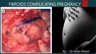
- 1. FIBROIDS COMPLICATING PREGNANCY By - Dr. Milan Kharel
- 2. Definition “A benign(non-caseous) tumor arising from the smooth muscles layer and accompanying connective tissue of the uterus” fibroid is chiefly composed of smooth muscle fibres & a small amount of connective tissue. The name fibroid is a misnomer, more appropriate term for this tumor of smooth muscle is Myoma or Leiomyoma.
- 3. Classification of fibroids • Intramural fibroids • Subserosal fibroids • Submucous fibroids Within Body of uterus Cervical Intraligamentary
- 4. INCIDENCE AND PREVALENCE Incidence in pregnancy : 1 in 1000. Prevalence : Highest in black women -18 percent Lowest in whites - 8 percent
- 5. EFFECTS OF FIBROID ON PREGNANCY May be none Abortion (submucous fibroid) Pressure symptoms due to impaction of (a) Bladder - retention of urine (b) Rectum - constipation Red degeneration Malpresentation (Four fold risk) Preterm labor and prematurity Non-engagement of the presenting part
- 6. EFFECTS ON LABOR May be unaffected Uterine inertia Dystocia Obstructed labor Postpartum hemorrhage due to atonicity or adherent placenta Difficult cesarean section (Eight fold risk)
- 7. EFFECTS ON PUERPERIUM Subinvolution Inversion of uterus Secondary PPH Sepsis Lochiometra and pyometra.
- 8. EFFECTS OF PREGNANCY ON FIBROID Changes in size – increases (?) Changes in position Changes in shape - becomes flattened Degenerative changes specially, red degeneration Torsion of pedunculated subserous fibroid Infection and polypoidal changes are more in puerperium.
- 9. EFFECTS OF PREGNANCY ON FIBROID Changes in size – increases (?) Fibroid is an estrogen dependent tumor. Pregnancy is a progesterone predominant state. So how does fibroid increases in size during pregnancy?
- 10. EFFECTS OF PREGNANCY ON FIBROID Actually, most fibroids do not increase in size during pregnancy. 69% - had no increase in fibroid volume throughout pregnancy 31% - noted increase in fibroid volume, greatest increase before 10th week of gestation and a reduction to baseline value 4 weeks after delivery
- 11. EFFECTS OF PREGNANCY ON FIBROID The main reason for the fibroid to increases in size during pregnancy is due to Increased vascularity Edema Hypertrophy and hyperplasia of the fibromuscular tissues.
- 12. RED DEGENERATION OF FIBROID
- 13. RED DEGENERATION • Red degeneration (carneous degeneration) occurs in a large fibroid mainly during second half of pregnancy and puerperium. Partial recovery is possible and as such called necrobiosis. Red degeneration is a result of softening of the surrounding supportive connective tissue, the capillaries tend to rupture and blood effuses out into the myoma causing a diffuse reddish discolouration of the same.
- 14. Clinical features of Red Degeneration These patients typically present with acute lower abdominal pain, with may be severe and is often associated with nausea, vomiting and even fever. Examination may show a tender palpable mass in the uterus. In severe cases, there may be signs of localized peritonism.
- 15. Naked eye appearance of the tumor shows dark areas with cut section revealing raw-beef appearance often containing cystic spaces. The odor is often fishy due to fatty acids. Color is due to the presence of hemolysed red cells and hemoglobin. Microscopically, evidences of necrosis are present. Vessels are thrombosed but extravasation of blood is unlikely. Gross Appearance:
- 16. Diagnosis 1. Ultrasound scanning is the primary modality of diagnosis, and typically shows a fibroid corresponding to the area of tenderness over the uterus. Most often, the degenerating fibroid has a mixed echogenic appearance. 2. MRI : Where sonography is inconclusive, abdominal MRI may be considered. In red degeneration, the typical finding is an uterine fibroid with increased T1 signal intensity.
- 17. Treatment Red degeneration is a self-limiting condition and is almost always managed conservatively. The pain should be managed symptomatically, with either NSAIDs or opioids. Where opioids are ineffective, a 24 to 48 hour course of indomethacin has been shown to be efficacious. Note that NSAIDs are best avoided during the third trimester, as there is an increased risk of premature closure of the ductus arteriosus and oligohydramnios
- 18. It is often difficult to diagnose a fibroid during pregnancy because of Marked softening Alteration in the shape (flattening) In early months, fibroid is diagnosed but pregnancy is missed whereas in later months, pregnancy is diagnosed but the fibroid is missed. DIAGNOSIS
- 19. Ultrasonography confirms the diagnosis with certainity. DIAGNOSIS USG showing uterine fibroid
- 20. If ultrasonographic findings are unclear, Magnetic Resonance Imaging can be done DIAGNOSIS MRI showing degenerating fibroid
- 21. In uncomplicated tumor, fibroid is often confused with Ovarian tumor Retroverted gravid uterus Non gravid half of uterus didelphys DIAGNOSIS
- 22. BASIC PRINCIPLE Avoid treatment whenever possible. TREATMENT
- 23. DURING PREGNANCY Uncomplicated : Usual antenatal care is followed. All cases to be assessed at 38 weeks to formulate the method of delivery. Acute pain following red degeneration Conservative management TREATMENT
- 24. DURING LABOR Fibroid situated above the presenting part: Usually results in uneventful vaginal delivery Fibroid situated below the presenting part: Spontaneous vaginal delivery may occur. If it fails, cesarean section is to be done. TREATMENT
- 25. Fibroid in lower uterine segment Cervical fibroid, even if it is small Fibroid impacted in pelvis Malpresentation Obstetric complications INDICATION FOR ELECTIVE CESAREAN SECTION Cesarean delivery for a large leiomyoma in the lower uterine segment
- 26. TECHNICAL ASPECTS ON ELECTIVE CESAREAN SECTION Myomectomy should be avoided during cesarean section Be alert for postpartum hemorrhage and retained placenta. Reverts to a smaller size during puerperium
- 27. DC Dutta’s – Textbook of Gynecology : 6th edition Shaw’s Textbook of Gynaecology : 15th edition DC Dutta’s – Textbook of Obstetrics : 6th edition