
Recommended
More Related Content
What's hot
What's hot (20)
Viewers also liked
Viewers also liked (20)
Similar to Dentistry News example
Similar to Dentistry News example (20)
Recently uploaded
Recently uploaded (20)
Dentistry News example
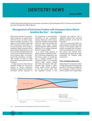
- 1. Dentistry news January 2009 Geistlich Biomaterials Workshop at the European Association of Osseointegration EAO 7th Annual Scientific Meet- ing, 8 to 20 September 2008, in Warsaw Management of Extraction Sockets with Anorganic Bone Matrix Geistlich Bio-Oss® – An Update After tooth extractions, the physio- the periosteum is recommended, concluded that patients had “a logical reduction of alveolar bone irrespective of the subsequent significant benefit from receiving and soft tissues can affect subse- measure. Reduction of the resorp- grafting materials at the time of quent restorative procedures. This tive process has been demonstrated extraction”6. applies to both implant-supported when the socket was grafted with Also in a canine study, GBOC was and conventional restorations. As anorganic bone matrix, without able to preserve the dimension and the buccal portion of the socket is (Geistlich Bio-Oss®, GBO) or with the contour of the alveolar ridge. The partially or totally composed of the addition of 0 % collagen (Geistlich authors concluded that “the place- tooth-related bundle bone, loss of Bio-Oss® Collagen, GBOC). ment of a biomaterial in an extrac- this tissue will inevitably reduce the In a canine study, grafting of tion socket may promote bone vertical and horizontal dimensions GBOC resulted in a significant reduc- modeling and compensate, at least of the alveolar ridge. To compen- tion of the bony invagination over temporarily, for marginal ridge sate for this resorption, a consider- the former socket, in comparison contraction”. able number of socket or ridge with a collagen sponge or the preservation techniques have been untreated control2. In a controlled Time of implant placement proposed. clinical study, sockets grafted Depending on the respective socket In dogs it was also shown that an with GBO and sutured with split- or ridge preservation technique the elevation of flaps can increase the thickness flaps showed a signifi- amount of soft tissue available for amount of buccal resorption3. cantly smaller vertical reduction the subsequent implant insertion Therefore an atraumatic extraction of the thin buccal plate than and/or guided bone regenerative technique without exposure of the ungrafted controls. It was 100% Benefit of Geistlich Bio-Oss® Collagen quot;Soft tissue punchquot; benefit Soft tissue volume Immediate implant Early implant placement Delayed implant placement placement Fig. . Impact of extraction site management technique on soft tissue volume. 2009 PAGE
- 2. DENTISTRY NEWS January 2009 measure will differ (Fig. ). The (red line in Fig. , recommendation a membrane. In the event of an appropriate technique is deter- alveolar dehiscence, a biomaterial Markus Hürzeler, see Fig. ). This mined, among other parameters, can be used in combination with a method is indicated when optimal by the esthetic demands of the collagen membrane (black line in soft tissue quality is required in the patient, and the time of implant Fig , technique Dietmar Weng). esthetic area. placement (Types to 4 according The therapeutic window can be to Hämmerle, 2004)4. broadened by placing the implant 2 Presentations at the EAO In particular, immediate implant to 4 months (Type 3), or more than placement can be accompanied by 4 months after extraction (Type 4). A specific workshop at the EAO pro- grafting of GBO particles into the An advanced approach in the vided more than 400 participants gaps between implant and bone. esthetically sensitive area is to with the latest research data in the When early implant placement 4 to combine a soft tissue punch tech- field of socket preservation. Two 8 weeks after extraction (Type 2) is nique with grafting of anorganic renowned clinicians, Prof. Dr. Markus planned, an advanced free gingival bone matrix (e.g. GBOC, blue line in Hürzeler and Dr. Dietmar Weng, graft technique can be used. The Fig. , by Ronald E. Jung5). Alterna- presented their clinical concepts graft, which is harvested with a tively, when the socket is intact, a based on these scientific results and punch technique, is sutured mini- biomaterial can be grafted without discussed their practical relevance mum-invasively into the supra- with the audience. closure with a soft tissue graft or alveolar soft tissues of the socket References . Araujo, M., Linder, E., Wennstrom, J. Lindhe, J., (2008) The influence of bio-oss collagen on healing of an extraction socket: An experimental study in the dog. Int J Periodontics Restorative Dent 28: 23–35. 2. Cardaropoli, G., Araujo, M., Hayacibara, R., Sukekava, F. Lindhe, J., (2005) Healing of extraction sockets and surgically pro- duced - augmented and non-augmented - defects in the alveolar ridge. An experimental study in the dog. J Clin Periodontol 32: 435–440. 3. Fickl, S., Zuhr, O., Wachtel, H., Bolz, W. Huerzeler, M., (2008) Tissue alterations after tooth extraction with and without surgical trauma: A volumetric study in the beagle dog. J Clin Periodontol 35: 356–363. 4. Hammerle, C.H., Chen, S.T. Wilson, T.G., Jr., (2004) Consensus statements and recommended clinical procedures regarding the placement of implants in extraction sockets. Int J Oral Maxillofac Implants 19 Suppl: 26–28. 5. Jung RE, Siegenthaler DW, Hammerle CH. Postextraction tissue management: a soft tissue punch technique. Int J Periodontics Restorative Dent 2004;24:545–553. 6. Nevins, M., Camelo, M., De Paoli, S., Friedland, B., Schenk, R.K., Parma-Benfenati, S., Simion, M., Tinti, C. Wagenberg, B., (2006) A study of the fate of the buccal wall of extraction sockets of teeth with prominent roots. Int J Periodontics Restorative Dent 26: 9–29. 2009 PAGE 2
- 3. www.geistlich.com The Master’s Choice 600 + publications 20 years experience 2 reliable products LEADING REGENERATION
- 4. DENTISTRY NEWS January 2009 Extraction Site Management Revisited – New Scientific Insights with High Practical Relevance the socket may also be an option. maintain the ridge dimensions. In In beagle dogs fresh extraction all the best results were obtained sites were treated with one of the with method #4 (GBO plus FGG). following procedures: Histological evaluation of the samples will follow. . Grafting with an anorganic bone matrix containing Clinical implications collagen (Geistlich Bio-Oss® Collagen GBOC) Staged implant procedures are 2. Grafting with GBOC, in combi- intended to (re-)build the soft tissue nation with an experimental architecture, and to re-establish collagen membrane placed interproximal bone after a certain buccaly into the socket degree of time-related resorption. 3. Socket seal with a free gingi- At the time of early implant inser- val graft (FGG) tion, after 6 to 8 weeks, minor 4. Untreated control guided bone regenerative tech- niques will compensate for recent Histomorphometrical analysis and previous loss of bone. As it is showed that neither method was much more stable and stays in the able to completely preserve the site for many years, the best mate- Markus Hürzeler buccal lamella. However, there was rial for this purpose is anorganic Private Practice in Munich, Germany significantly more bone volume bone matrix (Geistlich Bio-Oss®), Associate Professor at the Albert-Ludwig with the two GBOC procedures than not autogenous bone. University Freiburg, Germany with the FGG technique, or the Until the time of implant place- control. ment it is important to maintain as In a further study, preservation of In the esthetic area clinicians have much of the soft tissue as possible the buccal lamina was attempted to make a choice between fast and (cf. Fig. , page of this supple- with the following methods2: convenient immediate protocols, or ment). A thick free gingival graft more predictable, but long-term . Grafting of Geistlich Bio-Oss® from the palate, sutured with multi-step approaches. Patients (GBO) into the socket, and minimum-invasive sutures, will tend to prefer the immediate option, over-compensation of the ensure an optimal soft tissue qual- however in the great majority of buccal aspect with GBO and ity and volume (see case report on cases, this does not permit predict- Geistlich Bio-Gide® (GBG) the following page). As it will not able esthetic results. Therefore in 2. Flapless expansion of the prevent the loss of the buccal our practice we tend to place buccal lamina lamella, grafting of a biomaterial implants “early” after 6 to 8 weeks. 3. Grafting of GBO into the into the extraction socket is not To prevent soft tissue involution socket, plus buccal over- necessary in connection with an during this time, we suture a free augmentation with a connec- early implant protocol. However, in gingival graft to seal the socket. tive tissue graft (CTG) and an connection with delayed or late This measure optimizes soft tissue FGG protocols grafting of Geistlich quality for subsequent procedures. 4. Grafting of GBO, plus an FGG Bio-Oss® or Geistlich Bio-Oss Unpublished data from our group Collagen® into the socket at the Again, volumetric results obtained suggest that, in connection with time of tooth extraction is a logical with a CAD technique showed that delayed implant placement proto- cols, grafting of biomaterials into approach. neither method was fully able to References . Fickl, S., Hinze, M., Zuhr, O., Wachtel, H., Bolz, W. Hurzeler, M., (2008) Hard tissue alterations after overaugmentation of the extractions socket. Clin Oral Implants Res 19: 94, poster #239. 2. Fickl, S., Zuhr, O., Wachtel, H., Bolz, W. Huerzeler, M., (2008) Hard tissue alterations after socket preservation: An experimen- tal study in the beagle dog. . Clin Oral Implants Res 19: –8. 3. Hurzeler, M., Fickl, S., Zuhr, O. Wachtel, H., (2006) Clinical features and shortfalls of immediate implant procedures. Eur J Esthet Dent 1: 28–40. 2009 PAGE 4
- 5. DENTISTRY NEWS January 2009 Fig. 2. Separation of gingival attachment and Fig. 3. Clinical examination shows that the buccal Fig. . Tooth cannot be preserved due to an desmodontal ligament is performed with a lamina is not intact, therefore, immediate implant endodontic complication. micro-scalpel. placement is not possible on a predictable basis. Fig. 4. The supra-crestal internal aspect of the Fig. 5. A free gingival graft is removed from the Fig. 6. The graft is sutured into position with alveolus is deepithelialised with a coarse diamond anterior palate with a tissue punch. microsurgical sutures. instrument. Fig. 7. One week after extraction and soft tissue Fig. 8. Occlusal view of the situation one week Fig. 9. Six weeks after extraction the soft tissue grafting, the site heals without complications. after extraction. volume and condition are perfect for implant surgery. Fig. 2. The final restoration shows a natural Fig. 0. Soft tissue volume is well preserved even Fig. . Intraoperative view at the time of implant appearance and good soft tissue integration. in the horizontal dimension. inservtion illustrates loss of the buccal lamina. An autogenous bone block was grafted, contoured with GBO, covered with GBG, and then left to heal for 5 months. 2009 PAGE 5
- 6. DENTISTRY NEWS January 2009 Simplify your Augmentation – What to Consider at the Time of Extraction sockets, Cardaropoli and coworkers to handle in comparison with early (2003) confirmed this observation2. placement after 6 to 8 weeks (cf. The loss was not prevented by Fig. , page of this supplement). immediate implant placement, Depending on the defect situa- either. tion, one the following procedures If no effective extraction site man- is performed4: When there is no agement is performed there will be buccal fenestration or bony dehis- too little soft tissue available for cence, only GBO is grafted into the subsequent GBR measures. On the socket. One or several gelatine other hand, 6 to 8 weeks after sponges are placed over the graft extractions, no hard bone will be material to the level of the soft available for precise flap surgery tissue margin, and fixed with required in connection with early criss-cross sutures. implant placement. As the imma- When there is a dehiscence or fen- ture intraalveolar bone tissue estration, a collagen membrane cannot be clearly discerned from (Geistlich Bio-Gide®, GBG) is first connective tissue, the socket has to placed between the periosteum be cleaned all the way to the bony and the bone on the buccal aspect, walls. Now the defect situation does without raising a flap (cf. case report not differ much from the fresh alve- on the following page). Then GBO is olus. Futhermore the “bundle bone Dietmar Weng grafted into the socket, the GBG effect” will have resorbed major Private Practice in Starnberg, Germany membrane placed over the graft parts of the thin buccal bone walls. material, and the membrane cov- Although part of the buccal bone ered with gelatine sponges. In both will inevitably be reduced after cases, at the time of implant place- Clinical implications extractions this loss can be com- ment, a sufficient amount of buccal pensated for by socket grafting. For bone will be present due to the In our practice, we prefer either this reason, when no immediate socket preservation procedure. immediate or delayed implant pro- implant can be placed, we routinely In the event of an immediate cedures. In the case of a delayed place a bone substitute material implant placement, grafting and placement the socket is immedi- into the fresh socket. After 4–5 implant insertion do not need to be ately grafted with anorganic bone months of healing a delayed separated. All gaps between implant matrix (Geistlich Bio-Oss®, GBO). implant is inserted into the well and socket walls, especially on the After 4-5 months the implant is preserved site. This separation of buccal aspect, are filled with GBO to placed. If however, implant place- the time points of augmentation allow for new bone formation ment has to be performed at a later and implant placement reduces beneath the fragile bundle bone. If time point, there will be no dis- problems with wound healing, facil- there is a slight dehiscence, GBO advantages in terms of volume itates soft tissue management, and and a GBG membrane are placed shrinkage, since GBO is slowly simplifies surgical demands. over the implant. Again one or sev- resorbable. The main benefit is the As early as in 967, Carlsson and eral gelatine sponges will protect separation of augmentation (socket coworkers demonstrated that after graft) and implant placement. The the site from debris and bacteria. If 40 days the buccal bone of human implant is placed into maturing the case was well selected, the extraction sockets will be almost bone, and the soft tissues are also outcome of this procedure will be completely resorbed3. In canine more mature and therefore easier successful and predictable. References . Araujo, M.G., Wennstrom, J.L. Lindhe, J., (2006) Modeling of the buccal and lingual bone walls of fresh extraction sites following implant installation. Clin Oral Implants Res 17: 606–64. 2. Cardaropoli, G., Araujo, M. Lindhe, J., (2003) Dynamics of bone tissue formation in tooth extraction sites. An experimental study in dogs. J Clin Periodontol 30: 809–88. 3. Carlsson, G.E., Thilander, H. Hedegard, B., (967) Histologic changes in the upper alveolar process after extractions with or without insertion of an immediate full denture. Acta Odontologica Scandinavica 25: 2–43. 4. Weng, D., (2006) “Simplify your augmentation“ - Was bei der Extraktion zu beachten ist, damit die Implantation einfach wird. Implantologie 14: 355–363. 2009 PAGE 6
- 7. DENTISTRY NEWS January 2009 Fig. . Tooth 6 has to be extracted due to Fig. 2. After atraumatic extraction the exact Fig. 3. On the buccal side, a collagen membrane endodontic problems. Hard and soft tissue amount of bone loss can be investigated with a (Geistlich Bio-Gide®) is placed between the recession seen on the buccal side. periodontal probe. periosteum and the bone surface (blunt preparation). No incisions were made. Fig. 5. On the palatal side the free end of the Fig. 6. A gelatine sponge is positioned over the Fig. 4. The socket is filled with non-resorbable membrane is pushed between the periosteum and collagen membrane and fixed with a cross suture. anorganic bone matrix (Geistlich Bio-Oss®) in order the bone surface. to restore the original contour of the alveolar ridge. Fig. 8. After 0 days the gelatine sponges have Fig. 7. A second gelatine sponge is placed on the Fig. 9. After 3 weeks the fibrin has almost disintegrated. Fibrin covers the site and isolated first cross suture and held in place with a second disappeared, healing is uneventful. particles of bone substitute material are visible. cross suture. Fig. . A flap is raised during implant placement. Fig. 2. Due to the successful preservation of the Fig. 0. After 3 months, the external ridge The buccal contour is completely intact. ridge the implant could be placed without volume proves well preserved. additional augmentation procedure. 2009 PAGE 7
- 8. DENTISTRY NEWS January 2009 Concluding remarks Irrespective of the time point of implant placement, biomaterials from Geistlich have proven to compensate for the loss of bone and soft tissue after dental extractions. Geistlich Bio-Oss® Collagen should particularly be taken into account for socket and ridge preservation. It has been shown in animal and clinical research that the management of extraction sockets with this material will preserve the alveolar hard and soft tissue dimensions to a considerable percentage. Recent still unpublished clinical research will provide further quantitative and three-dimensional data. Last but not least, a great number of presentations and case reports by leading clinicians have underlined the high relevance of socket preservation concepts with biomaterials in daily practice. The presentations of Markus Hürzeler and Dietmar Weng at the EAO provided the audience with well documented and successful concepts in relation to the time of implant placement. _______________________________________________________________________________ Please send me more information on Geistlich Bio-Oss® First name Surname Geistlich Bio-Gide® Extraction Sockets _______________________________________________________________________________ Street Town/ postal code I would like to order the following reprints Weng 2006 Araujo 2008 Fickl 08 Jung 04 _______________________________________________________________________________ Nevins 06 Phone e-Mail Thanks for faxing or mailing us your order: biomaterials@geistlich.ch, Fax +4 4 492 56 39 © Geistlich Biomaterials, CH-60 Wolhusen, www.geistlich.com Contents Management of Extraction Sockets with Anorganic Bone Matrix Geistlich Bio-Oss® – An Update ………………………………… Extraction Site Management Revisited – New Scientific Insights with High Practical Relevance …………………………………… 4 Simplify your Augmentation – What to Consider at the Time of Extraction ………………………………………………………… 6 Concluding remarks …………………………………………………………………………………………………………………… 8 Imprint This publication is sponsored by Geistlich Pharma AG. The views and opinions expressed within the publication is not the responsibility of Wiley-Blackwell. The Dentistry News is a new publication offering the dental industry the opportunity to transport workshop presentations, new scientific findings, press releases etc. quick and easy to selected target group of specialists subscribing to Wiley-Blackwell journals. The publication is a loose insert of 4-8 pages which is distributed with selected journals. Wiley-Blackwell Copenhagen Martin Steiniche Nielsen Direct Phone: +45 77 33 33 89 Global Corporate Sales E-mail: mnielsen@wiley.com 2009 PAGE 8