
Recommandé
Contenu connexe
Similaire à +IR cases 1.ppt
Similaire à +IR cases 1.ppt (20)
Dernier
Dernier (20)
+IR cases 1.ppt
- 2. Angiomyolipoma • Findings: – hypervascular renal mass – multiple foci of contrast pooling = aneurysms – Confirm diagnosis by detecting fat on CT • ddx: – NONE! – This is an Aunt Minnie!
- 4. Leriche syndrome • Findings: – occlusion of the proximal left common iliac artery – reconstitution distally by multiple collateral arteries • causes: – atherosclerosis – traumatic – radiation
- 6. Splenic vein thrombosis & portal-portal anastamosis (arc of Barkow) • Findings: – venous phase of splenic artery injection – splenic vein thrombosis – large collateral vessel from spleen to portal vein = portal-portal shunt • causes: – pancreatitis – malignancy – portal hypertension
- 8. Budd-Chiari • Findings: – markedly inhomogeneous enhancement of liver parenchyma – transjugular wedged right hepatic venogram show a complex “spider web” network of tortous venous collaterals • ddx: – NONE! – This is an Aunt Minnie!
- 10. Buerger’s disease • Findings: – mulitple abrupt segmental occlusions with “corkscrew collaterals” – otherwise normal background appearance of vessels – a.k.a. Thromboangitis obliterans – inflammatory arteritis of unknown etiology – small and medium sized artieries of extremities – young me, cigarette smokers
- 12. Celiac axis occlusion & medial arcuate ligament • Findings: – opacification of the celiac vessels from an SMA injection – collateral flow via the inferior pancreatico- duodenal arteries via the GDA – characteristic appearance of celiac artery stenosis • ddx: – atherosclerosis
- 14. Great vessel stenoses TAKAYASU • Findings: – Smooth narrowing of the multiple great vessels – Aoritc involvement, too • ddx: – Takayasu's – Bechet syndrome – Giant cell arteritis – Radiation vasculitis – Williams syndrome
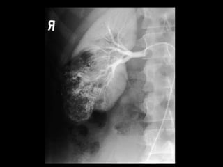
- 16. HCC & arterial – portal shunting • Findings: – focal increased vascularity in the left lobe = vascular tumor – prominent and early opacification portal veins = arterial - portal shuting – large round filling defect in main portal ven = thrombus / tumor invasion • ddx: – NONE! – This is an Aunt Minnie!
- 18. Hypothenar hammer syndrome • Findings: – contour irregularity of the distal ulnar artery – multiple distal occlusions of digital branches • ddx: – NONE! – This is an Aunt Minnie!
- 20. Klatskin tumor • Findings: – percutaneous cholangiogram shows dilated intrahepatic biliary tree – obstruction likely at the confluence of the biliary system • ddx: – choledocolithiasis – other invasive tumor – portal lymphadenopathy
- 22. Persistent sciatic artery • Findings: – anomalous arteries of the left leg: • CFA is normal but profunda and SFA are smaller than normal • large vessel arising from IIA continues into the leg • ddx: – NONE! – This is an Aunt Minnie!
- 24. Posterior knee dislocation & popliteal artery occlusion • Findings: – complete posterior dislocation of the knee – angiogram is diagnostic for popliteal artery injury • thrombosis • dissection • transection – try to cross with wire -- if unsuccessful -- surgery
- 26. Right aortic arch & aberrant left subclavian • Findings: – right sided arch with non- mirror image branching (four great vessels) • 1) Lt common carotid • 2) Rt common carotid • 3) Rt subclavian • 4) Lt subclavian • ddx: – NONE! – This is an Aunt Minnie!
- 29. Splenic artery aneurysm • Findings: – KUB shows LUQ curvilinear signet ring calcifications – celiac angiogram is diagnositc • causes: – portal hypertension – pancreatitis – pregnancy – fibromuscular dysplasia – (NOT hypertension)
- 31. Takayasu’s dz & bilateral subclavian steal • Findings: – large carotid arteris and occluded subclavian arteries – reconstitution of flow from vertebral arteries on delayed image • other causes: – atherosclerosis – trauma – radiation
- 33. Venous thoracic outlet obstructon • Findings: – tight stenosis of the left subclavian vein – multiple collaterals reconstitute left brachiocephaic vein • causes – cervical rib – anomalous muscles – post-traumatic changes • Rx: – surgical (NOT stent)
- 36. Traumatic aortic injury & active extravasation • Findings: – Widened mediastinum – Obscuration of the aortic arch – Aortagram is diagnostic • Abrupt caliber change • Irregular luminal contour • Active extravasaton • ddx: – NONE! – This is an Aunt Minnie!
- 38. Marfan-like syndrome & Type B aortic dissection • Findings: – Angiogram shows widened aorta and opacification of two channels – Lt subclavian and vertebral arteries have a beaded, aneurysmal appearance • ddx: – Atherosclerosis – Hypertension – Pregnancy – Ehlers-Danlos syndrome
- 40. Polyarteritis nodosa • Findings: – Selective angiogram of the right kidney shows multiple tiny aneurysms of the parenchymal arteries • ddx: – Mycotic aneurysms
- 42. Replaced right hepatic artery • Findings: – Right hepatic artery originates from the SMA – Proper hepatic artery may also arise from SMA – Normal variant – Left hepatic artery may arise directly from common hepatic or gastric • ddx: – NONE! – This is an Aunt Minnie!