1. 3 types:
Cardiac - involuntary; in heart only
Smooth - involuntary; found in airways,
arteries, GI tract, urinary bladder, uterus
Skeletal - voluntary; half of body's weight,
requires neuronal stimulation to contract
Contractions:
Isometric - Increased tension within muscle
but no movement
Isotonic - Shortens muscle and produces
movement
Flexion: bending a joint
Extension - straightening a joint
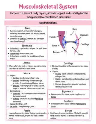
Musculoskeletal System
Purpose: To protect body organs, provide support and stability for the
body and allow coordinated movement
Key Definitions
Bone
Function: support, protect internal organs,
voluntary movement, blood cell production and
mineral storage
Classified as cortical (compact and dense) or
cancellous (spongy)
Bone Cells
Osteoblasts - synthesize collagen; the basic bone-
forming cells
Osteocytes - mature bone cells
Osteoclasts - assist in the breakdown of bone
tissue
Joints
Place where the ends of 2 bones are in proximity
and move in relation to each other
Cartilage
Flexible tissue that is the main connective tissue
in body
3 types:
Hyaline - most common; contains mostly
collagen fibers
Elastic - more flexible; contains collagen and
elastic fibers
Fibrous - tough, shock absorber; contains
mostly collagen fibers
Muscle
Ligaments
Dense connective tissue connecting bones to
bones
Tendons
Dense connective tissue connecting muscles to
bones
Fascia
Layers of connective tissue that surround muscles,
nerves, blood vessels, organs and holds them in
place
Bursae
Small sacs of connective tissue filled with synovial
fluid; located in joints to decrease pressure &
friction
Bone
Bone
Muscle
Tendon
Bursa
Ligament
Cartilage
2. Musculoskeletal Assessment
Muscle Strength Scale
0 = No muscle contraction
1 = A barely detectable contraction
2 = Active movement of body part without gravity
3 = Active movement of body part against gravity
4 = Active movement of body part against gravity
and some resistance
5 = Active movement of body part against full
resistance without evident fatigue
Assess
Range of motion
Goniometer - measures ROM of joint
Muscle strength
Look for normal spinal curvatures
Asymmetry
Joint swelling / tenderness
Look for abnormalities
Atrophy - ↓size/strength of muscle
Ankylosis - Stiffness and fixation of joint
Kyphosis - exaggerated thoracic curvature
Swayback - exaggerated lumbar curvature
Scoliosis - asymmetric elevation of shoulders
Older Adults
Inquire about exercise practices; type and
frequency
Determine age-related changes of
musculoskeletal system on functional status (ADL,
etc)
↑risk of falls due to ↓muscle mass and
strength and changes in patient's balance
Bone resorption increases and bone formation
decreases with age which leads to osteopenia and
osteoporosis
30% of muscle mass lost by age 70
Tendons and ligaments less flexible with leads to
rigid movement
Joints often have osteoarthritis
Older Adults - instruct
Use ramps in buildings and at street corners
instead of steps
Eliminate scatter rugs at home
Use a walker or cane
Avoid excessive weight gain
Get regular and frequent exercise
Use shoes with good support
Avoid walking on uneven ground and wet floors
Avoid sudden change in position to prevent
dizziness, falls, etc
R
I
C
E
→To prevent further injury
est
ce
ompression
levation
→To reduce inflammation and pain
→To prevent edema and encourage
fluid return
→To mobilize excess fluid and prevent
further edema
3. Injuries resulting from prolonged force or
repetitive movements and awkward postures
Tendons, ligaments and muscles are strained
causing tiny tears that become inflamed
At risk: musicians, dancers, those who use
mouse/keyboard often, competitive athletes
S/S: pain, weakness, numbness
Tx: stop activity causing RSI, heat/cold, NSAIDs,
rest, PT
Repetitive Strain Injury
Caused by compression of the median nerve in
the wrist
May be caused by hormones - often occurs
during PMS, pregnancy, menopause
At risk: those with DB, PVD, rheum arthritis
S/S: weakness, pain, numbness, impaired
sensation in thumb, index and middle fingers
Tx: stop repetitive motions, fix ergonomics,
splint, PT, corticosteroids, surgery
Carpal Tunnel Syndrome
Rotator cuff = 4 muscles in the shoulder
May be gradual, degenerative process or from
injury while falling
Often due to repetitive motions
S/S: shoulder weakness, pain and ↓ROM;
positive drop arm test
Tx: rest, ice and heat, NSAIDS, corticosteroids,
PT, surgery
Rotator Cuff Injury
Menisci - cartilage in the knee, AC and other
joints
Usually caused by rotational stress when knee is
in flexion and foot is planted or fixed
At risk: athletes (basketball, football, soccer)
S/S: pain upon flexion, unstable, 'pops' in knee
Tx: ice, immobility, crutches, PT, surgery;
instruct importance of warming up b4 exercise
Meniscus Injury
Most commonly injured knee ligament
Usually occurs in athletes while pivoting or
landing from jump
S/S: hear 'pop,' then pain, swelling, knee
unstable
Tx: rest, ice, NSAIDs, elevate, crutches, knee
brace, PT, surgery; full recovery 6-8 months
Anterior Cruciate Ligament (ACL) Injury
Inflammation of the bursa from repeated or
excessive trauma/ friction, or from gout,
rheumatoid arthritis or infection
Affects hands, elbows, shoulders, knees, hip
S/S: warmth, pain, swelling, limited ROM in
affected part
Tx: rest, splint, ice, NSAIDs, corticosteroids
Bursitis
Sprain - an injury to ligaments surrounding a
joint usually caused by wrenching or twisting
motion in ankle, wrist or knee joint
Strain - excessive stretching of a muscle, its
facial sheath or a tendon
S/S of strain or sprain: pain, edema, ↓fnx in
injured area, contusion; usually occur during
vigorous activities
Tx: RICE to ↓local inflammation & pain; ice &
elevate 24-48 hrs post injury; full function
returns in 3-6 weeks
Dislocation
Complete displacement of the joint
Results from severe injury of the ligaments
surrounding the joint
Usually in thumb, elbow, shoulder, hip, kneecap
Subluxation - partial displacement of the joint
S/S - deformity, local pain, tenderness, loss of
fnx of injured area, swelling near joint
Tx: requires prompt attention; orthopedic
emergency b/c may include vascular injury;
realignment 1st action, then immobilize to allow
to heal
Sprains and Strains
Musculoskeletal Trauma
Soft tissue injury - damage to any skin, muscle, tendon or ligament
4. A break or crack in a bone caused by traumatic injury or disease such as cancer
or osteoporosis
Open (Compound) or Closed (Simple)
Open - Skin broken, bone exposed, soft tissue injury
Closed - Skin remains intact
Complete or Incomplete
Complete - Break goes completely through the bone
Incomplete - fracture goes partly across bone shaft
Displaced or Nondisplaced
Displaced - 2 ends of broken bone separated and out
of alignment
Comminuted - 3 or more fragments
Oblique - fractured at a slant
Nondisplaced - bone fragments aligned
Transverse - fracture straight across bone
Spiral - fracture in spiral direction down bone
Greenstick - one side of bone bent, other side
splintered; incomplete; common in pediatrics
Fracture
Classifications
Types of Fractures
Colles' fracture - in distal radius (forearm); most common; > 50 years old; risk w/osteoporosis
Humeral shaft - shaft of humerus (long bone in arm); common among young and middle-aged
Pelvic fracture - small percentage; associated with↑mortality rate
Hip - common in older adults; > 95% resulting from fall
Stable vertebral fracture - car crashes, falls; fragments unlikely to cause spinal cord damage
Signs and Symptoms
Immediate, localized pain, tenderness
↓function in affected part
Unable to bear weight on affected part
Edema and swelling
Deformity (abnormal position)
Contusion/bruising
Crepitation - grating or crunching of bone
fragments; audible sounds
Comminuted
Oblique
Transverse
Spiral
Greenstick
5. Treatment
Traction
Aligns the bone with a constant steady
pulling action
Electrical Bone Growth Stimulation
To facilitate the healing process
Increases Ca uptake of bone, activates
intracellular Ca stores, increases
production of bone growth factors
Electrodes in band applied to skin 10-12
hrs/day (sleeping)
Meds
Muscle relaxants - Soma, Robaxin
Tetanus shot if open fracture
Bone-penetrating Abx (Kefzol)
Nutrition
Protein 1g/kg BW
Vits B, C, D; Ca, Ph, Mg
Goal:
Realign bone fragments (via closed or
open reduction)
Immobilize to maintain realignment
Restoration of normal function
Closed reduction
Nonsurgical, manual realignment
Under local or general anesthesia
Traction, cast, splint, or brace used after
Open reduction
Correction of alignment through surgery
Wires, screws, pins, plates internal or
external
Traction, cast, splint, or brace used after
Facilitates early ambulation
Intervention
Traction -
Wts need to hang freely (not on floor)
Monitor pin sites for infection
Encourage pt to participate in ROM
activities (as allowed)
Casts
Keep elevated above heart level
1st 2 days ice packs
Monitor for hot spots, pain, foul odor,
swelling, 6 Ps
Monitor skin integrity - use moleskin
around top edge of cast
Keep dry!
Immediately after injury - immobilize
w/splint
Apply pressure w/sterile dressing if open
Elevate extremity
Apply ice to ↓swelling
NPO until evaluated by surgeon
X-ray
Pain meds - monitor to see if pain relieved
Monitor for:
Compartment syndrome (see next page)
Fat embolism (see next page)
Asses 6 Ps (see next page)
Prep for surgery if needed
Post op:
Monitor vitals and Assess 6 Ps
Watch dressing for bleeding or excessive
drainage
6. Complications of Fractures
Infection -
Open fractures and soft tissue injuries have ↑incidence of infection
May require aggressive surgical debridement
May have IV Abx 3-7 days post op phase
Compartment syndrome -
Swelling causes ↑pressure within muscle compartment
Fascia surrounding muscle has limited ability to stretch
Continued swelling decreases function of blood vessels and nerves and decreases blood
flow to muscle
2 causes:
Decreased compartment size from restrictive dressings, splints, casts, traction
Increased compartment contents from bleeding, inflammation, edema
Usually associated with:
Trauma, large bone fractures, extensive tissue damage and crush injury
MUST be treated within 6 hours or nerve damage will result!
S/S: Look for the 6 Ps:
Pain - out of proportion to injury and not managed with meds -- early sign!
Paresthesia - numbness/tingling -- early sign!
Poikilothermia - affected limb cooler than non-affected limb
Pallor - coolness, loss of normal color in distal extremity
Paralysis - loss of function in extremity; late sign
Pulselessness - decreased or absent peripheral pulse; late sign
Intervention:
Regular neuro assessments on all patients with fractures
Notify HCP of pain and paresthesia as these are the 1st signs!
Keep extremity at heart level and NOT below
Monitor UO: look for dark reddish-brown color, may be from damaged muscles
Fat Embolism Syndrome -
Systemic fat globules from fractures travel to tissues, lungs and other organs after a
traumatic skeletal injury
Usually seen with long bone, rib, tibia, pelvis fractures
Need to recognize early on!
Usually occurs 24-48 hours post injury
S/S: chest pain, tachypnea, cyanosis, dyspnea, tachycardia, ↓PaO2, changes in mental
status, restlessness, confusion, petechiae
Tx: fluid resuscitation, correct acidosis, blood transfusion
Intervention: Encourage coughs/deep breathing, O2 for hypoxia
7. Joint
Capsule
Articular cartilage (hyaline cartilage found in synovial
joints) begins to deteriorate due to tissue damage
Normally soft, smooth, white cartilage becomes less
elastic, dull, yellow and granular
Joint space decreases →bones begin to grate against
each other →bone erosion follows along with
osteophyte (bone spur) formation
Pieces of cartilage and bone break off and float
around in the joint space
Osteoarthritis
Progressive joint disorder that develops due to the deterioration of
articular cartilage.
AKA Degenerative Arthritis and Degenerative Joint Disease
Pathophysiology
*Most common type of arthritis*
Hyaline - most
common - end of
bones (joints),
ribs, nose
Fibrous -
intervertebral
discs, knee
Elastic - external
ear, epiglottis
3 Types of
Cartilage:
A condition damages cartilage
(gout, rheumatoid arthritis)
An event damages cartilage
or causes joint instability (e.g.
ACL knee injury)
Low estrogen at menopause
Obesity (increases stress on
joints)
Repetitive motions (e.g.
occupations that require
frequent kneeling and
stooping have high risk of
knee OA)
Genetics
Causes / Risk Factors
Bone
Bone
Bone
Bone
Healthy Joint
Osteoarthritis
Bone ends
rub together
Thinned
cartilage
Synovial
Membrane
Synovial
Fluid
Cartilage
Bone
spur
Broken
pieces of
cartilage and
bone
8. *Joint pain * - Main sign - gets worse with use
Early stages gets better w/rest
Later stages pain still at rest
Pain leads to disability and loss of function
Early morning stiffness - disappears after 30
minutes
Crepitation - grating caused by bones rubbing or
loose cartilage
Deformity, bone spur formation and tenderness at
specific joints
Heberden's Node (joint closest to fingernail)
Bouchard's Node (joint in middle of finger)
Affects hands, knees, hips and spine
Asymmetrical
Signs and Symptoms
NOT a normal part of aging process
Begins between 20 - 30 yrs and the majority of adults affected by age 40. Symptoms
appear after age 50 - 60 yrs
Men affected more than women before age 40
Women affected more than men between 40 -70 yrs, then both the same after 70 yrs
No cure - Focus on managing pain and joint inflammation and maintain/improve joint function
Diagnosis
No single test
Analyze symptoms and rule
out other disorders such as
rheumatoid arthritis and gout
X-ray to see:
Dense bone
Osteophytes
Decreased joint space
Treatment / Intervention
Rest when joints acutely inflamed
Use splints/brace if needed during acute
inflammation but not longer than 1 week
Knee OA: avoid standing/kneeling/squatting
for long periods
Assistive devices to decrease joint stress
Heat/cold to decrease pain and stiffness
Ice for acute inflammation
Heat best for stiffness
Nutrition - educate on weight loss if
overweight
Alternative therapies:
Acupuncture, Massage, Tai Chi
Surgery - arthroscopy for knee OA
Exercise - necessary to preserve articular
cartilage health
Low-impact (walking, water aerobics),
weight training, ROM exercises
Avoid high-impact
Meds
Tylenol for mild pain
Capsaicin cream
Bengay, Arthricare (contain camphor,
eucalyptus oil, menthol)
Topical Salicylates (Aspercreme)
NSAIDs
Intraarticular injections:
corticosteroids (temp relief 1-2 mos)
9. Starts with initial immune response to antigen
The antigen triggers formation of an abnormal immunoglobulin G (IgG)
The body reacts with autoantibodies known as rheumatoid factor (RF) which land on
the synovial membranes and cartilage in joints
Inflammation results which triggers release of neutrophils (Stage 1 Synovitis)
Proteolytic enzymes released →damage to cartilage and thickening of synovial lining
Pannus (layer of vascular fibrous tissue) forms (Stage 2)
Pannus grows and damages bone & cartilage (Stage 3)
Ankylosis develops (fusion of bone) (Stage 4)
Rheumatoid Arthritis
Chronic autoimmune disease that causes inflammation in the joints.
Pathophysiology
Exact cause unknown- most
likely a combination of genetics
and environmental trigger
(smoking, infection, etc)
Cause
Bone
Bone
Healthy Joint
Synovial
Membrane
Synovial
Fluid
Cartilage
Joint
Capsule
Stage 1: Synovitis
Synovial membrane
inflamed & fluid
thickened
Bone & cartilage
gradually eroded
Stage 2:
Pannus
Pannus forms,
cartilage
eroded, bones
exposed
Stage 3: Fibrous
Ankylosis
Stage 4: Bony
Ankylosis
Fibrous
connective
tissue
invades joint
Bones fused
10. Occurs at any age, peaking between 30-50 yrs
Occurs in women 3x more than men
Onset slow, insidious
Fatigue, anorexia, wt loss, generalized
stiffness
Stiffness becomes localized
Joints become painful, stiff with limited ROM
Joints soft and inflamed (hot, swollen, tender)
Symmetrical (Bilateral)
Typically in small bones of hands, wrists, feet
But may also be in elbows, shoulders, knees,
hips, ankles and jaw
Morning stiffness lasting 60+ minutes
Rheumatoid nodules - develop in 50% of pt
Firm masses subcutaneous (usually on
fingers and elbows)
Signs and Symptoms
Treatment
No cure
Meds (main tx):
DMARDs (Disease-modifying antirheumatic
drugs), helps slow disease progression; may
be prescribed more than 1 at a time:
Methotrexate (Trexall) -
Side effect: bone marrow
suppression; hepatotoxicity (rare);
Needs frequent lab monitoring
Sulfasalazine (Azulfidine) - May cause
neutropenia
Hydroxychloroquine (Plaquenil)- may
cause vision problems; needs vision
check-ups regularly
Leflunomide (Avava)- teratogenic -
cannot be pregnant and need adequate
contraception
Late stages - Deformity and disability
May spread to other parts of body:
Cataracts, vision loss
Pleurisy
Pleural effusion
Pericarditis
Pericardial effusion
Cardiomyopathy
Sjogren's syndrome:
dry mouth/eyes
photosensitivity
Felty syndrome:
enlarged spleen
↓WBC count
Depression
↓self-care capabilities
BRMs (Biologic Response Modifiers) -
for moderate to severe cases who do
not respond to DMARDs
Enbrel, Remicade, Humira, Cimzia,
Simponi
Immunosuppressants -
Azathioprine/cyclosporine (side
effects: liver dz, infections)
Corticosteroids - Prednisone (oral for
a limited time, injection into joint for
acute relief)
Celebrex, NSAIDs/salicylates
Surgery - may be needed to relieve
severe pain and increase fnx of
severely damaged joints
Synovectomy - remove joint lining
Arthroplasty - total joint
replacement
11. Diagnosis
Blood tests to check:
+RF (Rheumatoid factor)
ESR (Erythrocyte Sedimentation Rate)
CRP (C-Reactive Protein)
ANA (Antinuclear antibody) titers
Anti-CCP (Antibodies to citrullinated
peptide)
Check synovial fluid for:
↑WBC
MMP-3 (enzyme)
X-ray to show joint deterioration (will see
later in disease process)
Intervention
Flare-ups
Inflamed joints - rest and ice
Splints
Heat for stiffness
Monitor for anemia (pale, fatigued, SOB on
exertion, palpations)
May need supplements: Fe, folic acid, Vit
B12
Monitor for GI bleed (look for dark, tarry
stool)
Educate:
On disease; no cure, how to manage
Importance of balanced diet and
maintaining healthy weight
Exercise - importance of; as tolerated;
low-impact; ROM exercises
Schedule rest and activity so no over-
exertion
Meds to take and side effects
Importance of follow-up visits
Difference between Osteoarthritis and Rheumatoid Arthritis
Deterioration of articular hyaline cartilage
of bones
Non-symmetrical
Hands/knees/hips/spine
Not systemic (joints only)
Cause: Wear and tear/overuse/injury
Older adults
No systemic inflammation
Osteophytes
AM stiffness < 30 minutes
Autoimmune disease that causes
inflammation of the synovium in joints
Symmetrical
Fingers/wrists/feet
Systemic
Cause: Unknown
Any age
Systemic inflammation
No osteophytes
AM Stiffness > 60 minutes
Osteoarthritis Rheumatoid Arthritis
12. There are 2 types of bone:
Compact: rigid, outer bone
Spongy (cancellous): porous, inner bone
Bones are in a constant state of remodeling
Bone is deposited by osteoblasts (built up)
Bone is resorbed by osteoclasts (broken down)
Usually this remodeling is in a state of equality
In osteoporosis, bone resorption > bone deposition
This inequality causes bone (specifically spongy bone)
to become very porous and thus bone density
decreases, making the bone weak
Parathyroid Hormone - When blood Ca levels are low, the PTH gland secretes PTH.
This causes osteoclasts to break down stores of Ca in the bone and enter the blood.
PTH also increases SI reabsorption of Ca and decreases kidney excretion of Ca.
Calcitonin - When Ca levels are too high, the thyroid creates calcitonin to decrease
the activity of osteoclasts
Growth hormone - Stimulates osteoblasts to build bone
Estrogen - keeps bones strong by preventing bone resorption by the osteoclasts
Testosterone - converts to estrogen and thus helps keep bones strong
Osteoporosis
Chronic, progressive bone disease characterized by decreased bone mass
and deterioration of bone tissue, leading to increased bone fragility
Pathophysiology
Hormones involved in bone health
Known as the
'Silent Thief'
because it slowly
steals a person's
bone health
Women consume less calcium
Women have less bone mass due to smaller frames
Bone resorption begins earlier in women and
becomes more rapid at menopause
Pregnancy and breast feeding deplete a woman's
skeletal reserves (unless Ca intake is adequate)
Women live longer (higher chance of developing it)
Osteoporosis is more common in women
than in men because:
Stages of Osteoporosis
Normal Bone Osteopenia
Osteoporosis
Severe
Osteoporosis
13. Early sign - pain in back, neck, hip
or spontaneous fracture
Osteoporosis most common in
spine, hips, wrists
Loss of height and humped
thoracic spine (kyphosis -
dowager's hump) - happens over
time from vertebral fracture
and wedging
Signs and Symptoms
> 65 years old
Female (8x more common than men)
↓BW (BMI < 19)
White/Asian ethnicity
Cigarette smoking
Sedentary lifestyle
Family history
Estrogen-deficient (surgical or age-
related menopause)
Risk Factors
Low intake of Ca and/or Vit D Deficiency
> 2 alcohol drinks/day
↓testosterone in men
Long-term use: Corticosteroids (major
contributor), thyroid replacement, heparin,
long-acting sedatives, aluminum-containing
antacids, anticonvulsants (phenobarbital,
Dilantin, Depakote, Tegretol),
glucocorticoids (for > 3 months)
During menopause
there is rapid bone
loss when
estrogen decline is
greatest, then
bone loss slows to
equal men's loss
Bone mineral density (BMD) test
All women over 65+ yrs should get tested
Measured by:
Quantitative ultrasound (QUS) - for
heel, kneecap, shin
Dual-energy x-ray absorptiometry
(DXA) - *Gold standard - for spine,
hips, forearm - (No Ca supplements
24 hr before test)
Results listed as T-score and
compared to healthy 30-yr old
0 = normal for healthy young adult
+1 to -1 is normal
-1 to -2.5 = osteopenia
-2.5 or lower = osteoporosis
Diagnosis
Inflammatory Bowel Disease
Intestinal malabsorption
Kidney disease
Rheumatoid arthritis
Hyperthyroidism
Alcoholism
Cirrhosis of the liver
Hypogonadism
Diabetes
Diseases associated with
Osteoporosis
14. Treatment/Intervention
Treatment focuses on: proper nutrition, calcium supplements, meds, exercise, fall prevention
Loss of bone cannot be significantly reversed but further loss can be prevented
Ca supps hard to absorb in single doses > 500 mg
Take in divided doses to ↑absorption
Ca Carbonate - has 40% elemental Ca
Take w/meals because stomach acid needed to
dissolve and absorb
Ca Citrate - has 20% elemental Ca
Not dependent on stomach acid to absorb
Better for pt on PPIs or H2 Blockers
Ca Lactate and Ca Gluconate NOT recommended
Vit D important for Ca absorption and function and
bone formation
Get it from sun 20+ min/day
Supplemental D recommended for post-
menopausal women or those homebound, in LTC
or northern climates
High calcium foods
Milk, cheese
Yogurt
Turnip Greens
Cottage cheese
↓alcohol intake and quit smoking
Nutrition
Ice cream
Sardines, salmon
Spinach
Tofu, almonds
Calcium Supplementation
Biphosphonates -
Fosamax - Daily or weekly oral tablet
Boniva - once-per-month oral tablet
Actonel - daily, weekly, monthly dep on dose
Reclast - once yearly IV infusion to treat
osteoporosis or every 2 yrs for prevention
Main side effect is GI upset:
Take with full glass of water in morning
on empty stomach with no other meds;
sit upright for 30-60 min and nothing to
eat for 1 hr
Rare side effect is osteonecrosis (bone
death) of jaw so pt should be evaluated by
dentist before starting meds
Calcitonin - interacts with osteoclasts
IM, subcutaneous, intranasal (alternate
nostrils)
Side effects - nausea, facial flushing, nasal
dryness
Monitor for hypocalcemia
Evista - selective estrogen receptor modulator
(SERM) - mimics estrogen -
Side effects - leg cramps, hot flashes, blood
clots (monitor for DVT)
Forteo - form of PTH, increases action of
osteoblasts; for severe osteoporosis
Side effects - leg cramps/dizziness
Prolia - used for postmenopausal women with
osteoporosis who are at a high risk for fractures
Subcutaneous injection every 6 months
Meds
Important for building and maintaining bone
mass
Best exercises are weight bearing
Walking, hiking, weight training, tennis, etc
Rec 30 min 3 x/week
Exercise
Treatment begins when:
T-score less than -2.5 OR
-1 to -2.5 with add'l risk factors OR
History of hip or vertebral fracture
Goal for Ca intake:
1000 mg/day for women 19-50 yrs and men 19-70 yrs
1200 mg/day for women 51+ yrs and men 71+ yrs
Educate patient:
Rooms clutter-free
Non-slip socks, shoes
Avoid throw rugs
Assistive devices
Fall Prevention
15. Gout
Type of acute arthritis characterized by hyperuricemia and deposits of
uric acid crystals in one or more joints. AKA Gouty Arthritis
Uric acid (UA) is the major end product of purine catabolism; excreted by kidneys via urine
Hyperuricemia - 2 types
Primary - hereditary error of purine metabolism leading to overproduction or retention
of UA
Secondary -
Related to another disorder such as: acidosis/ketosis, diabetes, renal insufficiency,
atherosclerosis
Caused by certain drugs: Thiazide diuretics, B-blockers, ACE inhibitors, niacin,
aspirin, cyclosporine
Gout can be acute or chronic
Decreased excretion of UA by
kidneys (most common)
Increased in UA production
High intake of
foods/beverages containing
purines (small factor)
Causes Obesity **
Excessive alcohol consumption
Prolonged fasting (↑ketoacids
inhibit UA excretion)
CKD
Metabolic syndrome
Dehydration
Physical stress on body
More common in men
Pt on cyclosporine
Risk Factors
Diagnosis
Synovial fluid aspiration to look for
monosodium urate crystals; also helps
decompress a swollen joint capsule;
* main diagnostic test
24-hr urine test to see if disease is from
decreased renal excretion or
overproduction of UA
High Purine Foods/Beverages
Red meat
Organ meats (liver, kidney, sweetbread)
Beer
Shellfish (sardines, herring, mussels)
Fructose drinks (fruit juice, soda)
Venison, goose
Anchovies
16. Multiple joint involvement
Visible deposits of sodium urate crystals
(tophi)
Tophi - white/yellow nodules under skin;
appear years after onset
Joints become damaged; cartilage
destruction may lead to secondary OA
Excessive UA excretion may lead to
kidney or urinary tract stone formation
Marked by painful flares lasting days to weeks with long periods of no symptoms
Sharp urate crystals form around the joints causing intense inflammation/pain/redness
Most commonly occurs in BIG toe (podagra), but also affects fingers, elbows, knees, wrists
Cool areas: crystals form more readily in cool areas on the body than warm areas
Usually starts in big toe in middle of night
Inflammation
Joints dusky or cyanotic
Sudden swelling
Severe pain - peaks within several hours
Area sensitive to touch
Random flare-ups, may have 1-2 episodes
and no more
Attacks end in 2-10 days with or w/o tx
Signs and Symptoms
Acute Chronic
Treatment
Maintenance meds (to prevent future attacks):
Drugs to lower urate level: Allopurinol
(Zyloprim, Aloprim); helps prevent attacks
Drugs to increase excretion of UA in urine:
Probenecid
Uloric for chronic gout
Krystexxa for those who can't take allopurinol
(given via IV)
Serum UA monitored regularly if on meds
Main treatment: Colchicine (anti-
inflammatory agent) within 12-24 hrs
of attack
Usually combined w/NSAIDs
NO Aspirin
Weight loss if needed
Avoid alcohol and foods high in purine
Corticosteroids or ACTH for acute
attack
Intervention
Hydrate 2-3 L/day
Bed rest with cradle or foot board
Educate:
Weight loss if needed
Discuss flare-ups and possible
contributor
Low-purine diet
Regular assessment of UA levels if on
meds
Avoid fasting
Acute attack:
Cold and warm compresses (alternating)
If on Colchicine:
Monitor for GI upset, neutropenia (slow
wound healing), toxicity (muscle pain,
easy bleeding)
Do not give with grapefruit juice
If on Allopurinol:
No vit C Supplements
Encourage regular eye exams