SlideShare a Scribd company logo
1 of 48
Cervical Spine Assessment &
Clearance in Emergency
Dr.PONNILAVAN
Injuries to c- spine are common and are among the few skeletal injuries that carry a high
likelihood of death.
They are often difficult to diagnose on initial imaging studies
A thorough understanding of the complex anatomy of c spine is essential for accurate
diagnosis of injury and for proper planning of Rx.
Anatomy of subaxial lower cervical spine is almost consistent, whereas anatomy of upper
cervical spine is unique at each level, hence the injuries of cervical spine is best described in
the two headings of craniovertebral and subaxial cervical spine injuries separately
• Goals for management of cervical injuries are
- prompt recognition of the problem and
- prevention of secondary neurological damage.
Missed diagnoses are common because of the difficulty in
identification of cervical spine trauma, particularly in polytrauma
patients with altered level of consciousness.
Estimate-
all trauma patients- 2 to 3%
serious head injuries- 10% sustain cervical spine injuries,
& of those, between 3 & 25% suffer extension of those injuries from
delay in diagnosis or unwarranted manipulation in the emergency
department.
When will u suspect a cervical spine
injury????
How to Obtain Radiographs
Radiographic clearance of
c- spine begins with the std
three-views.
- cross-table lateral, AP &
open-mouth views.
A swimmer’s view is added
when the initial lateral
projection fails to
demonstrate C7-T1 junction.
The swimmer’s lateral view
may be somewhat limited
secondary to the overlapping
shadows of the clavicle and
ribs.
Fuch view / closed
mouth odontoid view
Standard Fuch view
not to be used in a
trauma setting &
modified fuch can be
used
supine-oblique projections, in
which only the X-ray beam is
angled 45 degree from the
sagittal plane aimed at the
anterior margin of middle of
sternomastoid muscle, &
radiographic cassette is slid under
the scapulae without moving the
patient.
When an occult spinal injury is strongly suspected, i.e. due to the mode
of injury or because of suggestive findings on plain film, cervical CT
scanning is indicated.
Injury identified in one spinal region mandates plain film screening of
the remainder of the spine.
Interpretation of Radiographs
ATLS guidelines, assess the lateral
X-ray for alignment of four vertical
lines,
(i) anterior soft tissue line
(ii) anterior vertebral body line,
(iii) posterior vertebral body line,
(iv) a line joining the tips of spinous
processes.
Prevertebral soft tissue thickness varies
anterior to the C1 arch.
• This plane narrows to 2-3 mm anterior to
C2 to C4.
• At C5 and below, the thinner
retropharyngeal space widens to the
retrotracheal space, which approximates
the width of the vertebral body.
Webb et al described a tetrad of signs that should warn
the clinician of a possibility of unstable hyperflexion
cervical spinal injury, which includes
(i) Interspinous widening,
(ii) anterior subluxation exceeding 3 mm in adult or 4 mm in children,
(iii) tear drop fracture, and
iv)focal Kyphosis exceeding 11° or widening of the interspinous
distance relative to the adjacent levels.
• Conversely, a hyperextension injury may show anterior disc space
widening, focal lordosis, extension tear-drop fracture at C-2, and
posterior subluxation.
• In upper cervical region, anterior
atlantodens interval exceeding 3
mm in adult & 5 mm in children
indicate damage to transverse
ligament.
• In the elderly patients with
degenerative disc disease
presence of some degree of
retrolisthesis is common and may
be less likely to predict injury.
• X-ray is uncommon and is more
likely to indicate a hidden injury.
Flexion-Extension Radiographs, CT and MRI
• Flexion-extension views are not used to determine stability of known
cervical spine injuries.
• Dynamic films are best used when assessing a suspected hyperflexion
sprain when routine radiographs are equivocal.
• Their purpose is to assess the integrity of the posterior ligament
complex.
Degree of flexion-extension must be limited to the point of the
patient’s pain tolerance. These may be safely performed in awake and
alert patients in the emergency room.
Dynamic Films produce unacceptably high false positive and false
negative rates in an acute setting because pain and spasm limit cervical
spine motion.
This examination is best performed when the patient comfortably
exhibits a more normal arc of motion, usually after 1 to 2 weeks.
Multiple studies have shown that cervical spine clearance based on
plain radiographs alone misses spine fractures in 15 to 30% cases.
The limitations of plain radiography have led to the widespread use of
flexion-extension radiographs, CT, and MRI to evaluate for subtle
cervical spine injuries.
Computed tomography (CT) scans
- However, CT scans are limited their ability to demonstrate axially oriented
fractures (like type II odontoid fractures) or ligamentous injures.
- If CT scans are used as a substitute for a 3-view spine series, then coronal
and sagittal reconstructions are essential.
Thin slice CT scan (2mm or less) & multiplanar reconstructed images will
show fractures that are oriented purely in the axial plane, subluxation of
facet joints and vertebral bodies, as well as angular and rotational
abnormalities.
Sensitivity of CT scans for detection of fractures ranges from 97 to 100%
MRI scan
MRI scan provides direct visualization of the posterior ligament
complex and is therefore, the definitive imaging examination for
anterior subluxation.
Cost and limited availability preclude its use as a screening study.
Patients with significant neck pain but normal radiographs should be
evaluated with an MRI scan, or treated presumptively with a hard
cervical orthosis until flexion-extension views can be obtained at a later
date
For comatose patients
Passive flexion-extension views under fluoroscopic guidance has been
recommended.
• While controversial, this may be an acceptable method for clearing
the spine for ligamentous injury when MRI scanning is
contraindicated.
• Care must be exercised to assure that adequate motion occurs. This
maneuver may be performed with the patient log-rolled onto his side,
taking extra care to protect the spine
Case Scenario
A 12-month-old female infant
presented with nausea,
vomiting, and drowsiness to
emergency room after falling
from a height of less than
30 cm.
She had no neurological deficit
at presentation, and cervical
spine plain radiographs (a) and
CT with 3D reconstruction (b)
showed no abnormal findings.
c) 7 days after the injury the patient
developed right sided hemiparesis &
cervical MRI revealed increased
intensity (arrows) in the T2-weighted
images at the level of C6.
(d) Repeat cervical MRI one month
later shows that increased signal
intensity has disappeared.
The patient continued to improve
neurologically until 24 months after
the injury and returned to near-
normal
• SCIWORA, first described by Pang and Wilberger in 1982 occurs in
approximately 2-4% of spinal injures.
• They described the clinical profile of the SCIWORA syndrome in
• 55 children, of which there were
- 10 upper cervical (C1-C4),
- 33 lower cervical (C5- C8), and
- 12 thoracic cord injuries.
SCIWORA
Absence of radiological evidence confirming a spinal injury should not lead to a relaxation of
precautions until the patient is lucid & cooperative enough to move all limbs & report any
areas of excessive tenderness.
Spinal cord may be injured even though vertebral column is spared from disruption, because of
the inherent elasticity of the juvenile spine, which permits selfreduction but significant
intersegmental displacements when subjected to flexion, extension & distraction forces.
This vulnerability is most evident in children younger than 8 years.
Hendey et al
Using the database of the National Emergency X-Radiography
Utilization Study (NEXUS).
Studied the incidence & characteristics of patients with
SCIWORA
What is NEXUS??
NEXUS criteria
National Emergency X radiography Utilisation Group
• Remember Nexus by NSAID
N - Neurodeficit
S - Spine tenderness
A - Alertness
I - Intoxication
D - Distracting injuries
NEXUS criteria (5 N)
• No posterior midline cervical tenderness
• No evidence of intoxication
• Normal alertness level
• No focal neurological deficit
• No painful distracting injuries
If any one N is missing request for
a radiograph
SCIWORA
-defined as SCI demonstrated by MRI, when a complete, technically adequate plain
radiographic series revealed no injury.
All the SCIWORA cases were identified with adults.
There were over 3000 children enrolled, including 30 with cervical spine injury, but
none had SCIWORA.
The most common MRI findings among SCIWORA patients were
- central disc herniation,
- Spinal stenosis, and
- cord edema or contusion.
Central cord syndrome was described in 10 cases.
They concluded that SCIWORA was an uncommon disorder that occurred only in
adults.
Steroids (Methylprednisolone) in Spinal Cord
Injury
National Acute Spinal Cord Injury Studies (NASCIS) I and II published in
the 1990s demonstrated significant benefit in administering high doses
of methylprednisolone early after a spinal cord injury (within 8 h).
Recommended dose is 30 mg/kg IV over 15 minutes, followed by 5.4
mg/kg/h via continuous intravenous infusion over 24 hours.
• The NASCIS I & II trials have received significant criticism with regards
to both their design & possible benefit-to-risk ratio.
• No full consensus has been reached on the use of
methylprednisolone in person with acute cord injury. Management
should be guided based on local guidelines.
UPPER CERVICAL SPINE
• The craniocervical junction refers to the osseoligamentous and
neurovascular structures that extend from the skull base to C2.
• It is comprised of highly specialized bony articulations between the
occipital condyles, C1 and C2 and the complex ligamentous system
linking these bones into one functional joint.
All high energy injuries should be considered potential for upper
cervical instability.
Cranial nerve function
• should be a part of any examination in patients with possible head or
neck injuries.
• Abducens and hypoglossal nerves are the commonest involved in
craniocervical injuries.
• Patterns ranging from complete pentaplegia to incomplete injuries
such as cervicomedullary syndromes and brainstem disorders.
• In view of presence of subluxations or partially reduced injuries ,
clinicians should look for indirect clinical signs such as
-soft tissue swelling and hemorrhage during clinical and X ray
examinations.
• Fractures of upper cervical spine (CO-C2) often present without
neurologic deficit, since there is a proportionally greater space
available for the cord than in the lower cervical spine.
• Furthermore, if significant cord damage is caused by a high cervical
fracture, patients are frequently dead on the scene of accident.
• The most common patterns of injury seen in the upper cervical spine
are CO-C-1 disruption, C-1 ring fracture, C1-2disruption, C-2 ring
fracture, and odontoid process fracture
Subaxial cervical spine
Accurate classification of injuries speeds the delivery of appropriate
diagnostic & therapeutic intervention.
Compressive Flexion Injuries
Vertical Compression Injuries
Distractive Flexion Injuries
Compressive Extension Injuries
Distractive Extension Injuries
Lateral Flexion Injuries
The mechanistic classification proposed by Ferguson and Allen are
bases on the position of the neck at the time of injury and the
dominant mode of force application.
Furthermore, each injury pattern graded in terms of the degree of
injury (bony or ligamentous) to the involved motion segment.
A higher stage of injury is associated with greater amount of
displacement and a greater risk of neurological injury.
Injury designation is based on the mechanism of injury & review of
plan radiographs.
Crucial to the differentiation of injury patterns is recognition that
compressive load result in shortening of vertebral elements, while
distraction results in lengthening.
In the Ferguson-Allen classification, the PLL & structures anterior to it
are considered the anterior column of the spine, and the structures
posterior to the PLL are considered the posterior column of the spine.
It cannot be universally applied; patients often present with injuries
that represent a combination of injury patterns.
Timing of Surgery
Mx of acute SCI has traditionally concentrated on conservative care.
Pharmacologic interventions, in particular iv methylprednisolone
therapy, have shown modest improvements in clinical trials & are still
undergoing evaluation.
More recent interest has focused on the role of surgical reduction &
decompression, particularly early surgery.
• A review of the current evidence available in the literature suggests that there is
no standard of care regarding the role & timing of surgical decompression.
• In a recent study, Dimar et al produced thoracic SCI in rats and an epidural spacer
placed adjacent to the contusion to mimic the effect of persisting compression.
• The effect of decompression at 0, 2, 6, 24, and 72 hours after SCI was then
assessed by quantitative analysis of locomotor recovery, lesion volume, &
electrophysiology.
• Neurologic recovery was significantly dependent on time to decompression, with
significant differences seen in all experimental groups.
• This study provides the strongest experimental evidence to date of a clear
beneficial effect of spinal cord decompression after SCI
There is insufficient data to support overall treatment standards or
guidelines for timing of surgery in spinal cord injury.
There are, however, class II data indicating that early surgery (< 24
hours) may be done safely after acute SCI. Furthermore, there are Class
III data to suggest a role for urgent decompression in the setting of
1) bilateral facet dislocation and
2) incomplete spinal cold injury with a neurologically deteriorating
patient.
Whereas there is biologic evidence from experimental studies in
animals that early decompression may improve neurologic recovery
after SCI, the relevant time frame in humans remains unclear.
To date, the role of decompression in patients with SCI is only
supported by Class III and limited Class II evidence.
Accordingly, there is a strong rationale to undertake prospective,
controlled trials to evaluate the role and timing of decompression in
acute SCI.
Summary
• A total of 4,80,652 road accidents took place in India last year
resulting in the loss of 1,50,785 lives
• Often cervical injury occurs in high speed motor accidents
In RTA
• Maintain ABC first
• Strict log rolling and Spine immobilisation
• Other injury related specific treatment
Thank you

More Related Content

What's hot

Posterior Spine Fixation
Posterior Spine FixationPosterior Spine Fixation
Posterior Spine Fixation
Ghazwan Bayaty
 
Ligamentotaxis principle in the treatment of intra articular fractures of dis...
Ligamentotaxis principle in the treatment of intra articular fractures of dis...Ligamentotaxis principle in the treatment of intra articular fractures of dis...
Ligamentotaxis principle in the treatment of intra articular fractures of dis...
Sitanshu Barik
 

What's hot (20)

Acetabular defects
Acetabular defectsAcetabular defects
Acetabular defects
 
Aseptic loosening total hip arthroplasty
Aseptic loosening total hip arthroplastyAseptic loosening total hip arthroplasty
Aseptic loosening total hip arthroplasty
 
Acetabular fractures
Acetabular fracturesAcetabular fractures
Acetabular fractures
 
Distal Clavicle Fractures
Distal Clavicle Fractures Distal Clavicle Fractures
Distal Clavicle Fractures
 
Floating Knee
Floating KneeFloating Knee
Floating Knee
 
Supra condylar humerus fracture in children
Supra condylar humerus fracture in childrenSupra condylar humerus fracture in children
Supra condylar humerus fracture in children
 
Surgical Approaches to Hip Joint
Surgical Approaches to Hip JointSurgical Approaches to Hip Joint
Surgical Approaches to Hip Joint
 
MRI in orthopaedics
MRI in orthopaedicsMRI in orthopaedics
MRI in orthopaedics
 
Posterior Spine Fixation
Posterior Spine FixationPosterior Spine Fixation
Posterior Spine Fixation
 
Evolution of Intramedullary Nails
Evolution of Intramedullary NailsEvolution of Intramedullary Nails
Evolution of Intramedullary Nails
 
malunion.pptx
malunion.pptxmalunion.pptx
malunion.pptx
 
Cervical spine
Cervical spineCervical spine
Cervical spine
 
Plates and screws 11
Plates and screws 11Plates and screws 11
Plates and screws 11
 
Osteotomies around the hip
Osteotomies around the hipOsteotomies around the hip
Osteotomies around the hip
 
syndesmotic injury mechanism and treatment subject review
syndesmotic injury mechanism and treatment subject reviewsyndesmotic injury mechanism and treatment subject review
syndesmotic injury mechanism and treatment subject review
 
Tibial plateau fractures
Tibial plateau fracturesTibial plateau fractures
Tibial plateau fractures
 
Ligamentotaxis principle in the treatment of intra articular fractures of dis...
Ligamentotaxis principle in the treatment of intra articular fractures of dis...Ligamentotaxis principle in the treatment of intra articular fractures of dis...
Ligamentotaxis principle in the treatment of intra articular fractures of dis...
 
Principles of locking compression plates
Principles of locking compression platesPrinciples of locking compression plates
Principles of locking compression plates
 
Essex-Lopresti Longitudinal Instability of the Forearm
Essex-Lopresti Longitudinal Instability of the ForearmEssex-Lopresti Longitudinal Instability of the Forearm
Essex-Lopresti Longitudinal Instability of the Forearm
 
TENS
TENSTENS
TENS
 

Similar to Cervical spine clearance in polytrauma

Imaging of spinal trauma
Imaging of spinal traumaImaging of spinal trauma
Imaging of spinal trauma
Vishal Sankpal
 
Cervical Spine Clearance
Cervical Spine ClearanceCervical Spine Clearance
Cervical Spine Clearance
kk 555888
 
Pediatric c-spine injuries
Pediatric c-spine injuriesPediatric c-spine injuries
Pediatric c-spine injuries
aalthekair
 
classification and managenement of paediatric craniocervical junction injuries
classification and managenement of paediatric craniocervical junction injuriesclassification and managenement of paediatric craniocervical junction injuries
classification and managenement of paediatric craniocervical junction injuries
Dieu Merci KABULO
 
Posttraumatic spinal cord injury without radiographic abnormality
Posttraumatic spinal cord injury without radiographic abnormalityPosttraumatic spinal cord injury without radiographic abnormality
Posttraumatic spinal cord injury without radiographic abnormality
Ponnilavan Ponz
 

Similar to Cervical spine clearance in polytrauma (20)

Imaging of spinal trauma
Imaging of spinal traumaImaging of spinal trauma
Imaging of spinal trauma
 
imagingofspinaltrauMA
imagingofspinaltrauMAimagingofspinaltrauMA
imagingofspinaltrauMA
 
Ariunaa spine trauma
Ariunaa spine traumaAriunaa spine trauma
Ariunaa spine trauma
 
Cervical Spine Clearance
Cervical Spine ClearanceCervical Spine Clearance
Cervical Spine Clearance
 
Pediatric c-spine injuries
Pediatric c-spine injuriesPediatric c-spine injuries
Pediatric c-spine injuries
 
Role of magnetic resonance Imaging in acute spinal trauma
Role of magnetic resonance Imaging in acute spinal trauma Role of magnetic resonance Imaging in acute spinal trauma
Role of magnetic resonance Imaging in acute spinal trauma
 
Spinal cord injury without radiographic abnormalities (SCIWORA)
Spinal cord injury without radiographic abnormalities (SCIWORA)Spinal cord injury without radiographic abnormalities (SCIWORA)
Spinal cord injury without radiographic abnormalities (SCIWORA)
 
Recent advances in imaging of scoliosis final
Recent advances in imaging of scoliosis finalRecent advances in imaging of scoliosis final
Recent advances in imaging of scoliosis final
 
Lateral condyle of humerus fracture in children
Lateral condyle of humerus fracture in childrenLateral condyle of humerus fracture in children
Lateral condyle of humerus fracture in children
 
Spinal Cord Syndrome
Spinal Cord SyndromeSpinal Cord Syndrome
Spinal Cord Syndrome
 
supracondylar fractures in children -contraversies
supracondylar fractures in children -contraversiessupracondylar fractures in children -contraversies
supracondylar fractures in children -contraversies
 
Study of current pattern of Cervical Spondylotic Myelopathy and to evaluate t...
Study of current pattern of Cervical Spondylotic Myelopathy and to evaluate t...Study of current pattern of Cervical Spondylotic Myelopathy and to evaluate t...
Study of current pattern of Cervical Spondylotic Myelopathy and to evaluate t...
 
classification and managenement of paediatric craniocervical junction injuries
classification and managenement of paediatric craniocervical junction injuriesclassification and managenement of paediatric craniocervical junction injuries
classification and managenement of paediatric craniocervical junction injuries
 
Evaluation and management of cervical spine injury
Evaluation and management of cervical spine injuryEvaluation and management of cervical spine injury
Evaluation and management of cervical spine injury
 
USG guidance for supracondylar fracture of humerus
USG guidance for supracondylar fracture of humerusUSG guidance for supracondylar fracture of humerus
USG guidance for supracondylar fracture of humerus
 
Approach to problem fractures
Approach to problem fracturesApproach to problem fractures
Approach to problem fractures
 
Slide pfof
Slide pfofSlide pfof
Slide pfof
 
Day 2 | CME- Trauma Symposium | Master trauma panel perspective
Day 2 | CME- Trauma Symposium | Master trauma panel perspectiveDay 2 | CME- Trauma Symposium | Master trauma panel perspective
Day 2 | CME- Trauma Symposium | Master trauma panel perspective
 
Spinetrauma 2
Spinetrauma 2Spinetrauma 2
Spinetrauma 2
 
Posttraumatic spinal cord injury without radiographic abnormality
Posttraumatic spinal cord injury without radiographic abnormalityPosttraumatic spinal cord injury without radiographic abnormality
Posttraumatic spinal cord injury without radiographic abnormality
 

More from Ponnilavan Ponz (20)

Cubitus varus and valgus
Cubitus varus and valgusCubitus varus and valgus
Cubitus varus and valgus
 
Rickets
RicketsRickets
Rickets
 
Meniscal injury
Meniscal injury Meniscal injury
Meniscal injury
 
Poliomyelitis
PoliomyelitisPoliomyelitis
Poliomyelitis
 
Anatomy of cervical spine
Anatomy of cervical spineAnatomy of cervical spine
Anatomy of cervical spine
 
Congenital pseudoarthrosis of tibia
Congenital pseudoarthrosis of tibiaCongenital pseudoarthrosis of tibia
Congenital pseudoarthrosis of tibia
 
screws and plate
screws and platescrews and plate
screws and plate
 
Distal femoral fresh osteochondral allografts
Distal femoral fresh osteochondral allograftsDistal femoral fresh osteochondral allografts
Distal femoral fresh osteochondral allografts
 
External fixation
External fixation External fixation
External fixation
 
Im nail
Im nailIm nail
Im nail
 
Krukenberg surgery
Krukenberg surgeryKrukenberg surgery
Krukenberg surgery
 
Patellofemoral disorders
Patellofemoral disordersPatellofemoral disorders
Patellofemoral disorders
 
Avn
AvnAvn
Avn
 
Osteochondroma
OsteochondromaOsteochondroma
Osteochondroma
 
Dupuytren
Dupuytren   Dupuytren
Dupuytren
 
Chopart amputation
Chopart amputationChopart amputation
Chopart amputation
 
Acl reconstruction
Acl reconstructionAcl reconstruction
Acl reconstruction
 
Bladder innervation
Bladder innervationBladder innervation
Bladder innervation
 
maduramycosis
maduramycosis   maduramycosis
maduramycosis
 
Adult acquired flat foot deformity
Adult acquired flat foot deformityAdult acquired flat foot deformity
Adult acquired flat foot deformity
 

Recently uploaded

Call Girl In Pune 👉 Just CALL ME: 9352988975 💋 Call Out Call Both With High p...
Call Girl In Pune 👉 Just CALL ME: 9352988975 💋 Call Out Call Both With High p...Call Girl In Pune 👉 Just CALL ME: 9352988975 💋 Call Out Call Both With High p...
Call Girl In Pune 👉 Just CALL ME: 9352988975 💋 Call Out Call Both With High p...
chetankumar9855
 
🌹Attapur⬅️ Vip Call Girls Hyderabad 📱9352852248 Book Well Trand Call Girls In...
🌹Attapur⬅️ Vip Call Girls Hyderabad 📱9352852248 Book Well Trand Call Girls In...🌹Attapur⬅️ Vip Call Girls Hyderabad 📱9352852248 Book Well Trand Call Girls In...
🌹Attapur⬅️ Vip Call Girls Hyderabad 📱9352852248 Book Well Trand Call Girls In...
Call Girls In Delhi Whatsup 9873940964 Enjoy Unlimited Pleasure
 
Call Girls in Gagan Vihar (delhi) call me [🔝 9953056974 🔝] escort service 24X7
Call Girls in Gagan Vihar (delhi) call me [🔝  9953056974 🔝] escort service 24X7Call Girls in Gagan Vihar (delhi) call me [🔝  9953056974 🔝] escort service 24X7
Call Girls in Gagan Vihar (delhi) call me [🔝 9953056974 🔝] escort service 24X7
9953056974 Low Rate Call Girls In Saket, Delhi NCR
 

Recently uploaded (20)

Independent Call Girls In Jaipur { 8445551418 } ✔ ANIKA MEHTA ✔ Get High Prof...
Independent Call Girls In Jaipur { 8445551418 } ✔ ANIKA MEHTA ✔ Get High Prof...Independent Call Girls In Jaipur { 8445551418 } ✔ ANIKA MEHTA ✔ Get High Prof...
Independent Call Girls In Jaipur { 8445551418 } ✔ ANIKA MEHTA ✔ Get High Prof...
 
Jogeshwari ! Call Girls Service Mumbai - 450+ Call Girl Cash Payment 90042684...
Jogeshwari ! Call Girls Service Mumbai - 450+ Call Girl Cash Payment 90042684...Jogeshwari ! Call Girls Service Mumbai - 450+ Call Girl Cash Payment 90042684...
Jogeshwari ! Call Girls Service Mumbai - 450+ Call Girl Cash Payment 90042684...
 
Call Girl In Pune 👉 Just CALL ME: 9352988975 💋 Call Out Call Both With High p...
Call Girl In Pune 👉 Just CALL ME: 9352988975 💋 Call Out Call Both With High p...Call Girl In Pune 👉 Just CALL ME: 9352988975 💋 Call Out Call Both With High p...
Call Girl In Pune 👉 Just CALL ME: 9352988975 💋 Call Out Call Both With High p...
 
🌹Attapur⬅️ Vip Call Girls Hyderabad 📱9352852248 Book Well Trand Call Girls In...
🌹Attapur⬅️ Vip Call Girls Hyderabad 📱9352852248 Book Well Trand Call Girls In...🌹Attapur⬅️ Vip Call Girls Hyderabad 📱9352852248 Book Well Trand Call Girls In...
🌹Attapur⬅️ Vip Call Girls Hyderabad 📱9352852248 Book Well Trand Call Girls In...
 
(Low Rate RASHMI ) Rate Of Call Girls Jaipur ❣ 8445551418 ❣ Elite Models & Ce...
(Low Rate RASHMI ) Rate Of Call Girls Jaipur ❣ 8445551418 ❣ Elite Models & Ce...(Low Rate RASHMI ) Rate Of Call Girls Jaipur ❣ 8445551418 ❣ Elite Models & Ce...
(Low Rate RASHMI ) Rate Of Call Girls Jaipur ❣ 8445551418 ❣ Elite Models & Ce...
 
Coimbatore Call Girls in Thudiyalur : 7427069034 High Profile Model Escorts |...
Coimbatore Call Girls in Thudiyalur : 7427069034 High Profile Model Escorts |...Coimbatore Call Girls in Thudiyalur : 7427069034 High Profile Model Escorts |...
Coimbatore Call Girls in Thudiyalur : 7427069034 High Profile Model Escorts |...
 
Top Rated Pune Call Girls (DIPAL) ⟟ 8250077686 ⟟ Call Me For Genuine Sex Serv...
Top Rated Pune Call Girls (DIPAL) ⟟ 8250077686 ⟟ Call Me For Genuine Sex Serv...Top Rated Pune Call Girls (DIPAL) ⟟ 8250077686 ⟟ Call Me For Genuine Sex Serv...
Top Rated Pune Call Girls (DIPAL) ⟟ 8250077686 ⟟ Call Me For Genuine Sex Serv...
 
Top Rated Call Girls Kerala ☎ 8250092165👄 Delivery in 20 Mins Near Me
Top Rated Call Girls Kerala ☎ 8250092165👄 Delivery in 20 Mins Near MeTop Rated Call Girls Kerala ☎ 8250092165👄 Delivery in 20 Mins Near Me
Top Rated Call Girls Kerala ☎ 8250092165👄 Delivery in 20 Mins Near Me
 
Call Girls Service Jaipur {9521753030 } ❤️VVIP BHAWNA Call Girl in Jaipur Raj...
Call Girls Service Jaipur {9521753030 } ❤️VVIP BHAWNA Call Girl in Jaipur Raj...Call Girls Service Jaipur {9521753030 } ❤️VVIP BHAWNA Call Girl in Jaipur Raj...
Call Girls Service Jaipur {9521753030 } ❤️VVIP BHAWNA Call Girl in Jaipur Raj...
 
Top Rated Hyderabad Call Girls Chintal ⟟ 9332606886 ⟟ Call Me For Genuine Se...
Top Rated  Hyderabad Call Girls Chintal ⟟ 9332606886 ⟟ Call Me For Genuine Se...Top Rated  Hyderabad Call Girls Chintal ⟟ 9332606886 ⟟ Call Me For Genuine Se...
Top Rated Hyderabad Call Girls Chintal ⟟ 9332606886 ⟟ Call Me For Genuine Se...
 
Russian Call Girls Service Jaipur {8445551418} ❤️PALLAVI VIP Jaipur Call Gir...
Russian Call Girls Service  Jaipur {8445551418} ❤️PALLAVI VIP Jaipur Call Gir...Russian Call Girls Service  Jaipur {8445551418} ❤️PALLAVI VIP Jaipur Call Gir...
Russian Call Girls Service Jaipur {8445551418} ❤️PALLAVI VIP Jaipur Call Gir...
 
Coimbatore Call Girls in Coimbatore 7427069034 genuine Escort Service Girl 10...
Coimbatore Call Girls in Coimbatore 7427069034 genuine Escort Service Girl 10...Coimbatore Call Girls in Coimbatore 7427069034 genuine Escort Service Girl 10...
Coimbatore Call Girls in Coimbatore 7427069034 genuine Escort Service Girl 10...
 
Call Girls Jaipur Just Call 9521753030 Top Class Call Girl Service Available
Call Girls Jaipur Just Call 9521753030 Top Class Call Girl Service AvailableCall Girls Jaipur Just Call 9521753030 Top Class Call Girl Service Available
Call Girls Jaipur Just Call 9521753030 Top Class Call Girl Service Available
 
Call Girls Service Jaipur {9521753030} ❤️VVIP RIDDHI Call Girl in Jaipur Raja...
Call Girls Service Jaipur {9521753030} ❤️VVIP RIDDHI Call Girl in Jaipur Raja...Call Girls Service Jaipur {9521753030} ❤️VVIP RIDDHI Call Girl in Jaipur Raja...
Call Girls Service Jaipur {9521753030} ❤️VVIP RIDDHI Call Girl in Jaipur Raja...
 
💕SONAM KUMAR💕Premium Call Girls Jaipur ↘️9257276172 ↙️One Night Stand With Lo...
💕SONAM KUMAR💕Premium Call Girls Jaipur ↘️9257276172 ↙️One Night Stand With Lo...💕SONAM KUMAR💕Premium Call Girls Jaipur ↘️9257276172 ↙️One Night Stand With Lo...
💕SONAM KUMAR💕Premium Call Girls Jaipur ↘️9257276172 ↙️One Night Stand With Lo...
 
Call Girls in Gagan Vihar (delhi) call me [🔝 9953056974 🔝] escort service 24X7
Call Girls in Gagan Vihar (delhi) call me [🔝  9953056974 🔝] escort service 24X7Call Girls in Gagan Vihar (delhi) call me [🔝  9953056974 🔝] escort service 24X7
Call Girls in Gagan Vihar (delhi) call me [🔝 9953056974 🔝] escort service 24X7
 
9630942363 Genuine Call Girls In Ahmedabad Gujarat Call Girls Service
9630942363 Genuine Call Girls In Ahmedabad Gujarat Call Girls Service9630942363 Genuine Call Girls In Ahmedabad Gujarat Call Girls Service
9630942363 Genuine Call Girls In Ahmedabad Gujarat Call Girls Service
 
Call Girls Service Jaipur {8445551418} ❤️VVIP BHAWNA Call Girl in Jaipur Raja...
Call Girls Service Jaipur {8445551418} ❤️VVIP BHAWNA Call Girl in Jaipur Raja...Call Girls Service Jaipur {8445551418} ❤️VVIP BHAWNA Call Girl in Jaipur Raja...
Call Girls Service Jaipur {8445551418} ❤️VVIP BHAWNA Call Girl in Jaipur Raja...
 
8980367676 Call Girls In Ahmedabad Escort Service Available 24×7 In Ahmedabad
8980367676 Call Girls In Ahmedabad Escort Service Available 24×7 In Ahmedabad8980367676 Call Girls In Ahmedabad Escort Service Available 24×7 In Ahmedabad
8980367676 Call Girls In Ahmedabad Escort Service Available 24×7 In Ahmedabad
 
Most Beautiful Call Girl in Bangalore Contact on Whatsapp
Most Beautiful Call Girl in Bangalore Contact on WhatsappMost Beautiful Call Girl in Bangalore Contact on Whatsapp
Most Beautiful Call Girl in Bangalore Contact on Whatsapp
 

Cervical spine clearance in polytrauma

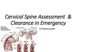
  • 1. Cervical Spine Assessment & Clearance in Emergency Dr.PONNILAVAN
  • 2. Injuries to c- spine are common and are among the few skeletal injuries that carry a high likelihood of death. They are often difficult to diagnose on initial imaging studies A thorough understanding of the complex anatomy of c spine is essential for accurate diagnosis of injury and for proper planning of Rx. Anatomy of subaxial lower cervical spine is almost consistent, whereas anatomy of upper cervical spine is unique at each level, hence the injuries of cervical spine is best described in the two headings of craniovertebral and subaxial cervical spine injuries separately
  • 3. • Goals for management of cervical injuries are - prompt recognition of the problem and - prevention of secondary neurological damage. Missed diagnoses are common because of the difficulty in identification of cervical spine trauma, particularly in polytrauma patients with altered level of consciousness.
  • 4. Estimate- all trauma patients- 2 to 3% serious head injuries- 10% sustain cervical spine injuries, & of those, between 3 & 25% suffer extension of those injuries from delay in diagnosis or unwarranted manipulation in the emergency department.
  • 5. When will u suspect a cervical spine injury????
  • 6. How to Obtain Radiographs Radiographic clearance of c- spine begins with the std three-views. - cross-table lateral, AP & open-mouth views.
  • 7. A swimmer’s view is added when the initial lateral projection fails to demonstrate C7-T1 junction. The swimmer’s lateral view may be somewhat limited secondary to the overlapping shadows of the clavicle and ribs.
  • 8. Fuch view / closed mouth odontoid view Standard Fuch view not to be used in a trauma setting & modified fuch can be used
  • 9. supine-oblique projections, in which only the X-ray beam is angled 45 degree from the sagittal plane aimed at the anterior margin of middle of sternomastoid muscle, & radiographic cassette is slid under the scapulae without moving the patient.
  • 10. When an occult spinal injury is strongly suspected, i.e. due to the mode of injury or because of suggestive findings on plain film, cervical CT scanning is indicated. Injury identified in one spinal region mandates plain film screening of the remainder of the spine.
  • 11. Interpretation of Radiographs ATLS guidelines, assess the lateral X-ray for alignment of four vertical lines, (i) anterior soft tissue line (ii) anterior vertebral body line, (iii) posterior vertebral body line, (iv) a line joining the tips of spinous processes.
  • 12.
  • 13. Prevertebral soft tissue thickness varies anterior to the C1 arch. • This plane narrows to 2-3 mm anterior to C2 to C4. • At C5 and below, the thinner retropharyngeal space widens to the retrotracheal space, which approximates the width of the vertebral body.
  • 14. Webb et al described a tetrad of signs that should warn the clinician of a possibility of unstable hyperflexion cervical spinal injury, which includes (i) Interspinous widening, (ii) anterior subluxation exceeding 3 mm in adult or 4 mm in children, (iii) tear drop fracture, and iv)focal Kyphosis exceeding 11° or widening of the interspinous distance relative to the adjacent levels.
  • 15.
  • 16. • Conversely, a hyperextension injury may show anterior disc space widening, focal lordosis, extension tear-drop fracture at C-2, and posterior subluxation.
  • 17. • In upper cervical region, anterior atlantodens interval exceeding 3 mm in adult & 5 mm in children indicate damage to transverse ligament. • In the elderly patients with degenerative disc disease presence of some degree of retrolisthesis is common and may be less likely to predict injury. • X-ray is uncommon and is more likely to indicate a hidden injury.
  • 18. Flexion-Extension Radiographs, CT and MRI • Flexion-extension views are not used to determine stability of known cervical spine injuries. • Dynamic films are best used when assessing a suspected hyperflexion sprain when routine radiographs are equivocal. • Their purpose is to assess the integrity of the posterior ligament complex.
  • 19. Degree of flexion-extension must be limited to the point of the patient’s pain tolerance. These may be safely performed in awake and alert patients in the emergency room. Dynamic Films produce unacceptably high false positive and false negative rates in an acute setting because pain and spasm limit cervical spine motion. This examination is best performed when the patient comfortably exhibits a more normal arc of motion, usually after 1 to 2 weeks.
  • 20. Multiple studies have shown that cervical spine clearance based on plain radiographs alone misses spine fractures in 15 to 30% cases. The limitations of plain radiography have led to the widespread use of flexion-extension radiographs, CT, and MRI to evaluate for subtle cervical spine injuries.
  • 21. Computed tomography (CT) scans - However, CT scans are limited their ability to demonstrate axially oriented fractures (like type II odontoid fractures) or ligamentous injures. - If CT scans are used as a substitute for a 3-view spine series, then coronal and sagittal reconstructions are essential. Thin slice CT scan (2mm or less) & multiplanar reconstructed images will show fractures that are oriented purely in the axial plane, subluxation of facet joints and vertebral bodies, as well as angular and rotational abnormalities. Sensitivity of CT scans for detection of fractures ranges from 97 to 100%
  • 22. MRI scan MRI scan provides direct visualization of the posterior ligament complex and is therefore, the definitive imaging examination for anterior subluxation. Cost and limited availability preclude its use as a screening study. Patients with significant neck pain but normal radiographs should be evaluated with an MRI scan, or treated presumptively with a hard cervical orthosis until flexion-extension views can be obtained at a later date
  • 23. For comatose patients Passive flexion-extension views under fluoroscopic guidance has been recommended. • While controversial, this may be an acceptable method for clearing the spine for ligamentous injury when MRI scanning is contraindicated. • Care must be exercised to assure that adequate motion occurs. This maneuver may be performed with the patient log-rolled onto his side, taking extra care to protect the spine
  • 25. A 12-month-old female infant presented with nausea, vomiting, and drowsiness to emergency room after falling from a height of less than 30 cm. She had no neurological deficit at presentation, and cervical spine plain radiographs (a) and CT with 3D reconstruction (b) showed no abnormal findings.
  • 26. c) 7 days after the injury the patient developed right sided hemiparesis & cervical MRI revealed increased intensity (arrows) in the T2-weighted images at the level of C6. (d) Repeat cervical MRI one month later shows that increased signal intensity has disappeared. The patient continued to improve neurologically until 24 months after the injury and returned to near- normal
  • 27. • SCIWORA, first described by Pang and Wilberger in 1982 occurs in approximately 2-4% of spinal injures. • They described the clinical profile of the SCIWORA syndrome in • 55 children, of which there were - 10 upper cervical (C1-C4), - 33 lower cervical (C5- C8), and - 12 thoracic cord injuries.
  • 28. SCIWORA Absence of radiological evidence confirming a spinal injury should not lead to a relaxation of precautions until the patient is lucid & cooperative enough to move all limbs & report any areas of excessive tenderness. Spinal cord may be injured even though vertebral column is spared from disruption, because of the inherent elasticity of the juvenile spine, which permits selfreduction but significant intersegmental displacements when subjected to flexion, extension & distraction forces. This vulnerability is most evident in children younger than 8 years.
  • 29. Hendey et al Using the database of the National Emergency X-Radiography Utilization Study (NEXUS). Studied the incidence & characteristics of patients with SCIWORA
  • 30. What is NEXUS?? NEXUS criteria National Emergency X radiography Utilisation Group
  • 31. • Remember Nexus by NSAID N - Neurodeficit S - Spine tenderness A - Alertness I - Intoxication D - Distracting injuries
  • 32. NEXUS criteria (5 N) • No posterior midline cervical tenderness • No evidence of intoxication • Normal alertness level • No focal neurological deficit • No painful distracting injuries If any one N is missing request for a radiograph
  • 33. SCIWORA -defined as SCI demonstrated by MRI, when a complete, technically adequate plain radiographic series revealed no injury. All the SCIWORA cases were identified with adults. There were over 3000 children enrolled, including 30 with cervical spine injury, but none had SCIWORA. The most common MRI findings among SCIWORA patients were - central disc herniation, - Spinal stenosis, and - cord edema or contusion. Central cord syndrome was described in 10 cases. They concluded that SCIWORA was an uncommon disorder that occurred only in adults.
  • 34. Steroids (Methylprednisolone) in Spinal Cord Injury National Acute Spinal Cord Injury Studies (NASCIS) I and II published in the 1990s demonstrated significant benefit in administering high doses of methylprednisolone early after a spinal cord injury (within 8 h). Recommended dose is 30 mg/kg IV over 15 minutes, followed by 5.4 mg/kg/h via continuous intravenous infusion over 24 hours. • The NASCIS I & II trials have received significant criticism with regards to both their design & possible benefit-to-risk ratio. • No full consensus has been reached on the use of methylprednisolone in person with acute cord injury. Management should be guided based on local guidelines.
  • 35. UPPER CERVICAL SPINE • The craniocervical junction refers to the osseoligamentous and neurovascular structures that extend from the skull base to C2. • It is comprised of highly specialized bony articulations between the occipital condyles, C1 and C2 and the complex ligamentous system linking these bones into one functional joint.
  • 36. All high energy injuries should be considered potential for upper cervical instability. Cranial nerve function • should be a part of any examination in patients with possible head or neck injuries. • Abducens and hypoglossal nerves are the commonest involved in craniocervical injuries. • Patterns ranging from complete pentaplegia to incomplete injuries such as cervicomedullary syndromes and brainstem disorders.
  • 37. • In view of presence of subluxations or partially reduced injuries , clinicians should look for indirect clinical signs such as -soft tissue swelling and hemorrhage during clinical and X ray examinations. • Fractures of upper cervical spine (CO-C2) often present without neurologic deficit, since there is a proportionally greater space available for the cord than in the lower cervical spine.
  • 38. • Furthermore, if significant cord damage is caused by a high cervical fracture, patients are frequently dead on the scene of accident. • The most common patterns of injury seen in the upper cervical spine are CO-C-1 disruption, C-1 ring fracture, C1-2disruption, C-2 ring fracture, and odontoid process fracture
  • 39. Subaxial cervical spine Accurate classification of injuries speeds the delivery of appropriate diagnostic & therapeutic intervention. Compressive Flexion Injuries Vertical Compression Injuries Distractive Flexion Injuries Compressive Extension Injuries Distractive Extension Injuries Lateral Flexion Injuries
  • 40. The mechanistic classification proposed by Ferguson and Allen are bases on the position of the neck at the time of injury and the dominant mode of force application. Furthermore, each injury pattern graded in terms of the degree of injury (bony or ligamentous) to the involved motion segment. A higher stage of injury is associated with greater amount of displacement and a greater risk of neurological injury.
  • 41. Injury designation is based on the mechanism of injury & review of plan radiographs. Crucial to the differentiation of injury patterns is recognition that compressive load result in shortening of vertebral elements, while distraction results in lengthening. In the Ferguson-Allen classification, the PLL & structures anterior to it are considered the anterior column of the spine, and the structures posterior to the PLL are considered the posterior column of the spine. It cannot be universally applied; patients often present with injuries that represent a combination of injury patterns.
  • 42. Timing of Surgery Mx of acute SCI has traditionally concentrated on conservative care. Pharmacologic interventions, in particular iv methylprednisolone therapy, have shown modest improvements in clinical trials & are still undergoing evaluation. More recent interest has focused on the role of surgical reduction & decompression, particularly early surgery.
  • 43. • A review of the current evidence available in the literature suggests that there is no standard of care regarding the role & timing of surgical decompression. • In a recent study, Dimar et al produced thoracic SCI in rats and an epidural spacer placed adjacent to the contusion to mimic the effect of persisting compression. • The effect of decompression at 0, 2, 6, 24, and 72 hours after SCI was then assessed by quantitative analysis of locomotor recovery, lesion volume, & electrophysiology. • Neurologic recovery was significantly dependent on time to decompression, with significant differences seen in all experimental groups. • This study provides the strongest experimental evidence to date of a clear beneficial effect of spinal cord decompression after SCI
  • 44. There is insufficient data to support overall treatment standards or guidelines for timing of surgery in spinal cord injury. There are, however, class II data indicating that early surgery (< 24 hours) may be done safely after acute SCI. Furthermore, there are Class III data to suggest a role for urgent decompression in the setting of 1) bilateral facet dislocation and 2) incomplete spinal cold injury with a neurologically deteriorating patient.
  • 45. Whereas there is biologic evidence from experimental studies in animals that early decompression may improve neurologic recovery after SCI, the relevant time frame in humans remains unclear. To date, the role of decompression in patients with SCI is only supported by Class III and limited Class II evidence. Accordingly, there is a strong rationale to undertake prospective, controlled trials to evaluate the role and timing of decompression in acute SCI.
  • 46. Summary • A total of 4,80,652 road accidents took place in India last year resulting in the loss of 1,50,785 lives • Often cervical injury occurs in high speed motor accidents
  • 47. In RTA • Maintain ABC first • Strict log rolling and Spine immobilisation • Other injury related specific treatment

Editor's Notes

  1. Traumatic injuries of c spine - common cause of morbidity & mortality all over the world. Most patients are young men who are the victims of vehicular accidents or injury may occur because of falls or sports injuries.
  2. Cervical spine injury should be suspected in all trauma cases with axial neck pain, h/o head injury, poly-trauma cases, and all unconscious cases. The cervical spine should be protected until it is cleared with appropriate clinical and radiological examination.
  3. Cervicothoracic view/ swimmers view c- spine radiographic evaluation cannot be said to be complete without visualizing the cervicothoracic junction.
  4. Some centers obtain
  5. According to the Advanced Trauma Life Support
  6. ANTERIOR SPINA; LINE POST SPINAL LINE SPINOLAELLAR LINE
  7. A 37-year old woman with motor vehicle trauma presented in the emergency room with persistent neck pain without neurologic deficit 3 weeks after her initial accident. The initial plain lateral radiograph indicates increased soft-tissue shadow in front of the upper cervical spine (arrow), which should have raised a suspicion of a spinal injury. (B) The diagnosis of an undisplaced hangman’s fracture was overlooked until a CT scan was obtained.
  8. The lateral radiograph of the cervical spine shows signs of flexion injuries at the C-7 vertebra. Note the loss of lordosis at C6-C7, the teardrop fracture from the anterior superior edge of the C7 vertebral body, and an increased angular gap (arrow) between the spinous processes of C6 and C7. These findings may be easily overlooked in patients with a short, thick neck and broad shoulders, in whom the lower cervical spine may not be clearly observed on a radiograph. Note absence of any fullness of the anterior soft tissue shadow.
  9. more reliable than plain radiographs in clearing the c spine in adult blunt trauma patients.
  10. - highly sensitive in the detection of disc and ligamentous injury, but less sensitive than CT in detection of posterior elements fractures or injuries to the craniocervical junction
  11. Spinal Cord Injury without Radiological Abnormality (
  12. The most common levels of injury are C4-5 and C5-6. cfi C6-7 vci C5-6,6-7 dfi Compressive extension injuries occur at all levels of the subaxial spine and may be associated with C1-2 injuries as well.