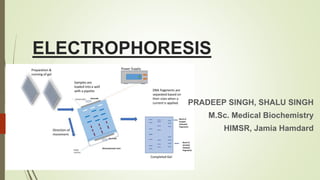
2. INTRODUCTION
Electro refers to the energy of electricity and phoresis from the Greek verb phoros ,
means to carry across.
The movement of charged particles through an electrolyte when subjected to an
electric field.
The positive charges particles (cations) move to cathode and negatively charged ones
(anions) move to anode.
3. An ampholyte , a molecule that is either positively charged or negatively
charged , takes on a positive charge (binds protons) in a solution acidic than
its pI and migrates towards cathode.
In a more alkaline solution, the ampholyte is negatively ionised (gives up
protons) and migrate towards anode.
5. Factors Affecting Electrophoresis
Net electrical charge of the molecule
Size and shape of the molecule
Electrical field strength
Properties of the supporting Medium
Temperature of operation
6. As, Felectrical= q.E
Ffrictional = v.f
q.E = v.f
The velocity, v, of a charged molecule in an electric field is given by
the equation:
v = q.E
f
7. ELECTROPHORETIC MOBILITY
Electrophoretic mobility (µ) of an ion is used, which is the ratio of the velocity
of the ion to field strength (v/E).
µ = v = q
E f
OR µ = q
6πrη
It is directy proportional to net charge and inversely proportional to molecular
size and viscosity of the electrophoretic medium.
Other factors affecting mobility include endosmotic flow and wick flow.
8. Instrumentation and reagents
Buffer Tank – to hold the buffer
Buffer
Electrodes made of either platinum or carbon
Power supply
Support Media
The entire apparatus is covered to minimize evaporation and protect the
system and is powered by a direct current supply .
9. Power supply
The current in the solution between the electrodes is conducted mainly by
the buffer ions, a small proportion being conducted by the sample ions
Ohm’s law expresses the relationship between current (I) , voltage (V) ,
Resistance
v = R
I
During electrophoresis the power (P,watts) ,generated in supporting medium
is given by
P= I2 R
Most of the power generated is dissipiated as Heat
H =I2RT
If a constant voltage is applied, the current increases during electrophoresis
owing to the decrease in resistance and the rise in current increases the heat
output still further.
10. Buffer
It is a multifunctional component in electrophoretic process as it-
Carries the applied current
Establishes the pH at which electrophoresis is performed
Determines the electrical charge on the solute
Buffer’s ionic strength influences the -
Conductance of the support
Thickness of the ionic cloud surrounding a charged molecule
Rate of migration
Sharpness of electrophoretic zones
11. With increasing buffer concentration , the ionic cloud increases in
size , and the molecule becomes hindered in its movement.
High ionic strength buffer yeilds sharper band separations, also
produce more joule heat due to increased current levels , an effect
that leads to heat labile proteins.
Ionic strength of a buffer composed of mono valent ions is equal to
its molarity
13. 1.) AGAR & AGAROSE GEL:
Agar is a mixture of polysaccharides extracted from sea weeds.
Agarose is a highly purified uncharged polysaccharide derived from agar.
Used in agarose gel electrophoresis for the separation of various substances.
Pore size in agarose gel is large enough for all proteins to pass through,
separation is based on charge-to-mass ratio of the protein.
14. APPLICATIONS:
Isolate & analyze the DNA molecules which are cut by restriction enzymes.
DNA sequencing.
Medical research, forensics.
ADVANTAGES:
Easily processed.
Sample recovery.
DISADVANTAGES:
Melting of gel due to electric current.
Resolution is less compared to polyacrylamide gels.
15. 2.)POLYACRYLAMIDE GEL:
It is thermostable, transparent, strong, relatively chemically inert,
& depending on conc. can be made in a wide range of pore sizes.
Separation is based on both charge-to-mass ratio & molecular
size(molecular sieving).
18. Filter paper such as Whatmann no1 is used.
Separation takes place in 12 to 14 hours.
ADVANTAGES:
It is economical & easy to use.
DISADVANTAGES:
Certain compounds such as proteins, hydrophilic molecules
cannot be resolved due to the adsorptive & ionogenic properties
of paper which results in tailing & distorting compounds.
Electro osmosis.
19. SDS - PAGE
Sodium Dodecyl sulphate- Polyacrylamide Gel Electrophoresis .
Most widely used method for analyzing protein
mixtures qualitatively.
Method is based on separation of proteins
according to size .
SDS is an anionic detergent.
20. Method
Samples to be run on SDS- PAGE are firstly boiled for 5 min. in sample buffer
containing β-mercaptoethanol and SDS
-Mercaptoethanol reduces any disulphide bridges present that are holding the
protein tertiary structure.
-SDS binds strongly to , and denatures the protein.
Each protein in the mixture is therefore fully denatured by this treatment and
opens up into a rod shaped structure with a series of negatively charged SDS
molecules along the polypeptide chain.
On average, one SDS molecule binds for every two amino acid residues .
21. The original native charge on the molecule is therefore completely swamped by
the negatively charged SDS molecules.
Sample buffer also contains an ionisable tracking dye, usually bromophenol blue ,
and sucrose or glycerol.
Once the samples are all loaded , a current is passed through the gel .
When the main separating gel has been poured between the glass plates and
allowed to set, a shorter stacking gel is poured on top of the separating gel and it
is into this gel that the wells are formed and the proteins loaded.
22. The purpose of this stacking gel is to concentrate the protein sample into a
sharp band before it enters the main separating gel.
This is achieved by utilising differences in ionic strength and pH between the
electrophoresis buffer and the stacking gel buffer and involves a
phenomenon known as isotachophoresis.
The stacking gel has a very large pore size (4% acrylamide), which allows the
proteins to move freely and concentrate,or stack, under the effect of the
electric field.
23. The band-sharpening effect relies on the electrophoretic mobility of ions present in
electrophoresis buffer.
Glycinate ions< Protein-SDS Complex< Cl- ions
The glycinate ions can move at the same speed as Cl only if they are in a region of
higher field strength.
Field strength is inversely proportional to conductivity, which is proportional to
concentration.
The pH of the stacking gel is 6.8, that of the separating gel is 8.8.
Typically separating gel is 15% polyacrylamide gel which is suited for separating
proteins in range of 10-100kDa.
24. Smaller the protein more easily it can pass through the pores of the gel,
whereas large proteins are successively retarded by frictional resistance due to
the sieving effect of the gels.
The bromophenol blue dye is totally unretarded and therefore indicates the
electrophoresis front.
When the dye reaches the bottom of the gel, the current is turned off.
The gel is removed from between the glass plates and shaken in an
appropriate stain solution .
washed in destain solution.
25. Observation
A pure protein should give a single band on an SDS–polyacrylamide gel,
unless the molecule is made up of two unequal subunits.
Since only submicrogram amounts of protein are needed for the gel, very
little material is used in this form of purity assessment .
Also a value for the relative molecular mass of the protein can be
determined on the same gel run with no more material being used.
26. NATIVE BUFFER GEL
Method useful in detection of a particular protein particularly an enzyme.
Polyacrylamidegels are usedbut SDS is absent.
Proteins are not denatured prior to loading .
All the proteins in the sample being analysed carry their native charge at the pH
of the gel (normally pH 8.7), proteins separate according to their different
electrophoretic mobilities and the sieving effects of the gel .
The enzyme of interest can be identified by incubating the gel in an appropriate
substrate solution such that a coloured product is produced at the site of the
enzyme.
An alternative method for enzyme detection is to include the substrate in an
agarose gel that is poured over the acrylamide gel and allowed to set.
27. Diffusion and interaction of enzyme and substrate between the two gels results in
colour formation at the site of the enzyme .
Often, duplicate samples will be run on a gel, the gel cut in half and one half
stained for activity, the other for total protein.
This way the total protein content of the sample can be analysed and the particular
band corresponding to the enzyme identified by reference to the activity stain gel.
28. GRADIENT GEL
A Polyacrylamide gel system
Instead of running a slab gel of uniform pore size throughout (e.g. a 15% gel) a
gradient gel is formed, where the acrylamide concentration varies uniformly
from, typically, 5% at the top of the gel to 25% acrylamide at the bottom of the
gel.
Procedure-The gradient is formed via a gradient mixer and run down
between the glass plates of a slab gel .
The higher percentage acrylamide (e.g. 25%) is poured between the glass
plates first and a continuous gradient of decreasing acrylamide concentration
follows.
Therefore at the top of the gel there is a large pore size (5% acrylamide) but as
the sample moves down through the gel the acrylamide concentration slowly
increases and the pore size correspondingly decreases.
29. Advantages
A much greater range of protein values can be separated than on a
fixed-percentage gel.
Proteins with very similar Mr values may be resolved, although they
cannot otherwise be resolved in fixed percentage gels.
30. IEF Gels
Ideal for the separation of amphoteric substances such as proteins .
Utilises horizontal gels on glass plates or plastic sheets.
Separation is achieved by applying a potential difference across a gel that
contains a pH gradient.
The pH gradient is formed by the introduction into the gel of compounds known
as ampholytes , which are complex mixtures of synthetic polyamino
polycarboxylic acids.
Ampholytes can be purchased in different pH ranges .
Commercially available ampholytes include Bio-Lyte and Pharmalyte.
31. Procedure
carrier ampholytes, covering a suitable pH range, and riboflavin are
mixed with the acrylamide solution.
mixture is then poured over a glass plate (typically 25 cm 10 cm),
which contains the spacer.
The second glass plate is then placed on top of the first to form the
gel cassette .
the gel polymerised by photopolymerisation by placing the gel in
front of a bright light.
The photodecomposition of the riboflavin generates a free radical,
which initiates polymerisation
32. This takes 23 h.
Once the gel has set, the glass plates are prised apart to reveal the gel stuck to one
of the glass sheets .
Electrode wicks, which are thick (3 mm) strips of wetted filter paper (the anode is
phosphoric acid, the cathode sodium hydroxide) are laid along the long length of
each side of the gel and a potential difference applied .
Under the effect of this potential difference, the ampholytes form a pH gradient
between the anode and cathode.
The power is then turned off.
samples applied by laying on the gel small squares of filter paper soaked in the
sample.
A voltage is again applied for about 30 min to allow the sample to electrophorese
off the paper and into the gel, at which time the paper squares can be removed
from the gel
33. Depending on which point on the pH gradient the sample has been loaded,
proteins that are initially at a pH region below their isoelectric point will be
positively charged and will initially migrate towards the cathode.
The surrounding pH will increase steadily as they proceed and the positive charge
ion protein will decrease correspondingly until protein arrives at a point where
the pH is equal to its pI.
Protein is now in zwitter ion form with no net charge, so further movement will
cease.
34. To achieve rapid separations (2-3 h) relatively high voltages (up to 2500 V) are
used.
As considerable heat is produced, gels are run on cooling plates (10 C) and power
packs used to stabilise the power output and thus to minimise thermal
fluctuations.
Staining-
The gel is therefore first washed with fixing solution (e.g. 10% (v/v) trichloroacetic
acid).
This precipitates the proteins in the gel and allows the much smaller ampholytes to
be washed out.
The gel is stained with Coomassie Brilliant Blue and then destained
35. 2D Gel electrophoresis
This technique combines the technique of IEF (first dimension), which separates
proteins in a mixture according to charge (pI), with the size separation technique
of SDS–PAGE (second dimension).
highly sophisticated analytical method for analysing protein mixtures.
To maximise separation, large format 2-D gels are used.
For large-format gels, the first dimension (isoelectric focussing) is carried
out in an acrylamide gel that has been cast on a plastic strip.
The gel contains ampholytes (for forming the pH gradient) together with
8M urea and a non-ionic detergent
36. The denatured proteins therefore separate in this gel according to their
isoelectric points.
The IEF strip is then incubated in a sample buffer containing SDS and then
placed between the glass plates of, and on top of, a previously prepared 10%
SDS–PAGE gel.
Electrophoresis is commenced and the SDS-bound proteins run into the gel
and separate according to size.
Using this method one can routinely resolve between 1000 and 3000 proteins
from a cell or tissue extract and in some cases workers have reported the
separation of between 5000 and 10 000 proteins.
37. Protein (Western) Blotting
The first step is to transfer or blot the pattern of separated proteins from
the gel onto a sheet of nitrocellulose paper.
Transfer of the proteins from the gel to nitrocellulose is achieved by a
technique known as electroblotting.
In this method a sandwich of gel and nitrocellulose is compressed in a
cassette and immersed, in buffer, between two parallel electrodes.
A current is passed at right angles to the gel, which causes the separated
proteins to electrophorese out of the gel and into the nitrocellulose sheet.
The nitrocellulose with its transferred protein is referred to as a blot.
This involves probing the blot, usually using an antibody to detect a specific
protein.
38.
39. Pulsed field Gel Electrophoresis
Discovered by Schwartz & Cantor that large molecules of DNA (yeast
chromosomes, 200-3000 kb) could be separated by pulsed field gel
electrophoresis (PFGE).
Electric field is not constant but changed repeatedly in direction & strength during
separation.
40.
41. APPLICATIONS:
Efficient method for estimation for genome size estimation.
Useful for fingerprinting & physical mapping of chromosome.
42. Capillary electrophoresis
It is a new technique that combines the high resolving power of electrophoresis
with the speed, versatility,& the automation of high-performance liquid
chromatography(HPLC).
Offers the ability to analyze very small samples & become widely used technique
in the analysis of amino acids, peptides, proteins, nucleic acids, & pharmaceuticals
44. Capillaries used are flexible, fused, silica tubes with a thin exterior covering of polyimide to provide strength
& flexibility.
Migration of solutes depends on charge & size ratios.
Sample volumes are loaded by hydrodynamic & electrokinetic injection.
Important feature of CE is electroosmotic flow.
Modes in which CE systems are operated include (1) Capillary zone electrophoresis, (2)Micellar
electrokinetic chromatography, (3)Capillary gel electrophoresis, (4)Capillary IEF, (5)Capillary ITP.
45.
46. APPLICATIONS:
Hemoglobin electrophoresis: abnormal hb detection & characterization.
Immunotyping.
Pharmaceutical application.
ADVANTAGES:
High resolving power & time saving.
Nanolitre sample injection.
DISADVANTAGES:
Sacrifice of biological-type of conditions.
Long equilibration time needed to obtain a reproducible surface.
47. MICROCHIP ELECTROPHORESIS
MCE results from miniaturization of CE & thus the separation process on the chip
is based on the same principle as the capillary.
FABRICATION: Microchips are constructed from substrates such as: glass,
silicon, polymeric materials.
glass chip
49. DETECTION: Made at the opposite end of the separation channel , most commonly by laser- induced
fluorescence(LIF).
50. APPLICATIONS:
For simultaneous separation of catecholamines & their cationic metabolites.
Separation method for complex sample mixtures.
ADVANTAGES:
Faster then conventional capillary electrophoresis.
Online coupling of various processes.
DISADVANTAGES:
Limited separation efficiency of zone electrophoretic measurements.
Imprecise injections.
Protein contain more ionisable amino and carboxylic group they act as ampholytes
When a potential difference (voltage) is applied across the electrodes, it generates a potential gradient, E, which is the applied voltage, V, divided by the distance, d, between the electrodes. When this potential gradient E is applied, the force on a molecule bearing a charge of q coulombs is Eq newtons. It is this force that drives a charged molecule towards an electrode. However, there is also a frictional resistance that retards the
movement of this charged molecule. This frictional force is a measure of the hydrodynamic size of the molecule, the shape of the molecule, the pore size of the medium in which electrophoresis is taking place and the viscosity of the buffer.
1)µ= electrophoretic mobility, q= net charge on the ion, r=ionic radius of the solute, η= the viscosity of the buffer in which migration is happening
2)When a potential difference is applied, therefore, molecules with different overall charges will begin to separate owing to their different electrophoretic mobilities. Even molecules with similar charges will begin to separate if they have different molecular sizes, since they will experience different frictional forces
3 electroendosmosis (also known as electroosmotic flow), which is due to the presence of charged groups on the surface of the support medium.
For this reason, workers often use a stabilised power supply, which provides constant power and thus eliminates fluctuations in heating.
- Particularly useful for monitoring protein purification .Also used to determine relative molecular mass of proteins
The rod-like structure remains, as any rotation that tends to fold up the protein chain would result in repulsion between negative charges on different parts of the protein chain, returning the conformation back to the rod shape.
Bromophen. Blue- allow electrophoretic run to be monitered
Sucrose- gives sample solution density thus allowing the sample to settle easily through the electrophoresis buffer to the bottom when injected into the loading well
negatively charged glycinate ions (in the electrophoresis buffer) have a lower electrophoretic mobility than do the protein–SDS complexes, which, in turn, have lower mobility than the chloride ions (Cl) of the loading buffer and the stacking gel buffer.
The destain solution removes unbound background dye from the gel, leaving stained proteins visible as blue bands on a clear background.
It is therefore not possible to predict the behaviour of a given protein in a buffer gel but, because of the range of different charges and sizes of proteins in a given protein mixture, good resolution is achieved.
Likewise, substances that are initially at pH regions above their isoelectric points will be negatively charged and will migrate towards the anode until they reach their isoelectric points and become stationary.
both of which denature and maintain the both of which denature and maintain the
solubility of the proteins being analysedsolubility of the proteins being analysed