Recommandé

Recommandé
Contenu connexe
Tendances
Tendances (19)
En vedette
En vedette (12)
Similaire à BMES poster 2016
Similaire à BMES poster 2016 (20)
BMES poster 2016
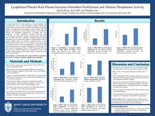
- 1. A critical-sized defect in bone represents a clinical challenge that necessitates the use of a bone regeneration strategy. Platelet-rich plasma (PRP) provides an easily accessible, patient-derived complex mixture of growth factors and cytokines. In early studies, PRP was applied as a platelet gel for bone healing. However, the mechanical integrity and degradation characteristics of platelet gels were problematic for bone regeneration. Recently, PRP has been incorporated into various gels, sponges, and electrospun scaffolds by first converting PRP into a bioactive lyophilized powder termed preparations rich in growth factors (PRGF). Including PRGF within a support scaffold allows for localized, sustained delivery of the platelet-derived factors. Previous in vitro studies using various bone cell models have shown that cell proliferation is increased by PRP treatment, but effects on osteoblast differentiation and function have been variable. Investigating effects of PRGF, rather than PRP, can provide a more controlled approach for evaluating how cells in the osteoblast lineage respond to the complex mixture of platelet-derived factors present in PRP. Study Objective: To determine the response of MG-63 osteoblast- like cells in 2-D culture to medium supplemented with PRGF. Lyophilized Platelet-Rich Plasma Increases Osteoblast Proliferation and Alkaline Phosphatase Activity Rachel Rone, Scott Sell, and Natasha Case Department of Biomedical Engineering, Parks College of Engineering, Aviation and Technology, Saint Louis University, Saint Louis, MO Introduction Materials and Methods Discussion and Conclusion Results • MG-63 human osteosarcoma cell line (ATCC® CRL-1427™) was used as an osteoblast model. • PRGF powder was created from human PRP that was isolated by standard techniques, subjected to freeze-thaw cycles to lyse platelets, and then lyophilized. • Cells were expanded and seeded (~10,000 cells/cm2 ) in high-glucose DMEM with 5% EquaFETAL bovine serum (Atlas Biologicals) and 1% penicillin/streptomycin. • Three days after seeding, cell layers were rinsed in serum-free medium and then cultured in medium containing 1% serum ± 0.5 mg/mL PRGF for 6 days, with a medium change on day 3. • For analysis, cell layers were scraped in lysis buffer (100 mM Tris- HCL, 1 mM MgCl2, 0.1% Triton X) and sonicated. • As a measure of osteoblast function , cell lysates were analyzed for alkaline phosphatase (ALP) activity based upon conversion of the substrate P-nitrophenyl phosphate. • Cell lysates were also analyzed for total protein content (BCA assay). 0 20 40 60 80 100 120 140 160 1% Serum 1% serum + PRGF Protein(mg) 0 1000 2000 3000 4000 5000 6000 7000 8000 1% Serum 1% Serum + PRGF ALPActivity (nmol/min/mgprotein) 0 20 40 60 80 100 120 140 160 ITS ITS + PRGF Protein(mg) 0 1000 2000 3000 4000 5000 6000 7000 ITS ITS + PRGF ALPActivity (nmol/min/mgprotein) 0 20 40 60 80 100 120 140 160 Day 0 Day 3 Day 6 Protein(mg) • Experiments were conducted in low-serum medium to reduce interfering effects of bovine serum on the response to PRGF. • MG-63 cells survived and proliferated in medium with 1% serum. • PRGF enhancement of MG-63 protein content was consistent with previous PRP studies in bone cells that showed increased cell proliferation with PRGF addition. • The increase in ALP activity by PRGF treatment, observed in both low-serum and ITS+-supplemented medium, indicated that one important aspect of osteoblast function was enhanced by platelet-derived factors. • Follow-up studies will evaluate PRGF effects on other aspects of osteoblast function. • Future experiments will evaluate the response of MG-63 cells seeded onto 3-D scaffolds in which PRGF is incorporated. CONCLUSION: PRGF, a lyophilized form of PRP, supports increased proliferation and alkaline phosphatase activity in MG- 63 osteoblast-like cells under low serum conditions. 0 1000 2000 3000 4000 5000 6000 1% Serum 1% Serum + PRGF ALPActivity (nmol/min/mgprotein) 0 50 100 150 200 250 1% Serum 1% Serum + PRGF Protein(mg) Figure 4. PRGF effect on ALP activity (experiment B). Significant increase with 6 days of PRGF treatment (p<0.01). Figure 5. PRGF effect on protein content (experiment B). Significant increase with PRGF (p<0.05). Acknowledgements • Technical assistance from K. Hixon, A. Dunn, and A. Martin • Funding provided by the Parks College of Engineering, Aviation and Technology Figure 6. PRGF effect on ALP activity (ITS+ experiment). 1% serum was replaced with a serum-free supplement containing insulin (ITS+). Significant increase with PRGF (p<0.001) at 6 days. Figure 7. PRGF effect on ALP activity (ITS+ experiment). The amount of protein was not different with PRGF addition to medium with ITS+. Figure 1. Establishing a low-serum culture model for testing effects of PRGF. Protein content was increased with culture time, indicating proliferation in medium with 1% serum. Figure 2. PRGF effect on ALP activity (experiment A). The addition of PRGF significantly increased (p<0.001) ALP activity in the cell layer at 6 days. Figure 3. PRGF effect on protein content (experiment A). The addition of PRGF also significantly increased (p<0.001) the amount of protein in the cell layer. * * * * *