

Recommandé
Contenu connexe
Tendances
Tendances (20)
Similaire à Paramyxoviruses
Similaire à Paramyxoviruses (20)
Plus de Dr. Rakesh Prasad Sah
Plus de Dr. Rakesh Prasad Sah (20)
Dernier
Dernier (20)
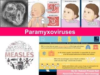
Paramyxoviruses
- 1. Paramyxoviruses By Dr. Rakesh Prasad Sah Associate Professor, Microbiology
- 2. Myxo = affinitiy to mucin Orthomyxo viruses Paramyxo viruses Myxoviruses •Smaller •Segmented RNA genome •Liable to Antigenic variation •Larger •Single piece of RNA genome •Not liable to Antigenic variation
- 3. Myxo = affinitiy to mucin Orthomyxo viruses Paramyxo viruses Myxoviruses •Smaller •Segmented RNA genome •Liable to Antigenic variation •Larger •Single piece of RNA genome •Not liable to Antigenic variation
- 4. Introduction • Paramyxoviruses resembles Orthomyxoviruses in morphology bt differ from other properties S.No. Property Orthomyxovirus Paramyxovirus 1 Size of virion 80-120nm 100-130nm 2 Shape Spherical; filamentous Pleomorphic 3 Genome Segmented; eight pieces* of negative sense single stranded RNA Single piece of negative sense, single stranded RNA 4 Nucleocapsid (diameter) 9nm 18nm 5 Site of ribonucleoprotein synthesis Nucleus Cytoplasm 6 Genetic recombination Common Absent 7 DNA-dependent RNA synthesis Necessary for multiplication Not required 8 Rate of Agenic change High Low 9 Haemolysin Absent Present *Influenza virus type C contains seven pieces of RNA
- 5. • Parainfluenza virus • Mumps virus • Measles virus • Respiratory syncytial virus (RSV) Important for human infections Parainfluenza & Mump Both have HA & NA Measles Has HA but no NA RSV & Metapneumovirus Do not have any of two
- 6. Morphology • Spherical enveloped particles • 100-300nm • Envelop lipoprotein & covered by projections • Projections HN (Haemagglutinin, neuraminidase) and F (fusion protein) 12-14nm long and 2-4nm wide. • Inner surface of envelope is lined by matrix (M) protein. • Nucleocapsid helical symmetry -veSSRNA and RNA-dependent RNA polymerase.
- 8. Parainfluenza Viruses • Causes respiratory infections in children and less often in adults • Five serotypes 1,2,3,4a and 4b. • Spherical 125-250nm, enveloped RNA viruses.
- 9. Pathogenesis Acquired by droplets and contact with respiratory secretions Incubation period 2-6days Are ubiquitous and produce URTI In infants and young children, cause severe respiratory disease laryngotracheobronchitis or croup (an infection of the upper airway, which obstructs breathing and causes a characteristic barking cough). Fever Cough Respiratory obstruction swelling of larynx and related structures Pneumonia or bronchiolitis occur especially with type 3. Type 4 only mild illness
- 11. Laboratory Diagnosis • Direct demonstration – Immunofluorescence: Viral Ags demonstrated in exfoliated cells aspirated from respiratory tract by immunofluorescence using monoclonal Abs. – ELISA
- 12. Laboratory Diagnosis • Isolation – Mouth washing samples posterior pharynx or nasopharynx and throat swabs inoculated in primary human or monkey kidney cells or in continuous cell lines such as H292. – Visible cytopathic effect is minimal except with type 2 induces syncytia formation. – By haemadsorption or guinea pig red cells or by use of specific immunofluorescent Ab. • Serology – Rising Ab titre by neutralisation, ELISA and CFT. • PCR
- 13. Mumps Virus Pathogenesis Mumps or parotitis ds of childhood Acquired from direct contact with infected saliva or aerosols from infected patients Mode of entry Respiratory tract I.P. 16-18 days Enters bloodstream and spreads to the salivary glands, testes, ovaries, pancreas, kidney and brain. Virus multiplies URT and local lymph nodes Shed in saliva six days before to one week after onset of clinical parotitis. Non-suppurative inflammation of the parotid glands (95%)
- 14. • Complications – Meningitis – Meningoencephalitis – Orchitis (common complication in – postpubertal male patients) – Oophoritis – Pancreatitis – Nephritis
- 15. Laboratory Diagnosis • Direct demonstration – Immunofluorescence secretions of throat and saliva demonstration of viruses. • Isolation – Virus isolated from • Saliva • Throat swab • CSF • Urine • Serology • IgM Ab • ELISA • PCR •Primary monkey kidney cells •H292 •Hep-2 cell cultures. Little CPE but can be demonstrated by haemadsorption (guinea pig red cells) By Immunofluorescence testing of infected cell cultures.
- 16. Prophylaxis • Live attenuated vaccine • Derived by passage in chick fibroblasts • Jeryl-Lynn strain of mump virus used for manufacturing of vaccine. • MOD subcutaneously in combination with attenuated measles and rubella vaccine (MMR vaccine). • Given to children aged 12-15 months. • Provides effective protection for at least 10years. • Contraindication – Pregnancy – Immunodeficiency – Hypersensitivity to egg protein.
- 17. Measles Virus • Highly infectious ds of childhood. • Spread by respiratory secretions. • Caused by measles virus. • Morphology – Resembles paramyxoviruses – Spherical 120-250nm – Helical nucleocapsid surrounded by lipoprotein envelop – Haemagglutinin (H) spikes but neuraminidase spikes absent. – Envelop has the F protein. – Matrix M protein located below lipoprotein envelope.
- 18. Pathogenesis Acquired by inhalation I.P. 10-12 days Viruses multiplies in lymhoid tissue of RT and invades bloodstream Primary viraemia Viruses spreads to R.E. system through blood. Virus enters to epithelial surfaces skin, mouth, RT, conjunctiva Multiplies there secondary viremia Koplik’s spots (on 12th day of infection) seen on buccal mucosa and are pathognomonic of measles. Characterised by high fever (on 10th day of infection), cough and conjunctivitis.
- 19. Pathogenesis With decline of acute symptoms in 1-2 days wide spread of maculopapular rash (on 14th day of infection) appears first on neck and then spreads to rest of body. Rash fades in a week and patient recovers by 10-14th days.
- 20. Pathogenesis Complications – Due to decrease in resistance of Respiratory epithelium • Secondary bacterial infections • Otitis media • Bronchopneumonia • Croup (an infection of the upper airway, which obstructs breathing and causes a characteristic barking cough) – Giant cell pneumonia in impaired CMI – Post-measles encephalitis and subacute slerosing panencephalitis (SSPE) a progressive neurological disorder of children and young adults that affects the central nervous system (CNS).
- 22. Lab Diagnosis • Specimens – Nasopharyngeal swab – Throat washings – Conjunctival swab – Blood and urine • Direct demonstration – Multinucleate Gaint cells Geimsa stained nasal secretions. Dacron, Rayon or polyester swab
- 23. Lab Diagnosis • Ag detection – Detected in exfoliated respiratory cells in nasal secretions by immunofluorescence. • Virus isolation – Cultured on primary human or monkey kidney cells CPE takes 7-10 days to develop multinucleated gaint cells (CPE) containing intranuclear and intracytoplasmic inclusion bodies suggestive of +ce of measles virus. • Serology – IgM Ab – ELISA – High titre of IgG Ab in CSF suggestive of SSPE. • PCR
- 24. Prophylaxis • Active immunisation – Live attenuated vaccine is used. – Schwartz strain, Moraten strain and Edmonston- Zagreb strain. – Prepared in chick embryo cell line. – Available in lyophilised form. – Reconstituted with D.W. and should be used within 4hrs. – Vaccine must be stored at -200C and is thermolabile. – Dose 0.5ml. – Route Subcutaneous route. – Is used in combination with Rubella (MR vaccine) with mumps and rubella (MMR vaccine) and with varicella (MMR-V vaccine).
- 25. • Indications – Under National Immunisation Programme of India, 0.5ml of MR (measles, rubella) administered subcutaneously at right upper arm at age of 9-12months along with vitamin A supplement. – 2nd dose 16-24 months. • Passive Immunisation – Measles immunoglobulin (IgG) 3 days to susceptible contacts for protection against measles. – Dose 0.25mg/kg body weight – Post exposure prophylaxis (PEP)
- 26. Respiratory Syncytial Virus • Pleomorphic • 150-300nm • Envelope lacks both HA & NA • Contains a surface glycoprotein G virus attaches to cell surfaces & • F (fusion) protein induces fusion of infected cells into large multinucleated syncytia so named RSV…. • Nucleocapsid 13nm diameter • Impt cause of bronchiolitis and pneumonitis in infants 6 months of age. • Infection in older children and adults rhinitis or common cold. • Is highly labile and inactivated at RT. • Lyophilisation for preservation. • Ag-enically stable.
- 27. Pathogenesis Highly contagious, transmitted by contact with contaminated hands and surface. Nosocomial infections nurseries and paediatric ward I.P. 4-6 days. Virus multiplies mucous membranes of nose and throat Spread into lower respiratory tract causing bronchiolitis and pneumonitis Viruses shed in respiratory secretions for several days or weeks.
- 30. Laboratory Diagnosis • Direct demonstration – Immunofluorescence in nasopharyngeal aspirates. – Viral Ag by ELISA • Virus isolation – HeLa, Hep-2 or monkey cell cultures – Characteristic giant cells and syncytia formation. – In 2-10 days. – Definitive identification by immunofluorescence. • Serology – ELISA for Ab • PCR