
Recommended
More Related Content
What's hot
What's hot (20)
Similar to Whole Genomic DNA extraction lecture
Similar to Whole Genomic DNA extraction lecture (20)
Recently uploaded
Recently uploaded (20)
Whole Genomic DNA extraction lecture
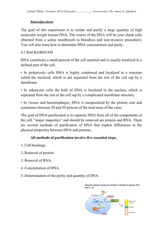
- 1. Ameer S. AlfatlawiDr.:.. Assist lecture……………Genomic DNA ExtractionLecture Three: Introduction: The goal of this experiment is to isolate and purify a large quantity of high molecular weight human DNA. The source of the DNA will be your cheek cells obtained from a saline mouthwash (a bloodless and non-invasive procedure). You will also learn how to determine DNA concentration and purity. 4.1 BACKGROUND DNA constitutes a small percent of the cell material and is usually localized in a defined part of the cell. • In prokaryotic cells DNA is highly condensed and localized in a structure called the nucleoid, which is not separated from the rest of the cell sap by a membrane. • In eukaryotic cells the bulk of DNA is localized in the nucleus, which is separated from the rest of the cell sap by a complicated membrane structure. • In viruses and bacteriophages, DNA is encapsulated by the protein coat and constitutes between 30 and 50 percent of the total mass of the virus. The goal of DNA purification is to separate DNA from all of the components of the cell. “major impurities” and should be removed are protein and RNA. There are several methods of purification of DNA that exploit differences in the physical properties between DNA and proteins. All methods of purification involve five essential steps. 1. Cell breakage. 2. Removal of protein. 3. Removal of RNA. 4. Concentration of DNA. 5. Determination of the purity and quantity of DNA.
- 2. Ameer S. AlfatlawiDr.:.. Assist lecture……………Genomic DNA ExtractionLecture Three: 4.1 CELL BREAKAGE • Cell breakage is one of the most important steps in the purification of DNA. • The usual means of cell opening, such as sonication, grinding, blending, or high pressure, cannot be used in DNA purification. These procedures apply strong forces to open cells that shear DNA into small fragments. • The best procedure for opening cells and obtaining intact DNA is through application of chemical (detergents) and/or enzymatic procedures. Advantage of used Detergents 1. It can solubilize lipids in cell membranes resulting in gentle cell lysis. 2. In addition, detergents have an inhibitory effect on all cellular DNases and can denature proteins The lysis of animal cells is usually performed using anionic detergents such as SDS (sodium deodecyl sulfate) or Sarcosyl (sodium deodecyl sarcosinate). 4.2 REMOVAL OF PROTEIN The second step in purification involves removing a major contaminant, namely protein, from the cell lysate. This procedure is called deproteinization. Removal of proteins from the DNA solution depends on differences in the physical properties between nucleic acids and proteins. 4.3.1 DEPROTEINIZATION USING ORGANIC SOLVENTS • The most frequently used methods for removing proteins explore the solubility differences between proteins and nucleic acids in organic solvents. • Nucleic acids are predominantly hydrophilic molecules and are easily soluble in water. Proteins, on the other hand, contain many hydrophobic residues making them partially soluble in organic solvents. • The organic solvents commonly used are phenol and/or chloroform containing 1 percent isoamyl alcohol.
- 3. Ameer S. AlfatlawiDr.:.. Assist lecture……………Genomic DNA ExtractionLecture Three: Phenol-Chloroform Method: 1. adding an equal volume of phenol-chloroform to an aqueous solution of lysed cells or homogenized tissue, 2. mixing the two phases, and allowing the phases to separate by centrifugation (Figure 4.2). 3. Centrifugation of the mixture yields two phases: the lower organic phase and the upper aqueous phase. 4. Nucleic acids are polar because of their negatively charged phosphate backbone, and therefore nucleic acids are soluble in the upper aqueous phase instead of the lower organic phase (water is more polar than phenol). 5. Conversely, proteins contain hydrophobic regions. 6. In the presence of phenol, the hydrophobic cores interact with phenol, causing the protein to swell and eventually to unfold or denature. 7. Chloroform denaturates proteins and lipids and makes DNA less soluble in the organic/phenolic phase 8. As a result, causing precipitation of proteins and polymers (including carbohydrates) to collect at the interface between the two phases. Figure 4.2 organic solvent deproteinization procedure.
- 4. Ameer S. AlfatlawiDr.:.. Assist lecture……………Genomic DNA ExtractionLecture Three: Limitations using phenol-chloroform: • it requires repeated time-consuming extractions when large amounts of protein are present. • Phenol-chloroform extractions, require vigorous mixing that contributes to shearing of large DNA molecules.
- 5. Ameer S. AlfatlawiDr.:.. Assist lecture……………Genomic DNA ExtractionLecture Three: 4.3.1 DEPROTEINIZATION USING ENZYMES A. Proteins can be removed from DNA preparations using a protease that can digest all proteins, i.e. a general-purpose protease. B. Two such enzymes are in use, proteinase K and pronase. C. These proteases are active in the presence of low concentrations of anionic detergent, high concentrations of salts, and EDTA and exhibit broad pH (6.0– 10.0) and temperature (50–67°C) optima. D. They can digest intact (globular) and denatured (polypeptide chain) proteins and do not require any co-factors for their activities. 4.1 REMOVAL of RNA The removal of RNA from DNA preparations is usually carried out using an enzymatic procedure. Two ribonucleases that can be used, namely ribonuclease A and ribonuclease T1. 4.2 CONCENTRATING THE DNA Precipitating with alcohol is usually performed for concentration of DNA from the aqueous phase A. Two alcohols are used for DNA precipitation: ethanol and isopropanol. B. Polar water molecules surround the DNA molecules in aqueous solutions. This interaction promotes the solubility of DNA in water. C. Ethanol is completely miscible with water. Ethanol molecules cannot interact with the polar groups of nucleic acids as strongly as water, making ethanol a very poor solvent for nucleic acids. D. Replacement of 95 percent of the water molecules in a DNA solution will cause the DNA to precipitate. E. To precipitate DNA at a lower ethanol concentration, the activity of water molecules must be decreased. This can be accomplished by the addition of salts to DNA solutions.
- 6. Ameer S. AlfatlawiDr.:.. Assist lecture……………Genomic DNA ExtractionLecture Three: DNA precipitation is customarily carried out with 70 percent ethanol (final concentration) in the presence of the appropriate concentration of sodium or ammonium salts. Advantages and disadvantages using each of these salts 1. convenience and low cost, 2. The use of sodium chloride is recommended if a high concentration of SDS has been used for lysing the cells. SDS remains soluble in ethanol in the presence of 0.2 M NaCl. 3. The disadvantage of sodium chloride is its limited solubility in 70 percent ethanol making it difficult to completely remove from the DNA samples. 4.1DETERMINATION OF THE PURITY AND QUANTITY OF DNA 4.6.1 QUANTITY OF DNA (CONCENTRATION) The most common technique to determine DNA yield and purity is measurement of absorbance. 1. Absorbance readings are performed at 260nm (A260) where DNA absorbs light most strongly, and the number generated allows one to estimate the concentration of the solution. 2. DNA concentration is estimated by measuring the absorbance at 260nm multiplying by the dilution factor, and using the relationship that an A260 of 1.0 = 50µg/ml pure dsDNA Concentration (µg/ml) = (A260 reading – A320 reading) × dilution factor × 50µg/ml Total yield is obtained by multiplying the DNA concentration by the final total purified sample volume. DNA yield (µg) = DNA concentration × total sample volume (ml) 4.6.2 PURITY OF DNA The most common purity calculation is the ratio of the absorbance at 260nm divided by the reading at 280nm (Proteins absorb maximally at 280 nm.) Good- quality DNA will have an A260/A280 ratio of 1.7–2.0. A reading of 1.6 does not
- 7. Ameer S. AlfatlawiDr.:.. Assist lecture……………Genomic DNA ExtractionLecture Three: render the DNA unsuitable for any application, but lower ratios indicate more contaminants are present. DNA purity (A260/A280) = (A260 reading – A320 reading) ÷ (A280 reading – A320 reading) * 1 ng/μl =1 μg/ml * For more accurate readings of the nucleic acid sample of interest, dilute the sample to give readings between 0.1 and 1.0. * A260 = 1, give concentration of nucleic acids as fallowing (Table 4.1): Table 4.1: Concentration per A260 Unit Nucleic Acid Concentration (μg/ml) per A260 Unit ds DNA 50 ss DNA 33 ss RNA 40 Table 4.2: A260/A280 ratio for given molecules Purity of Target A260/A280 Ratio DNA 1.8 RNA 2.0 Protein 0.6 Example of Calculation 4.1 A sample of dsDNA was diluted 60X. The diluted sample gave a reading of 0.65 on a spectrophotometer at OD260. To determine the concentration of DNA in the original sample, perform the following calculation: Concentration (µg/ml) = (A260 reading – A320 reading) × dilution factor × 50µg/ml dsDNA concentration = OD260 × dilution factor × 50 μg/mL dsDNA concentration = 0.65 × 60 × 50 μg/mL dsDNA concentration = 1.95 mg/mL